HISTOPATHOLOGY OF THE ADRENAL CORTEX
NORMAL HISTOLOGY
The adult adrenal cortex includes the outer 80% of the adrenal glands and a cuff of cortex surrounding the central vein. Three
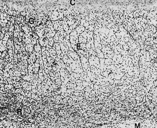
functional zones are present: glomerulosa, fasciculata, and reticularis (Fig. 71-5). The zona glomerulosa is responsible for aldosterone secretion and constitutes about 5% of the cortex. It is a discontinuous layer of nests of cells beneath the adrenal capsule. These cells are small, with a dense nucleus, a high nuclear/cytoplasmic ratio, and relatively low cytoplasmic lipid content. On electron microscopic examination, there are elongate mitochondria with abundant tubular cristae, a small amount of smooth endoplasmic reticulum (SER), and few lysosomes and microvilli.
functional zones are present: glomerulosa, fasciculata, and reticularis (Fig. 71-5). The zona glomerulosa is responsible for aldosterone secretion and constitutes about 5% of the cortex. It is a discontinuous layer of nests of cells beneath the adrenal capsule. These cells are small, with a dense nucleus, a high nuclear/cytoplasmic ratio, and relatively low cytoplasmic lipid content. On electron microscopic examination, there are elongate mitochondria with abundant tubular cristae, a small amount of smooth endoplasmic reticulum (SER), and few lysosomes and microvilli.
Functionally, the zonae fasciculata and reticularis form a unit, with both cell types having the capacity to secrete cortisol and androgens. Histologically, however, they are distinct. The zona fasciculata makes up about 70% of the adrenal cortex and consists of columns of cells extending from the inner reticularis zone to the glomerulosa (or to the capsule where the glomerulosa is absent). The cells are large, with a low nuclear/cytoplasmic ratio and abundant cytoplasmic lipid. During fixation, the lipid is removed, giving the cells a vacuolated appearance and hence the name clear cells. On electron microscopic examination, the cells form a continuum: from the outer zone, in which the cells have ovoid mitochondria with few internal vesicles, little SER, and few lysosomes, to the inner zone, in which the cells contain spherical mitochondria with numerous internal vesicles, more SER, and more lysosomes.
The zona reticularis comprises the inner 25% of the adrenal cortex. It consists of anastomosing columns of cells that vary widely in size and contain densely granular cytoplasm and sparse lipid (hence, the name compact cells). The nuclear/cytoplasmic ratio is intermediate to that between cells of the glomerulosa and cells of the fasciculata. Electron microscopic examination reveals ovoid to elongate mitochondria with tubular to vesicular cristae, abundant SER, and numerous lysosomes. In older persons, an increasing amount of lipofuscin may be seen, which imparts a darker color to these cells.
The reason for functional zonation of the adrenal cortex has been a matter of considerable debate. Embryologic evidence suggests that all cortical cells arise from the same precursor. Although cells from each of the three zones initially have distinct steroid secretory patterns in vitro, long-term cultures demonstrate that the histologic and functional distinctions between these cells disappear.2 The blood supply to the adrenal glands flows inwardly from the capsule. This creates a gradient of increasing steroid hormone concentration from the outer to the inner cortex. Intraadrenal steroid concentrations may modulate the relative activities of the steroidogenic enzymes.7,8 and 9 Such hormone gradients, which likely increase with increasing adrenal size, may explain not only the functional zonation of the adrenal cortex but also the development of the reticularis zone during adrenarche.
Electron microscopy has been useful in differentiating among cortical cell types, demonstrating transitional cells between the three zones of the adrenal cortex. It has not been useful, however, in elucidating pathologic changes in the adrenal cortex or in making the difficult distinction between benign and malignant neoplasms.
PATHOLOGY
ADRENAL NODULES: THE ADRENAL RESPONSE TO AGING
With advancing age, the adrenal glands of most persons begin to show microscopic nodular changes within the cortex. These changes begin as nests of clear cells located peripherally in the cortex or in cortical tissue surrounding the central vein. Larger nodules compress the adjacent cortex and eventually distort the adrenal capsule. They may contain foci of compact cells and areas of fibrosis, hemorrhage, and cyst formation (Fig. 71-6). These “yellow nodules” have been recognized in 3% of normotensive research subjects and may reach 2 to 3 cm in diameter. Their incidence appears to increase not only with age but also with the presence of vascular damage, as is seen with hypertension and diabetes. Pigmented (“black”) nodules seen at autopsy appear to represent a variant of yellow nodules and contain compact (zona reticularis–like) cells with increased amounts of lipofuscin.
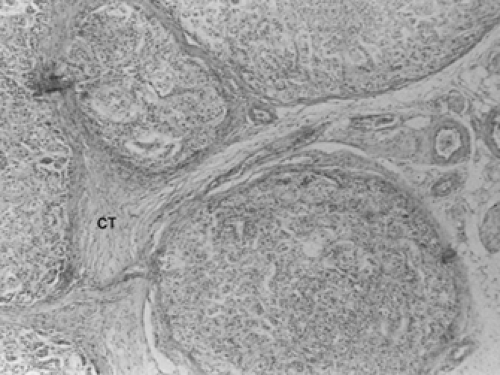 FIGURE 71-6. Adrenal nodules. Multiple nodules are shown, separated by fibrovascular connective tissue (CT). Normal cortical architecture is totally disrupted. (Courtesy of Dr. Stephen Boudreau.) |
These nodules are not neoplasms and, although they produce steroids in vitro, are not associated with adrenocortical hyperfunction. The unaffected cortex remains normal in appearance. Their main significance lies in their incidental detection on radiologic imaging. In the absence of clinical or biochemical evidence of hormonal hypersecretion, small asymptomatic adrenal nodules seen incidentally during abdominal imaging procedures are unlikely to represent clinically significant pathologic entities.10
ADRENOCORTICAL HYPERFUNCTION
Response to Stress.
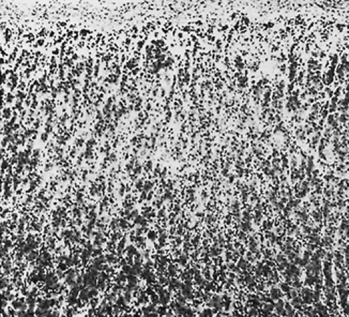
Adrenal glands examined at autopsies after prolonged illnesses have mean weights of about 6 g and may weigh as much as 9 g. This appears to be the result of prolonged corticotropin (ACTH) stimulation. Histologically, within several hours of ACTH stimulation, clear (fasciculata) cells at the fasciculata-reticularis junction begin to lose their lipid content and take on the light-microscopic and ultrastructural appearance of compact (reticularis) cells (Fig. 71-7). Eventually, this “lipid depletion” may involve the entire fasciculata, and compact cells may extend from the medulla to the glomerulosa or adrenal capsule.
Corticotropin-Dependent Cushing Syndrome.
Corticotropin-dependent Cushing syndrome includes both pituitary hypersecretion of ACTH (Cushing disease) and paraneoplastic (ectopic) ACTH secretion from a benign or malignant neoplasm. The pathologic changes in the adrenal glands form a continuum from the mild stress-induced changes to the extreme changes resulting from severe, prolonged ACTH hypersecretion commonly seen with the paraneoplastic ACTH syndrome. Cushing disease accounts for about 60% of endogenous (noniatrogenic) Cushing syndrome in adults and ˜35% in children (see Chap. 14, Chap. 75 and Chap. 83). The ACTH hypersecretion is generally mild but often prolonged. The adrenal glands are usually enlarged, each weighing 6 to 12 g. The cortex is widened, with a broadened zone of compact cells, suggesting a hyperplastic zona reticularis. Some of the reticularis may be replaced by lipid. The clear cells often are larger than normal, with increased lipid content. The zona glomerulosa is normal. Cortical micronodules, consisting of clusters of clear cells, also may be seen in the periphery or around the central vein (Fig. 71-8). The only difference between these micronodules and those seen with aging is that the remainder of the cortex is hyperplastic.
Stay updated, free articles. Join our Telegram channel

Full access? Get Clinical Tree


