Histiocytic Diseases


Histiocytic Diseases


Kenneth L. McClain

Carl E. Allen

John M. Hicks


INTRODUCTION

Histiocytic disorders are a group of diseases that are characterized by abnormal differentiation or function of “histiocytes.” Although histiocyte is an archaic term referring to tissue macrophages, the histiocytoses include diseases arising from all cells of the mononuclear phagocytic system, with diseases characterized by presumed lineage and biology into dendritic cell (DC) disorders, macrophage-related disorders, and malignant histiocytic disorders1 (Table 25.1). The specific disorders are further organized according to the phenotype of the pathologic cells, which may reflect cell of origin or terminal differentiation similar to physiologic myelomonocytic cells (Table 25.2).


LANGERHANS CELL HISTIOCYTOSIS


Langerhans Cell Histiocytosis Definition

Langerhans cell histiocytosis (LCH) is characterized by inflammatory lesions with infiltrating CD1a+/CD207+ pathologic DCs (Fig. 25.1). The clinical manifestations are highly variable, from single lesions with minimal impact, to life-threatening multisystem disease. Biopsies from patients with low-risk and high-risk LCH are generally indistinguishable.


Epidemiology

The incidence of LCH has been estimated to be 2 to 10 cases per million children under the age of 15 years.2 The male-to-female ratio is close to 1, with the median age at presentation being 30 months, although patients may present from birth through the ninth decade. A few identical and fraternal twins with early onset of LCH have been reported, which could be due to sharing common fetal circulation or shared genetics. There are rare case reports of nontwin siblings or multiple cases in one family. However, evidence of inherited single-gene defects in LCH is lacking. Solvent exposure in parents and perinatal infections have weak associations with LCH, and an increased frequency of family members with thyroid disease has been reported.3


TABLE 25.1 Classification of Histiocytic Disorders


Dendritic Cell Related


Langerhans cell histiocytosis


Juvenile xanthogranuloma/Erdheim-Chester disease


Macrophage related


Hemophagocytic syndromes


Primary hemophagocytic lymphohistiocytosis


Secondary hemophagocytic syndromes


Rosai-Dorfman disease


Malignant diseases


Monocyte-related leukemias


Extramedullary monocytic tumor (myeloid sarcoma)


Macrophage-related histiocytic sarcoma


Dendritic cell malignancy (malignant histiocytosis)


Historical Perspective

LCH is an enigmatic disorder with complicated history, although recent discoveries are beginning to clarify mechanisms of pathogenesis and clinical manifestations of the disease. The first reports of LCH appeared in the medical literature in the 1900s, describing children with combinations of lytic bone lesions, skin and mucosal lesions, and diabetes insipidus (DI), which was known as Hand-Christian-Schüller disease. More clinically severe presentations of children with histiocytic infiltration of bone marrow, spleen, liver, and lung were identified as Letterer-Siwe disease.4 In 1953, Dr. Lichtenstein reported the observation of common histology of many of the eponymous classifications of what we now recognize as LCH (Fig. 25.1), and proposed the hypothesis that they represent a common disorder, “histiocytosis X,” with “X” pending discovery of the cell of origin. Subsequently, Nezelof and colleagues observed the pentalaminar Birbeck granules that characterized epidermal Langerhans cells (LCs) in the pathologic cells of “histiocytosis X” lesions.5 After this report, “histiocytosis X” was rebranded Langerhans cell histiocytosis.


Etiology and Pathogenesis

Since the discovery of Birbeck granules in LCH lesions, debate over models of LCH pathogenesis for the next decades focused on functional dysregulation versus malignant transformation of epidermal LCs.4


Immune Dysregulation

LCH lesions include a median of 8% infiltrating pathologic DCs, but may be as low as 0.1%. The LCH DCs express high levels of T-cell costimulatory molecules and proinflammatory cytokines. The inflammatory infiltrate may include macrophages, eosinophils, and lymphocytes enriched for regulatory T cells.6,7 Dozens of cytokines, chemokines, and cytokine/chemokine receptors that constitute the “cytokine storm” in LCH lesions have been hypothesized to play roles in LCH pathogenesis. However, the mechanisms through which pathologic DCs in LCH orchestrate inflammation remain to be defined.


Neoplasia

Studies showing clonality in LCH were published in 1994 using polymorphisms of methylation-specific restriction enzyme sites on the X-chromosome regions coding for the human androgen receptor assay (HUMARA), as well as polymorphism for three other loci.8 Biopsies of lesions from both single system and multisystem disease were found to have a proliferation of LCs from a single clone. Despite clonality, pathologic DCs do not appear dysplastic, mitoses are rarely observed,7 and chromosomes are typically intact.9


TABLE 25.2 Immunophenotypes of Histiocytic Disorders


Disease


LCH


Malignant Histiocytosis


ECD/JXG


HLH


RDD


Phenotypic similarity


LC


IDC/FDC


DD/IDC


M/M


M/M


HLA-DR


++


+


+


+


CD1a


++


CD14


+/-


++


++


++


CD68


+/-


+/-


++


++


++


CD163


+


++


++


CD 207 (Langerin)


+++


+


Factor XIIIa


++


Fascin


++


++


+/-


+


Birbeck granules


+


Hemophagocytosis


+/-


+/-


+/-


Emperiopolesis


+


LCH, Langerhans cell histiocytosis; ECD, Erdheim-Chester disease; JXG, juvenile xanthogranuloma; HLH, hemophagocytic lymphohistiocytosis; RDD, Rosai-Dorfman disease; LC, Langerhans cell; IDC, interstitial dendritic cell; DD, dermal dendritic cell; M/M, monocyte/macrophage; FDC, follicular dendritic cell.


Adapted from Jaffe R. The diagnostic histopathology of Langerhans cell histiocytosis. In: Weitzman S, Egeler RM, eds. Histiocytic disorders of children and adults. Basic science, clinical features, and therapy. Cambridge University Press, 2005:14-39.


A major discovery in 2010 described recurrent somatic mutations in the BRAF gene in 57% of LCH lesions.10 The BRAF-V600E mutation leads to constitutive activation of the downstream kinases, MEK and ERK, and is observed in a wide spectrum of human cancers (e.g., melanoma, thyroid carcinoma, and hairy cell leukemia), as well as many benign neoplasias (e.g., nevi, colon polyps).11 In an institutional series, BRAF-V600E was associated with a twofold increase in risk of recurrence, but otherwise no clinical differences have been associated with the mutation, including age, survival, or clinical risk status.12 The presence of circulating cells carrying the BRAF-V600E mutation was associated with high-risk clinical status (liver, bone marrow, spleen involvement) and localized to myelomonocytic lineage (CD11c+ and CD14+ fractions). Hematopoietic cell progenitors with the BRAF-V600E mutation were also identified in patients with high-risk LCH. Therefore, a proposed model of LCH pathogenesis is one in which high-risk LCH arises from pathologic activation of ERK in hematopoietic progenitor cells, and low-risk disease arises from ERK activation in tissue-restricted precursors. This model is supported by the ability of enforced expression of BRAF-V600E in early myeloid (CD11c+) cells to drive a LCH-like phenotype in mice.12 The presence of BRAF-V600E in hematopoietic progenitor cells in bone marrow and myelomonocytic precursors in circulating blood in patients with high-risk LCH is also consistent with the classification of LCH as a myeloid neoplasia, and suggests that “histiocytosis X” may in fact be a more appropriate nomenclature than “Langerhans cell” histiocytosis.


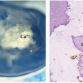
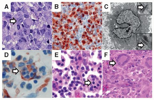
Figure 25.1 Histologic features of histiocytic disorders. A: H&E staining of a typical LCH lesion with characteristic histiocyte with reniform nucleus (arrow) and eosinophilic cytoplasm along with background inflammatory infiltrate (original magnification 400×). B: Langerin (CD207) immunostaining of pathologic dendritic cells within inflammatory lesions is diagnostic for LCH (original magnification 400×). C: Birbeck granules are identified by electron microscopy (arrows) in LCH lesions, as well as in normal epidermal Langerhans cells. This electron micrograph is typical for an LCH lesion with a Langerhans cell histiocyte with abundant cytoplasm and reniform nucleus (original magnification 7,000×; insets 20,000×). D: H&E staining of bone marrow aspirate demonstrates hemophagocytosis (arrow) in a patient with HLH. E: H&E staining of a typical lymph node biopsy from a patient with Rosai-Dorfman disease. The arrow highlights emperipolesis, where viable lymphocytes traffic through histiocytes (original magnification 400×). F: H&E staining of soft tissue juvenile xanthogranuloma with characteristic Touton giant cells (arrow) with nuclei arranged in a wreath-like pattern. Histiocytic cells are seen in the background (original magnification 400×).


Clinical Features

The most frequent presenting signs and symptoms of LCH include skin rash or painful bone lesions. Patients may also have fever, weight loss, diarrhea, edema, dyspnea, painless lymphadenopathy, polydipsia, or polyuria.

LCH is categorized as high-risk or low-risk based on specific organ involvement. High-risk organs include liver, spleen, and bone marrow. Low-risk organs include all other tissues; most common sites include skin, bone, lymph nodes, or the pituitary gland. Patients may present with disease at a single site or at multiple sites. Treatment recommendations for patients with LCH are based on high-risk or low-risk organ involvement and whether the presentation is as a single site or as a multisystem disease.


Pathologic Features

The typical LCH lesion comprises a mixture of characteristic pathologic DCs (histiocytes), T-cell lymphocytes, eosinophils, and macrophages (Fig. 25.1A). The hallmark LCH DC has an abundant eosinophilic-to-amphophilic cytoplasm and a nucleus that is deeply indented or grooved. The presence of eosinophils is quite variable. Occasional giant cells, likely composed of fused DCs, may also be seen. LCH can be distinguished from other DC disorders by histology with cytoplasmic and cell surface markers (Table 25.1). CD1a is physiologically expressed by epidermal LCs,
cortical thymocytes, and interdigitating DCs within the dermis and lymph nodes. LCH lesion DCs demonstrate a membranous-to-cytoplasmic pattern with CD1a. LCH DCs may also express langerin (CD207), a type II transmembrane protein. Langerin is normally located on the cell surface and is associated with the formation of characteristic intracytoplasmic Birbeck granules (identified by electron microscopy) thought to be involved in antigen processing. Langerin/Birbeck granules were initially considered specific to epidermal LCs, but CD207 has more recently been identified in other DC lineages.13 Expression of langerin is likely a later event in LCH DC differentiation as many studies describe variable expression of CD207+ among CD1a+ and BRAF-V600E+ histiocytes within LCH lesions (reviewed in Ref. 14).


Specific Organ Involvement


Skin

Seborrheic scalp rash is a common presentation of LCH, which may be confused with cradle cap in infants or severe dandruff in older children and adults. Red papular lesions resembling candida may also develop in the scalp, groin, abdomen, back, or chest (Fig. 25.2A). Ulcerative lesions behind the ears or involving the scalp, axillae, genitalia, or perianal region are often misdiagnosed as bacterial or fungal infections. Patients may also present with brown-to-purplish papules over any part of their body. In some patients, LCH skin lesions may disappear during the first year of life with no therapy, while in others skin lesions may be the most clinically evident manifestation of disseminated disease.15 In a report of 61 neonatal LCH cases from 1,069 patients in the Histiocyte Society database, almost 60% of neonates had multisystem disease and 72% had risk organ involvement.16


Oral Mucosa

Presenting symptoms of oral LCH include gingival hypertrophy and ulcers of the soft or hard palate, buccal mucosa, or on the tongue and lips. Early eruption of teeth occurs with LCH infiltration of the mandible or maxilla.17


Bone

The most frequent site of LCH is bone, usually a lytic lesion of the skull, which may be asymptomatic or painful (Fig. 25.2B).18 Other common sites of bone involvement include the femur, ribs, vertebra, and humerus. Proptosis from a LCH mass in the orbit may mimic rhabdomyosarcoma, leukemia, neuroblastoma, inflammatory pseudotumor, or benign fatty tumors of the eye. Some lytic skull lesions may have an accompanying soft tissue mass that impinges on the dura.


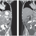
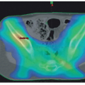
Figure 25.2 Clinical features of histiocytic disorders. A: Cutaneous Langerhans cell histiocytosis. An erythematous, papular rash resembling a Candida diaper rash. B: Skull is the most common site for LCH lesions in children. They can be isolated, part of multifocal bone disease, or part of multisystem disease. C: Lung lesions may arise as part of multisystem LCH, or in adults as isolated disease generally in the context of cigarette smoking. The computed tomographic (CT) image demonstrates typical cystic disease resulting from LCH lung lesions. D: Multisystem LCH can present with hepatosplenomegaly, as demonstrated by the MRI image. E: Neurodegenerative disease may be observed along with or following initial presentation of LCH. This MRI image illustrates the characteristic T2-weighted or FLAIR image with hyperintense signal in the cerebellum.

Central nervous system (CNS)-risk category of LCH bone lesions includes lesions of facial bones or anterior or middle cranial fossae (e.g., orbit, mastoid, temporal, sphenoid, ethmoid, or zygomatic). For reasons that remain to be defined, patients with lesions in these locations have a threefold increased risk for developing DI and an increased risk of LCH-associated neurodegenerative disease.


Lymph Nodes and Thymus

The cervical nodes are most frequently involved and may be soft or hard-matted groups with accompanying lymphedema. An enlarged thymus or involvement of the mediastinal nodes can mimic lymphoma or an infectious process and may cause asthma-like symptoms.19


Pituitary Gland

The posterior pituitary gland can be affected in LCH patients, causing central DI (see section Endocrine System). Anterior pituitary involvement often results in the failure of growth and sexual maturation.


Liver and Spleen

Hepatic involvement can cause hepatomegaly and liver dysfunction, leading to elevated liver enzymes, hypoalbuminemia with ascites, hyperbilirubinemia, and clotting factor deficiencies (Fig. 25.2D). Sonographic, computed tomographic (CT), or magnetic resonance images of the liver will show hypoechoic or low signal intensity along the portal veins or biliary tracts.20 Two of the most serious complications of hepatic LCH are cholestasis and sclerosing cholangitis. Biopsies typically do not include histiocytes, but demonstrate periportal lymphocytic infiltrates, bile duct inflammation, and loss of bile ducts. Seventy-five percent of children with sclerosing cholangitis will not respond to chemotherapy, and all of these patients require liver transplantation.21 Massive splenomegaly may lead to cytopenias, because of hypersplenism and respiratory compromise, although splenectomy is rarely indicated.


Lung

The lung was once considered a high-risk organ and is less frequently involved in children than in adults, in whom smoking is a key etiologic factor. Children with pulmonary LCH plus low-risk organs have a 5-year survival of 91% compared with 94% for those with only low-risk organ involvement.22 Chest x-rays may show a nonspecific interstitial infiltrate. A high-resolution CT of the chest is needed to visualize the cystic/nodular pattern of LCH that leads to the destruction of lung tissue (Fig. 25.2C). A spontaneous pneumothorax may be the first sign of LCH in the lung, although patients may present with tachypnea or dyspnea from widespread fibrosis and destruction of lung tissue. Declining diffusion capacity may also herald the onset of pulmonary hypertension. In young children with diffuse disease, there is significant potential for pulmonary tissue recovery with resolution of LCH.


Bone Marrow

Bone marrow involvement occurs most frequently in young children who have diffuse disease involving the liver, spleen, lymph nodes, and skin, and may be associated with marked thrombocytopenia or neutropenia. Others have only mild cytopenias and are found to have marrow involvement with LCH by sensitive immunohistochemical or flow cytometric analysis of the bone marrow.23 Of note, in many high-risk patients reported to have normal bone marrow by histology, BRAF-V600E mutations have been identified in hematopoietic precursor cells from bone marrow aspirate specimens.12 LCH patients with very high-risk disease may present
with marrow hemophagocytosis that mimics hemophagocytic lymphohistiocytosis.24


Endocrine System

Diabetes insipidus (DI) is the most frequent endocrine manifestation of LCH. Some patients present with an apparent idiopathic presentation of DI before other lesions are identified. A review of such patients found that 51% would have other lesions diagnostic of LCH within a year of identifying DI symptoms.25 A study of 589 LCH patients in France revealed that the 10-year risk of pituitary involvement was 24%.26 These investigators did not see a decreased prevalence of DI in chemotherapy-treated patients. However, Grois et al.27 reported a decline in the prevalence of DI from 40% to 20% with 6 months of treatment that included vinblastine and prednisone for patients with CNS-risk lesions (involvement of mastoid, orbit, temporal, or sphenoid bones).


Gastrointestinal System

A few patients with diarrhea, hematochezia, perianal fistulas, diarrhea, or malabsorption have been reported.28 Diagnosing gastrointestinal lesions in symptomatic patients may be difficult due to patchy involvement. Endoscopic evaluation, including multiple biopsies, may be required to identify foci of disease, along with CD207 immunostaining of all biopsies.


Central Nervous System—Diabetes Insipidus

Acute Manifestations of Diabetes Insipidus. DI caused by damage to the posterior pituitary is the most frequent initial sign of LCH (4%) in the CNS. Historically, DI was noted in 25% of all LCH patients, but in the LCH-III study the prevalence was 8% to 12%.29 As noted earlier, treatment of patients with bone lesions in CNS-risk sites of the skull and facial bones has been observed to reduce risk of progression to pituitary involvement and development of DI.27 Owing to the clinical risks, biopsies are rarely performed. Most often, the diagnosis is established by biopsy of skin, bone, or lymph node of a patient who also has pituitary imaging abnormalities or clinical signs and symptoms of DI. In the absence of additional LCH sites, and in the absence of clinical or laboratory evidence of an infectious etiology or alternative neoplastic disorder such as germinoma, one may consider a trial of empiric LCH therapy to minimize the potential for progressive pituitary damage.

Chronic Manifestations of Diabetes Insipidus. Fifty-six percent of LCH patients with DI will develop anterior pituitary hormone deficiencies (growth, thyroid, or gonadal-stimulating hormones) within 10 years of the onset of DI.30


Other Chronic CNS Disease Manifestations

LCH patients may develop mass lesions of the choroid plexus, grey, or white matter containing CD1a-positive LCs as well as CD8+ lymphocytes.31 In 1% to 4% of LCH patients, a chronic neurodegenerative syndrome may develop with clinical symptoms of dysarthria, ataxia, dysmetria, and behavior changes. Magnetic resonance imaging (MRI) shows hyperintensity of the dentate nucleus and white matter of the cerebellum on T2-weighted images, or hyperintense lesions of the basal ganglia on T1-weighted images, and atrophy of the cerebellum31 (Fig. 25.2E). Diagnostic imaging findings may precede the onset of symptoms by many years or be concurrent. A study of 83 LCH patients, with at least two brain MRIs for evaluation of craniofacial lesions, DI, and/or other endocrine deficiencies or neuropsychological symptoms, was recently published. Forty-seven of 83 patients (57%) had radiological neurodegenerative changes at a median time of 34 months from diagnosis. Of the 47 patients, 12 (25%) had clinical neurological deficits that presented 3 to 15 years after LCH diagnosis.32


Laboratory Features

Patients with high-risk disease may present with anemia and thrombocytopenia if the bone marrow is involved. Liver involvement may lead to hypoalbuminemia and elevations in liver enzymes and/or serum bilirubin concentrations. Intestinal involvement may also cause hypoalbuminemia. Lytic lesions of the bone are found on standard radiographs, CT, MRI, bone scans, or positron emission tomography (PET) scans. PET scans are useful for detecting lesions not found by bone scan or plain films, and comparison PET scans are particularly useful for providing evidence of healing 6 to 12 weeks after initiation of therapy.33 A biopsy of an affected organ, with staining of the LCH lesion DCs with antibody to CD207 and CD1a (Fig. 25.1B) rather than electron microscopy to identify Bibeck granules (Fig. 25.1C), is necessary for diagnosing LCH.34 For difficult cases, molecular analysis for the BRAF-V600E mutation may also be helpful, although many lesions will have an LCH DC infiltrate below the limit of detection for some sequencing methods, and in approximately 35% of LCH lesions, BRAF will not be mutated.12

Aug 25, 2016 | Posted by in ONCOLOGY | Comments Off on Histiocytic Diseases

Full access? Get Clinical Tree

Get Clinical Tree app for offline access