Hemostatic Challenges in Liver Disease


Hemostatic Challenges in Liver Disease


Charles S. Eby

Stephen H. Caldwell


The liver is the predominant site for synthesis and clearance of proteins involved in formation and lysis of fibrin, as well as its regulation, and the predominant source of thrombopoietin (TPO). Therefore, acute and chronic liver diseases cause major changes in multiple components of the hemostasis system. In the past, these changes have been viewed as shifting the balance toward bleeding based on traditional screening hemostasis tests and common bleeding problems in cirrhosis. However, the view is changing based on in vitro studies supporting suppression of both prothrombotic and regulatory pathways to produce a fragile, but rebalanced hemostasis state. The inability of traditional screening hemostasis tests to predict bleeding complications is perhaps most apparent in successful liver transplantation with minimal blood product replacement and minimal blood loss.1 Multiple observations have also identified evidence of a hypercoagulable predisposition in chronic liver disease patients who are at risk for thrombotic consequences. General application of this new paradigm to patients with severe liver disease awaits development and validation of novel laboratory methods and therapies in order to evaluate alternative strategies for assessment and management of hemostatic dysfunctions in patients with advanced liver diseases.

At this time, it is not possible to provide detailed, evidence-based guidelines for management of important clinical situations including prophylaxis and treatment of bleeding complications. Recognition of these knowledge gaps continues to stimulate clinical and laboratory research of hemostasis challenges in liver disease, and it is likely that in the coming years there will be more definitive management recommendations.


ACUTE LIVER FAILURE

Acute liver failure (ALF), defined as new-onset encephalopathy and coagulopathy following the development of jaundice in patients without preexisting liver disease,2 has an estimated incidence of approximately 2,000 cases/year3 and a mortality rate of approximately 33% in the United States, primarily due to cerebral edema, sepsis, and bleeding.2 The leading causes of ALF in the underdeveloped world are viral hepatitis A, B, and E, while in Europe and the United States, acetaminophen toxicity, idiosyncratic drug reactions, and indeterminate causes are the main etiologies.4

At presentation, most ALF patients have diminished procoagulant factors evident by international normalized ratio (INR) 1.5 to 5.0 in 81.1%, INR > 10 in 4.8%, platelet count <120,000/µL in 86.1%, and <30,000/µL in 2.6%, based on data from the Acute Liver Failure Study Group (ALFSG) prospective cohort study of 1,074 patients.5 Decreased hepatic synthesis of procoagulant, anticoagulant, and fibrinolytic proteins and accelerated consumption of hemostasis factors and platelets are consistent features of ALF regardless of underlying cause.6, 7, 8, 9 Plasminogen activator inhibitor-1 (PAI-1), von Willebrand factor (vWF), and factor VIII are elevated, likely reflecting the endothelial response to inflammatory cytokines.6, 7, 10 Despite considerable hemostatic derangements, spontaneous bleeding complications are uncommon (<10%) in ALF patients,5 and prophylactic plasma and platelet transfusions are inappropriate since they increase vascular volume and may exacerbate cerebral hypertension.11

Assessment of coagulopathy plays a role in determining if ALF patients should be listed for liver transplantation. The King’s College criteria for transplantation include INR > 6.5, while the Clichy-Villejuif criteria include factor V activity <20%,4 and initial factor VII activity <20% is associated with increased mortality.12 A high model of end-stage liver disease (MELD) score (in part dependent on INR, Table 126.1) appears to be a sensitive predictor of death without transplant, but requires validation.13 Indications for liver biopsy (rarely done) and intracranial pressure monitoring, and blood component transfusions prior to these invasive procedures, are controversial.11, 14 “Safe” INR and platelet count targets are not evidence based, and aggressive transfusion practices may worsen intracranial pressure.5, 14 Based on favorable anecdotal reports, the ALFSG recommends that cautious treatment with fresh frozen plasma (FFP) or cryoprecipitate (avoiding potentially detrimental volume overload) followed by recombinant activated factor VIIa (rFVIIa) be limited to highly invasive procedures such as insertion of intracranial pressure monitors.11 Notably, one recent study showed that ALF patients often maintain normal thromboelastography (TEG) in spite of prolonged INR consistent with the concept of a rebalanced system in liver disease.15

In bleeding patients who are also at high risk for thrombotic complications, plasma exchange could be an alternative therapy to improve hemostasis. A European randomized trial compared three daily plasma exchanges to routine management in 182 ALF patients. Fifty-nine percent of plasmapheresed patients survived to leave the hospital compared to 48% in the control group.16 However, confirmation of these preliminary findings is necessary before considering plasma exchange to be a standard strategy in ALF.


CHRONIC LIVER DISEASE

Causes of chronic liver injury include hepatitis B and C; alcohol; nonalcoholic steatohepatitis; autoimmune, inherited disorders; and cryptogenic disease. Chronic liver disease has long been associated with disturbances in hemostasis, and
both pro- and antithrombotic effects are recognized.17 As liver disease progresses, conventional indices of the clotting cascade worsen and have been incorporated into liver disease prognostic scores including the classical Child-Pugh and the MELD scores (Table 126.1). The latter is now used to guide organ allocation in liver transplant candidates.18 The relationship between abnormal coagulation indices and progressive liver fibrosis and cirrhosis is robust across various forms of chronic liver disease.


Table 126.1 Prognostic scoring systems in cirrhosis


Model of End-Stage Liver Disease (MELD) Score


9.57 × ln (creatinine mg/dL) + 3.78 × 3.78 × ln (bilirubin mg/dL) + 11.2 × ln (INR) + 6.43


Child-Pugh-Turcotte Score


Parameter


1 Point


2 Points


3 Points


Bilirubin (mg/dL)


<2.0


2.0-3.0


>3.0


Albumin (g/dL)


>3.5


2.8-3.5


<2.8


PT prolong (s)


<4.0


4.0-6.0


>6.0


Ascites


None


Easily controlled


Poorly controlled


Encephalopathy


None


Grades 1-2


Grades 3-4


Child A: 5-6 Points—well compensated


Child B: 7-9 Points—significant functional compromise


Child C: 10-15 Points—decompensated


REBALANCED COAGULATION PATHWAYS IN STABLE CIRRHOSIS

The concept of a rebalanced but fragile hemostatic system in chronic liver disease has become established over the past few years (FIGURE 126.1).1,19,20,21 In stable cirrhosis, the relative deficiency of hepatocyte-derived protein C and S, and antithrombin (AT), and increased factor VIII activity counterbalance the deficit in procoagulant factors. This concept is supported by in vitro methods that measure cumulative thrombin generation. Using small amounts of tissue factor and phospholipid to activate the extrinsic pathway, thrombin formation is monitored by measuring hydrolysis of a fluorescent-labeled peptide substrate to determine total thrombin generation or endogenous thrombin potential (ETP). Thrombin generation decreases as the severity of cirrhosis worsens.20 However when endogenous protein C activation (aPC) is incorporated into the thrombin generation test, the ratio of (ETP + aPC)/(ETP unmodified) was significantly higher in the cirrhotic groups compared to healthy controls.20, 22, 23 A higher ratio suggests a prothrombotic imbalance. Overall, the data suggest that protein C anticoagulant dysfunction is greater than the acquired coagulopathy in stable cirrhotic patients, producing a procoagulant state similar to congenital protein C deficiency or factor V Leiden (FVL) heterozygosity24 despite prolonged prothrombin time (PT)/INR. Importantly, in vitro thrombin generation results have not yet been correlated with clinical outcomes so clinical recommendations cannot yet be formalized, although this work provides strong evidence for questioning long-standing practices such as INR cutoffs as indications for plasma transfusion.

Therefore, conventional coagulation tests such as PT/INR and activated partial thromboplastin time (aPTT) do not accurately reflect the bleeding risk in compensated cirrhosis patients. Reduced hepatic synthesis of AT accompanies progressive liver disease and may also contribute to a prothrombotic imbalance. However, while AT infusion prior to liver transplantation corrects the deficiency, it does not reduce thrombin generation.25 Presently, there is no evidence to support a clinical benefit from AT replacement under any circumstance in patients with endstage liver disease.


REBALANCED PRIMARY HEMOSTASIS IN STABLE CIRRHOSIS

Platelets serve as the phospholipid scaffold for the clotting cascade and undergo complex quantitative and qualitative changes in cirrhosis.26 Prevalence estimates of thrombocytopenia in patients with chronic liver disease vary widely from 6% to 64%.27 Severe thrombocytopenia requiring prophylactic platelet transfusions or corrective intervention is uncommon27, 28 and typically occurs in patients with decompensated liver disease (the term “decompensated” is a common way to describe cirrhotic patients requiring more aggressive care and often hospitalization for a variety of complications related to chronic liver failure and portal hypertension—see also Decompensated Cirrhosis section).

The major causes of thrombocytopenia in cirrhosis include splenic sequestration, decreased survival, and decreased production. However it can be challenging to identify and prioritize the likely causes for individual patients. Using radiolabeled platelets, investigators determined that thrombocytopenic, cirrhotic patients sequestered 50% to 90% of platelets in their spleen in an exchangeable pool while maintaining near-normal total body platelet mass and mild-to-moderate shortened platelet lifespans.29,30 Splenic sequestration is a complex process, not simply dependent upon portal hypertension, as evidenced by
inconsistent improvement in platelet counts after transjugular intrahepatic portosystemic shunting or surgical decompression procedures.31 Reduction of splenic tissue, by splenectomy32 or partial splenic embolization,33 results in higher platelet counts, albeit with potential morbidity. Shortened platelet survival due to immune-mediated platelet destruction may be a factor in patients with severe thrombocytopenia out of proportion to the stage of hepatic dysfunction, and in some cases, there have been responses to steroids or other immunosuppression therapies.34, 35 However, there is no value in routinely measuring platelet-associated immunoglobin G in thrombocytopenic patients with chronic liver disease due to a lack of specificity of the assay. Causes for decreased platelet production include suppression of megakaryopoiesis due to decreased hepatic production of TPO, or viral illness (HCV, HIV), alcohol, or medication (alpha interferon) toxicity.36 Observations of platelet kinetics following orthotopic liver transplantation (OLT) indicate that TPO biology is disrupted in thrombocytopenic patients with advanced liver disease. A day after OLT, TPO concentrations increase followed by increased reticulated platelets and
resolution of thrombocytopenia.37 In addition, pharmacologic interventions with recombinant TPO, TPO mimetics, and IL-11 increase platelet production.28 However, TPO concentration in patients with chronic liver disease correlates poorly with platelet count,28 partly explained by analytical problems for measuring TPO.38 Therefore, it is of little value to measure TPO concentrations in thrombocytopenic patients with liver disease.


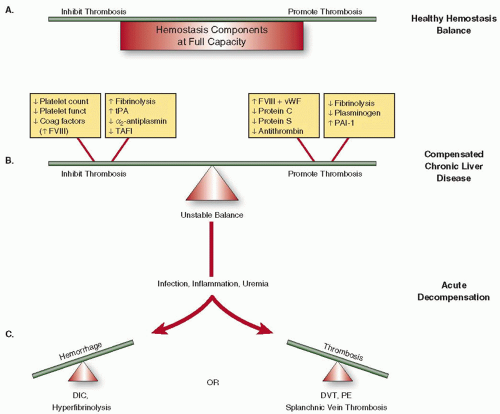
FIGURE 126.1 Changes in hemostasis balance with progressive liver disease. In healthy individuals (1A), cellular and protein components of prohemostasis and antihemostasis pathways have adequate capacities to adapt to acute physiologic stresses. In patients with compensated liver disease (1B), reductions in most coagulation/fibrinolysis factors and platelet number/function are offset by reductions in anticoagulant/antifibrinolysis factors and increased FVIII and vWF, resulting in a tenuous rebalancing of hemostasis. In patients with decompensated liver disease (1C), comorbidities further deplete most pro- and antihemostasis functions producing a fragile and imbalanced state and increased risk of hemorrhage and thrombosis.


FIGURE 126.2 Gastric varices with platelet plug (“nipple”) sign indicated by arrow. A scant amount of blood can be seen to be oozing from the base of the plug. (Image courtesy of Dr. Andrew Wang.)

In vitro evidence supports thrombocytopenia as a contributor to defective hemostasis in patients with liver disease. Tripodi et al.39 compared thrombin generation between healthy controls and patients with cirrhosis in the presence of soluble thrombomodulin and platelets. When platelet counts were normalized to 100,000/µL, ETP was similar in both groups. However, when platelet-rich plasmas were adjusted to patient and control whole-blood platelet counts, respectively, there was a positive correlation between platelet count and ETP.

Platelet dysfunction is especially important in mucosal and wound bleeding and possibly even in early stages of pressuredriven variceal bleeding, based on the well-known significance to endoscopists of the platelet plug sign (FIGURE 126.2).40 There is ample experimental evidence of acquired platelet adhesion, activation, and aggregation defects in liver disease patients.1, 41, 42 However in vitro model systems designed to assess platelet adhesion and aggregation in whole blood from liver disease patients under physiologic flow conditions suggest that primary hemostasis is not demonstrably defective, perhaps due to compensation from increased vWF activity.26, 43 Patients with advanced cholestatic liver diseases (primary biliary cirrhosis and primary sclerosing cholangitis) appear to have fewer bleeding complications and higher rates of portal vein thrombosis (PVT) compared to patients with hepatocellular injury.42 Preliminary in vitro studies suggest enhanced platelet activity may be involved.44, 45


FIBRINOLYSIS IN STABLE CIRRHOSIS

Multiple alterations to components of the fibrinolytic system occur with advanced liver disease46, 47 (Table 126.2). A lack of clinically validated functional tests to assess both fibrinolysis and its regulation hampers patient assessment.48 Consequently, there is debate as to whether the dominant process is excess plasmin generation, due to increased tissue plasminogen activator (tPA) activity, decreased thrombin activatable fibrinolysis inhibitor (TAFI)46, 47 or diminished α2-antiplasmin causing hyperfibrinolysis and elevated D-dimer, or decreased hepatic synthesis and clearance of fibrinolytic proteins and fibrin degradation products.49 It is reasonable to propose that compensated cirrhotic patients have a fragile, poorly regulated fibrinolysis system, which can be tipped over into overt hyperfibrinolysis by increased thrombin generation due to activators from ascites, infection, malignancy, or hemorrhage.21, 47

Clinically important secondary hyperfibrinolysis is estimated to occur in 5% to 10% of cirrhosis patients, typically during unstable, decompensated periods.50, 51, 52, 53, 54 In one series of 86 consecutive admissions to an inpatient liver unit, 36% of cirrhotic patients had a shortened euglobulin lysis time (<120 minutes) and 22% had signs of mucocutaneous bleeding.55 The condition should be suspected with spontaneous or delayed postprocedure bleeding and is accompanied by acute decreases in fibrinogen and platelet count, increased D-dimer, and prolonged PT and aPTT, or detected by TEG in the absence of significantly abnormal INR (personal observation). The clinical presentation and associated changes in hemostasis parameters are compatible with disseminated intravascular coagulopathy, although end-organ thrombotic injury is uncommon. Patients can present with spontaneous mucosal bleeding or multiple large hematomas due to primary hyperfibrinolysis and without evidence of an underlying precipitating event or multiorgan compromise. Typical laboratory results include acute hypofibrinogenemia and markedly shortened euglobulin lysis time, worsening anemia, but stable platelet count, PT, and aPTT.56 Recognition of this clinical entity is important as more specific therapy is available (see Specific Agents section).


Table 126.2 Typical changes in activation and regulation proteins of fibrinolysis in patients with advanced hepatic dysfunction and cirrhosis


tPA


↑↑


PAI-1


a2-Antiplasmin


TAFI


Plasminogen


Fibrin degradation products/D-dimer


Sources: Colucci M, et al. Deficiency of thrombin activatable fibrinolysis inhibitor in cirrhosis is associated with increased plasma fibrinolysis. Hepatology 2003;38(1): 230-237; Lisman T, et al. Thrombin-activatable fibrinolysis inhibitor deficiency in cirrhosis is not associated with increased plasma fibrinolysis. Gastroenterology 2001;121(1):131-139; Tripodi A, Mannucci PM. The coagulopathy of chronic liver disease. N Engl J Med 2011;365(2):147-156.


DECOMPENSATED CIRRHOSIS

A number of complications that influence the hemostatic system balance can occur in stable cirrhosis patients. “Decompensation” in this situation refers to the development of overt problems associated with worsening portal hypertension and liver failure such as ascites and encephalopathy, often in the setting of jaundice, which commonly necessitate hospitalization—a process more recently referred to as “acute on chronic liver failure.”57 Bleeding is common and sometimes precipitates the decompensation. In this situation, bewildering arrays of complex processes are evident, but three principle conditions affect hemostatic balance and bleeding risk: infection, renal failure, and vasomotor dysfunction.58, 59, 60

Bacterial infection, for example, subacute bacterial peritonitis, urinary tract infection, or cellulitis, is a common cause of decompensation in cirrhosis, and more subtle bacterial effects such as changes in intestinal permeability and endotoxemia impact the hemostatic system.61 One mechanism is release of endogenous heparinoids that inhibit factor Xa.62, 63 Renal failure, a common problem in decompensated cirrhosis (hepatorenal syndrome), contributes to platelet dysfunction.64 In this setting, hemodialysis can be viewed as a hemostatic intervention by reversing uremic platelet dysfunction and reducing volume overload.64, 65 Cirrhotic endothelial dysfunction is most evident by the occurrence of “hyperdynamic circulatory dysfunction,” an arteriolar vasodilated state characterized by stable but low mean arterial blood pressure and increased cardiac output that can progress to the hepatorenal syndrome.66 Experimentally, the condition is associated with impaired vascular smooth muscle contractility and impaired vasoconstriction in cirrhosis.67 Increased nitric oxide (NO) synthesis by activated or injured endothelial cells may have a role in cirrhosis-related altered vasodilatation since NO and vWF increase with progressive liver disease and elevations of both are associated with endotoxemia.68, 69 Other indirect signs of endothelial stimulation or damage in cirrhosis include progressive elevation of soluble thrombomodulin and tissue factor.70


LABORATORY ASSESSMENT OF BLEEDING RISK

Assessment of hemostatic status in patients with advanced liver disease is crucial for management decisions, including priority for liver transplantation, bleeding risk assessment, and monitoring response to treatment. However, most routine hemostasis tests are seriously flawed for lack of convincing evidence for prediction of bleeding tendency, incomplete interrogation of complex, regulated pathways, methods based on nonphysiologic conditions, and interlaboratory imprecision. This leaves clinicians in the difficult position of either using arbitrarily determined hemostasis test “cutoffs”71 or relying on clinical assessment of bleeding risk when deciding whether or not to use potentially harmful hemostatic therapies.

The hemostasis system in most patients with compensated, advanced liver disease appears to be functionally, but precariously, rebalanced (see FIGURE 126.1). Much of the data correlating hemostasis test results with bleeding complications are derived from percutaneous liver biopsy (PLBx) experience in relatively stable patients. Major bleeding complications after PLBx have been consistently low,72 with rates of 0.5% to 0.6% in large contemporary series.73, 74 Platelet count, occasionally a platelet function screening test, and INR thresholds are typically used to assess bleeding risk, and when exceeded, mandate alternative biopsy techniques or blood component transfusions. However, the supporting evidence for hemostasis parameter limits is insufficient.72, 75

Only gold members can continue reading. Log In or Register to continue

Stay updated, free articles. Join our Telegram channel

Jun 21, 2016 | Posted by in HEMATOLOGY | Comments Off on Hemostatic Challenges in Liver Disease

Full access? Get Clinical Tree

Get Clinical Tree app for offline access