In patients with kidney diseases, platelet defects and platelet-vessel wall abnormalities are the main determinants of hemorrhagic tendency, but other contributing factors including the abnormal production of nitric oxide (NO), uremic toxins, and anemia account for a multifactorial pathogenesis of bleeding
(Table 127.1).
Platelet Abnormalities
Severe thrombocytopenia is very rare and is limited to renal failure associated with disseminated intravascular coagulation, hemolytic uremic syndrome (HUS)/thrombotic thrombocytopenic purpura (TTP), eclampsia, and renal allograft rejection. Thrombocytopenia may also be caused during hemodialysis by the anticoagulant heparin.
3The majority of studies on uremic platelet were reported during the 1980s and 1990s and identified numerous biochemical abnormalities (reviewed in ref.
4,
5) including a decreased capacity to synthesize thromboxane A
2 by the prostaglandin-forming enzyme cyclooxygenase,
6,
7 a reduction in platelet granule ADP and serotonin content,
8 an increase in cyclic AMP (cAMP),
9 dysregulation of adenylate cyclase,
10 and impairment of Ca
2+dependent platelet function.
11,
12 A storage pool defect is supported by finding subnormal dense granule content,
8,
13 impaired release of α-granule proteins and β-thromboglobulin,
14 and reduced release of platelet ATP in response to thrombin.
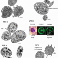
13 Moreover, platelet cytoskeletal dysfunction due to either a reduction in cytoskeletal proteins or a decrease in actin incorporation after thrombin stimulation has been reported.
15 All the above abnormalities and the defective platelet aggregation in response to various stimuli contribute to impaired platelet adhesion and aggregation in injured vessels.
Platelet adherence to foreign surfaces is significantly impaired in patients with end-stage renal disease.
16,
17,
18 Defective function of the platelet glycoprotein (GP)IIb-IIIa complex receptor may account for the decreased binding of von Willebrand factor (vWF) and fibrinogen to stimulated uremic platelets
19 and the reduced vWF-dependent adhesion and thrombus formation.
16,
17,
18 Dialysis improved the binding capacity of GPIIb-IIIa complex, suggesting a role of uremic toxins or receptor occupancy by fibrinogen fragments present in uremic blood.
19,
20Although quantitative and qualitative vWF abnormalities have not been consistently reported in uremic patients,
17,
21,
22 a functional defect in the vWF-platelet interaction cannot be excluded based on evidence that cryoprecipitate (a plasma derivative rich in vWF) and desmopressin (a synthetic derivative of antidiuretic hormone that releases autologous vWF from storage sites) significantly shorten the bleeding time in these patients.
23,
24 Increased levels of the platelet inhibitors prostacyclin (PGI
2) and NO have also been reported in uremia.
25,
26 The beneficial effect of conjugated estrogens on the bleeding times of uremic patients might be mediated by changes in the NO synthesis pathway since the NO precursor l-arginine reversed the effect of estrogens on bleeding.
27 Dialysis partially correct platelet dysfunction, suggesting that uremic toxins such as the guanidinosuccinic acid involved in NO generation
26 may play a role in platelet abnormalities.
Role of Anemia
Anemia is a constant feature of acute and chronic renal failure and is the main determinant of the prolonged bleeding time in uremic patients. There is evidence that the bleeding time is inversely related to the hematocrit in uremia
28,
29 as well as in other types of anemia. The cloning of the human erythropoietin gene and the production of recombinant human erythropoietin (rhEPO) has provided clinicians with a powerful tool to correct the anemia associated with renal failure, thereby eliminating dependency upon transfusion.
Dialysis
Dialysis improves platelet functional abnormalities and reduces, but does not eliminate, the risk of hemorrhage. Hemodial ysis, because of the interaction between blood and artificial surfaces, may induce chronic platelet activation that
can lead to platelet exhaustion and dysfunction. It has been found that plasma levels of the potent NO inducers TNF-α and IL-lβ increase during dialysis.
30,
31 These cytokines are generated
in vivo by monocytes during hemodialysis with complement-activating membranes. Thus, while dialysis removes uremic toxins, it also affects platelet activation and NO synthesis. Heparin, used to prevent filter clotting, can occasionally induce platelet activation and thrombocytopenia.
3
Role of Medications
Antiplatelet agents such as aspirin, as well as anticoagulants, increase the frequency of major bleeding in individuals with chronic renal insufficiency. Aspirin, at a dose of 100 mg/day, has been shown to significantly prolong the bleeding time in uremic patients when compared to controls
32 and anticoagulant therapy increases rates of bleeding in patients with end-stage renal disease as compared to the general population.
33,
34,
35 A recent study
36 showed that the risk for major bleeding episodes per year of exposure was 0.8%, 3.1%, 4.4%, and 6.3% in patients undergoing hemodialysis while taking neither warfarin nor aspirin, taking warfarin alone, taking aspirin alone, or taking the combination of warfarin and aspirin, respectively.
Given this bleeding risk, randomized, controlled trials are necessary to evaluate the efficacy and the safety of these agents in the secondary prevention of cardiovascular events in this high-risk population. Other medications that may increase the risk of bleeding in uremia include β-lactam antibiotics that accumulate in uremic patients
37 and act by perturbing platelet membrane function and interfering with ADP receptors.
38 The prolonged bleeding time and the abnormal platelet aggregation are related to the dose and duration of treatment and are promptly reversible after drug discontinuation. Third-generation cephalosporins may also inhibit platelet function and lead to marked disturbance of blood coagulation.
39 Nonsteroidal antiinflammatory drugs (NSAIDs) such as indomethacin, ibuprofen, naproxen, phenylbutazone, and sulfinpyrazone also inhibit platelet cyclooxygenase and disturb platelet function, but, in contrast to aspirin, the inhibitory effect of these compounds is readily reversed as the blood concentration of the drugs falls upon cessation of administration.
40
Clinical Manifestation
The clinical presentation of uremic bleeding ranges from mild and clinically unimportant bleeding (petechiae, purpura, and epistaxis) to severe hemorrhage (gastrointestinal bleeding, intracranial bleeding, and hemorrhagic pericarditis). Gastrointestinal bleeding occurs with the greatest frequency and has been observed in up to one-third of uremic patients. Low-grade gastrointestinal bleeding may be even common. Upper gastrointestinal bleeding accounts for 3% to 7% of all deaths in patients with end-stage renal disease.
41 A prospective study
42 found that acute gastrointestinal hemorrhage in patients with impaired renal function was associated with an increase in mortality and a 37% increase in length of hospital stay as compared to nonrenal patients.
42 The causes of bleeding are usually peptic ulcers, hemorrhagic esophagitis, gastritis, duodenitis, gastric telangiectasias, and diverticular disease.
43,
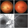
44 Other bleeding complications reported in chronic uremia are subdural hematoma, spontaneous retroperitoneal bleeding, spontaneous subcapsular hematoma of the liver, intraocular hemorrhage, and although now rare, hemorrhagic pericarditis with cardiac tamponade.
1 


