Head and Neck Cancer
INTRODUCTION
Cancers of the head and neck are composed of a spectrum of malignant neoplasms. Most commonly, however, “head and neck cancer” refers to epithelial carcinomas, squamous cell cancers, and their variant histologic subtypes that arise from the mucosal surfaces of the upper aerodigestive tract and constitute over 85% of the cancers encountered in this region. Those caring for patients with head and neck neoplasms must be conversant with the variable natural histories and approaches to treatment for the many different malignant tumors that arise within this region.
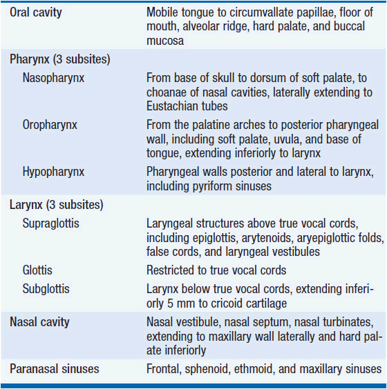
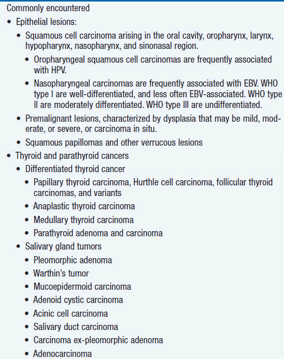
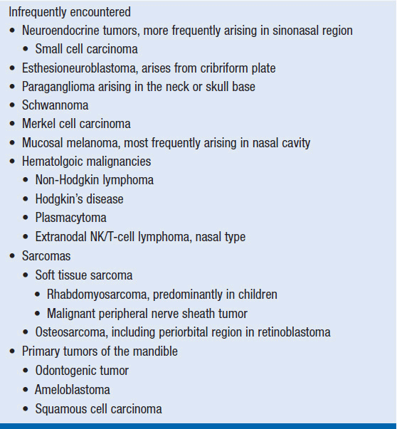
Cancers of the head and neck are traditionally divided into nine distinct anatomic regions from which mucosal cancers originate (Table 67-1). Other neoplastic conditions can arise within these regions and associated areas, such as the base of skull, orbit, and neck itself, including primary tumors of the major or minor salivary glands, the skin, the thyroid or parathyroid glands, and nonepithelial tissues of the neck. Sarcomas and hematologic malignancies are also encountered in the head and neck region. Representative histopathologies encountered in clinical practice are recorded in Table 67-2. This chapter will focus on the most common cancer of the region, namely squamous cell carcinoma of the head and neck (SCCHN).
INCIDENCE AND PREVALENCE
The American Cancer Society estimates that there will be 56,000 new cases of head and neck cancer in the United States in 2013 (excluding thyroid cancers). While head and neck cancer is one of the more curable adult malignances, the impact of this disease on individuals and society is significant, as it is not measured solely by the low absolute mortality, but also by the acute and chronic functional, cosmetic, and psychological morbidities experienced by patients. Head and neck cancer remains a difficult disease associated with significant rates of both morbidity and mortality.
Surveillance Epidemiology and End Results (SEER) data show that the incidence of head and neck cancer decreased from 1976 to 2009 in a pattern that approximates declines seen for lung cancer. These parallel changes follow the decline in tobacco use. However encouraging these data, several sobering facts must be noted.
For one, significant race-based differences exist in the incidence of head and neck cancer and its treatment outcomes. Not only do head and neck cancers occur with greater frequency in African-Americans compared to Whites, but also there are even greater differences in survival. For example, one large analysis of more than 20,000 patients treated for head and neck cancer in Florida from 1998 to 2002 showed that African-American race and poverty are independent predictors of poor prognosis, even when controlling for demographics, comorbidities, clinical characteristics, and treatment approach (1).
A second remarkable trend that is currently transforming the field of head and neck cancer is a dramatic increase in the incidence of oropharyngeal squamous cell carcinomas. In fact, SEER registries have shown an annual increase in oropharyngeal cancers of 0.80% in the period from 1973 to 2004 in the United States. Further analysis of SEER data indicates that this trend is due to a major increase in the prevalence of human papillomavirus (HPV) in oropharyngeal cancers, even while smoking-related cancers are declining. This increase in oropharyngeal cancers is most notable in white men, and in people from the most recent birth cohorts studied, that is, people born in 1940s and 1950s. From 1988 to 2004, the incidence of HPV-associated oropharyngeal cancers climbed 225%, whereas HPV-negative oropharyngeal cancers declined in the same period by 50% (2). Should this trend continue, it is estimated that the number of HPV-associated oropharyngeal cancers will outstrip those of cervical cancers by the year 2020, and account for the majority of head and neck cancers by 2030. The reason behind the increase in incidence is most likely changing sexual behaviors in our population, with an increase in oral sex and oral HPV exposure over time. In fact, the prevalence of HPV infection in the U.S. population at present is approximately 7%. Prevalence is greater in men and independently associated with both the number of lifetime sexual partners and smoking (3). It is currently not known if routine HPV vaccination will be effective in the primary prevention of HPV-associated oropharyngeal cancers. Still, it is hoped that HPV vaccination, as recommended for females aged 9–26 years and males aged 9–21 to reduce anogenital malignancy and genital warts, will further mitigate the impact of HPV-related malignancy by reducing the incidence of HPV-associated oropharyngeal cancers in our population. Embedded in the bad news of the increasing incidence of HPV-associated oropharyngeal cancers is the good news that these cancers are, stage-for-stage, more curable than HPV-negative oropharyngeal cancers. To date, the largest study examining the outcomes of HPV-associated versus HPV-negative oropharyngeal cancers was performed by Ang and colleagues (4). They examined outcomes of patients enrolled in the Radiation Therapy Oncology Group 0129 study investigating accelerated-fractionated radiotherapy versus standard-fractionated radiotherapy with concurrent cisplatin chemotherapy in patients with locally advanced SCCHN, and found that patients with HPV-associated oropharyngeal cancers had better 3-year overall survival (82%) than those with HPV-negative disease (57%).
RISK FACTORS FOR HEAD AND NECK CANCER
Tobacco and alcohol remain important risk factors for this disease. The local effects of tobacco and alcohol carcinogens are both dose-dependent and synergistic. Ever-smokers are at an increased risk for SCCHN even at relatively low exposures, whereas among never-smokers, alcohol increases the risk of SCCHN only at high doses (i.e., three or more drinks/day). Importantly, tobacco use also influences outcomes of HPV-associated SCCHN, increasing the risk of recurrence, distant metastasis, and death in this otherwise good-prognostic disease. Smokeless tobacco abuse results in a local exposure to carcinogens that promotes cancers of the oral cavity and to a lesser extent, the oropharynx, though the effect of smokeless tobacco alone is difficult to isolate as this form of tobacco is often used in combination with other products, such as betel nut, quid, slaked lime, and areca nut.
In addition to HPV, exposure to Epstein–Barr virus (EBV) is also associated with head and neck cancer. For both viruses, transient infection without long-term sequelae is common. Persistence of the viral genome and induction of malignant transformation is a rare event. While HPV is primarily associated with SCCHN of the oropharynx, (i.e., the tonsil and base of tongue tissues), EBV is associated with nasopharyngeal cancers. Nasopharyngeal cancer is rare in the United States and Europe, whereas nasopharyngeal cancer is endemic in Southern China, and seen with an intermediate incidence in Southeast Asia, the Mediterranean Basin, and the Arctic. EBV is a herpesvirus that is nearly ubiquitous worldwide. Most people infected with EBV will not, of course, develop cancer. Nonetheless, EBV viral proteins can have growth-transforming activity, and it is thought that disruption of the viral protein/host immune balance by modulation of the immune response can lead to the development of EBV-associated nasopharyngeal cancer.
Leukoplakia and erythroplakia are premalignant oral cavity lesions associated with a risk for transformation to in-situ and invasive cancers. Leukoplakia is a white, lacey-appearing lesion that may be posttraumatic and nondysplastic, with a low probability of malignant transformation. Persistent leukoplakia does, however, have malignant potential and should be sampled, preferably by excisional biopsy. Erythroplakia is a red lesion that harbors a greater malignant potential. This lesion should be surgically removed upon discovery. Not all patients with oral premalignant lesions will go on to develop an oral cancer. Clinical features that increase the risk of cancer include older age, lesions on the lateral or ventral tongue, and high-grade dysplasia. Molecular features associated with malignant transformation include increased epidermal growth factor receptor (EGFR) gene copy number and other genetic changes identified by gene expression profiling.
Lastly, one of the most important risk factors for developing SCCHN is having already had one. Compared to the general population, head and neck cancer survivors, particularly those with histories of tobacco and alcohol use, are at a significantly greater risk of developing a second aerodigestive tract primary malignancy, which occurs at a rate of approximately 2% per year. This has implications on both screening and risk modification for the long-term follow-up of SCCHN survivors.
MOLECULAR BIOLOGY AND NATURAL HISTORY
 MOLECULAR BIOLOGY
Several recent advances in the molecular biology of SCCHN have led to new understandings of the pathogenesis of the disease, as well as new treatment strategies. EGFR is overexpressed in the majority of SCCHNs. Activation of this receptor tyrosine kinase (RTK) upregulates signaling pathways involved in proliferation, angiogenesis, invasion, and metastasis. As predicted by these mechanisms, EGFR overexpression correlates with poor prognosis and is thus an attractive therapeutic target. Indeed, two therapeutic strategies are available for targeting EGFR. Small molecule EGFR inhibitors, such as gefitinib and erlotinib, that target the intracellular RTK ATP-binding domain have been studied in SCCHN, yielding disappointing results, likely due to the absence of EGFR-activating mutations in SCCHN. Monoclonal antibodies, including cetuximab, which bind to EGFR’s extracellular ligand-binding domain, have also been studied. While modest single-agent activity has been demonstrated in recurrent/metastatic SCCHN, an international phase III study comparing radiotherapy alone to cetuximab plus radiotherapy for definitive treatment of locally advanced SCCHN showed a 20-month improvement in median overall survival with the addition of cetuximab to radiotherapy (5). Cetuximab added to palliative chemotherapy for recurrent/metastatic SCCHN also improves outcomes compared to chemotherapy alone (6).
Further strides in our understanding of the biologic underpinnings of SCCHN were made by whole-exome sequencing of more than 100 SCCHN tumors carried out by two groups (7, 8). Several key findings were shared by these two studies. First, both confirmed the critical roles played by the previously identified tumor suppressor pathways governed by p53 and RB/INK4/ARF. Mutation in the tumor suppressor gene, TP53, was the most common alteration found, occurring in more approximately 50% of tumors. Rb pathway involvement was evidenced by frequent inactivation of CDKN2A, which encodes the cell cycle regulators p16/INK4 and p14/Arf/INK4B. CDKN2A mutations were found in approximately 10% of tumors, and additional tumors harbored copy number losses. Both groups further identified novel mutations in NOTCH1 in approximately 15% of tumors. While activating mutations in NOTCH family members have been implicated in hematologic malignancies, these studies detected inactivating mutations, suggesting a tumor suppressor role for the gene in SCCHN. Notch signaling has been linked to multiple functions, including terminal differentiation in squamous epithelium. While molecular targeting of tumor suppressor pathways remains an unmet challenge in oncology, approaches that target activating mutations in other signaling pathways have met with greater success. To this end, alterations in the PI3K signaling pathway have also been identified in SCCHN. Six to eight percent of tumors were found to have PIK3CA mutations, while PTEN, which encodes a negative regulator of the pathway, is subject to frequent loss of heterozygosity in SCCHN. This PI3K pathway activation in SCCHN has exciting clinical implications given the multiple agents that inhibit this pathway currently being investigated in clinical trials.
 NATURAL HISTORY
Squamous cell cancers of the head and neck are occasionally discovered during routine dental examination. More commonly, they are discovered after weeks to months of nonspecific, vague symptoms arising from the primary site, such as sore throat, dysphagia, otalgia, nasal congestion, or bleeding. Alternatively, many patients will present with isolated cervical adenopathy. Such patients are often treated empirically for infection, and discovered to have head and neck cancer only later when the adenopathy does not resolve and the primary physician or a surgical consultant consequently performs a detailed examination of the head and neck region or biopsy of the neck mass.
The AJCC TNM staging system is used to define the extent of disease, and the majority of SCCHNs are locally advanced stage III or IVa/b at presentation. Distant metastatic disease at initial presentation is unusual, and symptomatic disease related to such is rarely seen. The AJCC TNM staging system has great clinical value, but it is understandably complex given the many anatomic subdivisions of the head and neck region, each with its own unique definition of T stage. N stage and the combined overall TNM stage are consistent for all mucosal sites, except in the case of nasopharyngeal cancer.
A site-specific review of the common presenting symptoms, natural history, and treatment of head and neck cancer is beyond the scope of this text. In general, squamous cell cancers of the head and neck region are locally aggressive and carry a moderate risk of spread to regional lymph nodes, but have a low probability of spread to more distant sites. This natural history has several site-specific exceptions. For example, certain primary sites are more likely associated with regional disease at presentation (e.g., the nasopharnyx, hypopharynx, base of tongue, and supraglottic larynx). In contrast, other sites have a relatively low risk for regional spread (e.g., paranasal sinuses and small true vocal cord primaries).
STANDARD EVALUATION AND TREATMENT
 EVALUATION
It is important to obtain a surgical evaluation for patients with signs or symptoms relating to possible cancer in head and neck region. Depending on the setting, the initial interaction may be with a general surgeon or an otolaryngologist. Regardless of the specialty, the ultimate evaluation, diagnosis, staging, and follow-up must be undertaken by an otolaryngologist with sufficient training and expertise to achieve the following tasks.
A complete physical examination should include inspection of all visible mucosal surfaces of the head and neck including indirect mirror or fiberoptic laryngoscopy, as well as palpation of the floor of mouth, tongue and tonsil regions, and neck. Obvious tumor masses should be identified, along with potential second primary lesions and premalignant lesions. Suspicious lesions should be defined by primary site, extent of local involvement, and overall size. Associated cervical adenopathy should be assessed and if enlarged, the regional extent of disease should be quantified for staging purposes. Independent of whether surgical resection will be deemed appropriate, the potential for resection of suspected primary site and neck lesions should be determined. For example, lesions of the lateral tongue are often resectable while those of the nasopharynx or posterior pharyngeal wall are not. Neck lesions fixed to deep tissues or surrounding the carotid artery are not resectable.
In addition to physical examination, staging workup often includes computed tomography (CT) and/or magnetic resonance imaging (MRI) of the neck to characterize the extent of the disease. Patients with large T4 lesions or significant (>N1) lymph node involvement should also undergo chest CT to screen for distant metastases. PET/CT imaging is frequently performed, but is not always considered to be an essential component of the initial workup. One current important limitation to this modality is the lack of IV contrast available for the CT portion of the study, which is critical to evaluating the extent of both the primary lesion and nodal disease.
Examination under anesthesia is a key aspect of definitive staging for the majority of SCCHNs. Examination under anesthesia may include laryngoscopy, esophagoscopy, and bronchoscopy (a.k.a. triple endoscopy). During this procedure, biopsies are obtained to pathologically confirm a primary diagnosis, to define the extent of primary site disease, and to identify additional premalignant lesions or second primary cancers. An accurate description of anatomical involvement is critical to clinical staging as well to determining the expected morbidity of initial surgical resection.
Of special consideration is the evaluation of a neck mass of unknown origin. Supraclavicular neck disease should raise the question of a primary thoracic or thyroidal cancer, or perhaps an intra-abdominal cancer in the case of a left-sided supraclavicular lesion. Higher neck lesions are more likely to represent spread from a primary lesion in head and neck, including a possible cutaneous primary. Pathological confirmation of a squamous malignancy metastatic to a lymph node can usually be achieved by fine needle aspiration (FNA). A neck mass suspicious for regional metastatic disease can be approached by FNA if a potential primary mucosal abnormality of the upper aerodigestive tract or a salivary gland is not identified by a thorough physical examination. FNA specimens may also be sent for HPV and EBV analysis to potentially localize the primary site to the oropharynx or nasopharynx, respectively. Should an FNA prove inconclusive, an excisional nodal biopsy may be appropriate. In this case, consideration may be given to an immediate neck dissection at the time of this biopsy if frozen section indicates SCCHN or another nonlymphomatous malignancy. Such a neck dissection may assist in the control of extracapsular spread and additional malignant adenopathy, if present. However tempting, an incisional nodal biopsy should be avoided as it may compromise subsequent treatment and is associated with a lower probability of disease control.
 PRINCIPLES OF TREATMENT
General Principles
Once a patient’s primary diagnosis is established and initial staging studies are complete, secondary referrals are made to radiation and medical oncology, if not already done. The application of modern multidisciplinary treatments requires early input for all potential care providers. A team approach to care is clearly superior, and the team commonly includes head and neck cancer nurses, a speech and swallow therapist, a nutritionist, a social worker, and maxillofacial prosthodontist, among others.
As head and neck cancers differ greatly in locoregional stage and primary site, a detailed review of treatment options is beyond the scope of this chapter. However, a standard approach to treatment can be related. In general, all patients with untreated locoregional head and neck cancer are considered potentially curable if there is no evidence for distant metastatic disease. Treatment decisions begin with an understanding of primary site extent and a decision as to whether the lesion is resectable (removable with a high probability of negative surgical margins). If a lesion is resectable, careful consideration must be given to the specific morbidity that would result from curative surgical resection. With few exceptions, primary lesions that are resectable with acceptable morbidity may benefit from primary resection; however, this varies by site and stage. Lesions that are classically unresectable (e.g., sinonasal lesions involving the skull base and lesions of the nasopharynx, posterior pharyngeal wall or encasing the carotid artery), or for which primary surgery would have significant morbidity (e.g., base of tongue necessitating total glossectomy) are typically approached with primary radiation plus concurrent chemotherapy. Exceptions to this general approach exist. For example, cancers of the hypopharynx and larynx are usually resectable, but chemoradiation is often offered as primary therapy in an effort to avoid laryngectomy and preserve the natural voice. In this case, laryngectomy is reserved for persistent or recurrent disease after radiation or in the primary setting of advanced disease with significant laryngeal destruction precluding acceptable posttreatment function. The oropharynx is another “resectable” site that is often suitable for an organ preservation approach, especially as HPV-related SCCHN is uniquely sensitive to radiation, although recent advances in transoral laser microsurgery and robotic surgery have renewed interest in upfront surgery for oropharyngeal cancer.
Regional nodal disease from head and neck cancer is strategically approached as a separate entity. With rare exception, all patients with defined nodal disease should receive radiation therapy, either before or after resection of the involved nodes. Traditionally, patients underwent neck dissection when nodal disease was advanced and not fixed to underlying structures. Exceptions include patients with nasopharyngeal cancers, which are uniquely sensitive to radiation, and patients with large unresectable primary site lesions who will be treated with definitive chemoradiation because of the extent of primary site involvement.
Advances in the multidisciplinary care of patients with chemotherapy have altered the historic paradigms for treating patients with head and neck cancer. For example, concurrent chemoradiotherapy has replaced traditional surgery and postoperative radiation for advanced lesions of the sites with high associated surgical morbidity, such as the hypopharynx, larynx, and oropharynx.
Management of metastatic cervical lesions has also changed with advances in chemoradiotherapy. Currently, the decision to resect regional nodes is often deferred pending reevaluation after definitive nonsurgical treatment with chemoradiotherapy. Patients whose regional disease has completely regressed on physical examination and imaging after combined chemoradio-therapy appear to have excellent outcomes with minimal to no added benefit from additional neck surgery. Should residual disease in the neck remain after chemoradiotherapy, salvage neck dissection should be undertaken.
Management of Limited head and Neck Cancer
About one-third of patients present with limited T1 or T2 cancers without associated lymph node involvement or distant disease (stage I–II disease). In general, these patients can be treated by a single modality, either surgery or radiation therapy alone, and have a projected 5-year survival of 70%–90%. Radiation doses range from 6600 to 7200 cGy, depending upon the site and stage. Comprehensive radiation treatment to the primary site and regional lymphatics carries a high risk of chronic morbidities such as mild to moderate xerostomia, dental decay, and mild dysphagia; and a low risk for more severe complications such as osteonecrosis of the mandible, chondronecrosis of the larynx, or accelerated atherosclerosis of the carotid artery.
Definitive surgical resection of limited cancers is preferred in several settings. For example, limited oral cavity lesions can often be resected with minimal impact on speech and swallowing. In this setting, patients often undergo primary site resection with a staging neck dissection for identification and treatment of possible occult spread to regional nodes. Indications for postoperative radiation include close or positive primary site margins, perineural spread, lymphovascular disease, or regional nodal involvement on the staging neck dissection. Positive primary site margins and/or extracapsular extension are clearly established high-risk features that warrant the addition of chemotherapy to adjuvant radiation.
Management of Locoregionally Advanced Cancer
More than half of all patients with head and neck cancer present with locoregionally advanced disease (stage III or IV disease with a T3 or T4 primary lesion and/or regional nodal metastases). In the absence of distant metastatic disease, such patients are also treated with curative intent. In the following sections, we outline options for combined modality regimens for locoregionally advanced disease.
Surgery followed by postoperative radiation ± chemotherapy When surgery is performed for stage III/IV disease, evidence from two randomized trials provides the basis for optimal postoperative treatment (9, 10). Both trials compared standard postoperative radiation to radiation plus 3 cycles of concurrent cisplatin in patients at high-risk for recurrence (i.e., positive margins at the primary site, perineural or lymphovascular spread of disease, multiple involved regional lymph nodes, or extracapsular extension of disease in the neck). While the toxicity of concurrent chemotherapy with radiation was greater than that seen with radiation alone, chemoradiotherapy was associated with improved locoregional control in both studies, improved overall survival in one trial, and a trend toward improved survival in the second. At this time, postoperative radiation is standard for all patients with resected stage III or IV head and neck cancer. Patients at high risk for locoregional relapse despite surgery should be considered for postoperative chemoradiotherapy. Ongoing clinical trials seek to identify additional radiation sensitizers (chemotherapy, anticancer antibodies, or small molecule inhibitors) that will enhance the effects of radiation to degrees similar or greater than cisplatin, but with less toxicity.
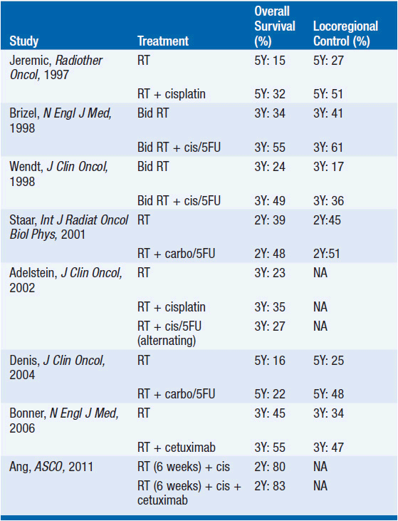
Definitive radiation with concurrent chemotherapy For the patient with locally advanced SCCHN, an alternative approach to primary site surgery is that of definitive radiation with concurrent chemotherapy. In this setting, primary site preservation is one of the therapeutic endpoints, and surgery is reserved for the salvage of recurrent or persistent disease after chemoradiotherapy. Multiple randomized trials have proven the validity of this approach (Table 67-3). Definitive standard fractionation radiation with concurrent high-dose cisplatin administered every 21 days is the regimen most frequently studied in SCCHN. However, additional chemotherapy regimens are commonly used during the typically 7-week course of radiation, such as cisplatin administered weekly, cisplatin with 5-flurouracil (5FU), carboplatin with 5FU, and weekly carboplatin and paclitaxel.
Stay updated, free articles. Join our Telegram channel

Full access? Get Clinical Tree


