Hamartomatous Polyps and Polyposis Syndromes


Hamartomatous Polyps and Polyposis Syndromes


Rhonda K. Yantiss


Hamartomatous polyps of the colorectum develop sporadically or in patients with hamartomatous polyposis syndromes, such as Peutz-Jeghers syndrome, juvenile polyposis, and phosphatase and tensin homologue (PTEN) hamartoma tumor syndrome. Although hamartomatous polyps are benign lesions comprising a variable admixture of nonneoplastic epithelial and stromal elements, syndromic polyps are associated with cancer risk, and, thus, clinicians and pathologists endeavor to identify at-risk patients and their family members. Recent advances in molecular pathology have provided important insight into the pathogenesis of hamartomatous polyposes and their relationships to cancer. However, application of pathologic, clinical, and molecular criteria still fails to identify all patients with hamartomatous syndromes. Despite recent advances in molecular techniques, a substantial number of patients with multiple gastrointestinal hamartomas lack detectable mutations, and up to 50% of patients do not have afflicted family members. With the exception of Peutz-Jeghers polyps, most gastrointestinal hamartomas are sporadic and do not reflect the presence of an underlying syndrome. Furthermore, most nonneoplastic polyps of the gastrointestinal tract are hyperplastic or inflammatory lesions that may be indistinguishable from hamartomas, particularly juvenile polyps. Pathologists play an important role in the detection and classification of hamartomatous polyposis syndromes, as well as distinguishing them from inflammatory conditions that require specific therapies. The purpose of this chapter is to discuss the clinicopathologic features and differential diagnoses of the most common hamartomatous polyps of the colorectum.


SPORADIC HAMARTOMAS

Sporadic hamartomas show a spectrum of morphologic features and contain nondysplastic epithelium, lamina propria, lymphoid tissue, and submucosal elements in various combinations. They may comprise some, or mostly, mesenchymal elements, in which case they show overlapping features with ganglioneuromas, Schwann cell hamartomas, lipomas, and perineuriomas. Polyps that consist of abundant inflamed stroma and cystically dilated glands are generally considered to represent inflammatory-type polyps in adults, whereas similar-appearing polyps among children are usually classified as juvenile polyps, as discussed subsequently. Most other sporadic hamartomas are asymptomatic, subcentimeter polyps that are incidentally discovered during screening or surveillance colonoscopy. They contain well-organized elements and show subtle abnormalities, such as distorted, thick-walled blood vessels, abnormal fat deposits, and irregularly distributed ganglion cells (Figure 5.1). Indeed, they may be overlooked and interpreted to represent normal mucosa in polypectomy specimens.


Peutz-Jeghers Syndrome


Clinical and Endoscopic Features

Virtually all Peutz-Jeghers hamartomatous polyps occur in the setting of the Peutz-Jeghers syndrome. Morphologically similarly polyps are rarely encountered as sporadic lesions, especially in the small intestine. Indeed, limited data suggest a relationship between sporadic Peutz-Jeghers

polyps and a variety of malignancies typically associated with the syndrome, suggesting that “isolated” Peutz-Jeghers polyps simply represent a forme fruste of the syndrome.1 Thus, detection of polyps that show morphologic features of Peutz-Jeghers hamartomatous lesions should prompt evaluation for a heritable syndrome and assessment for possible malignancy, particularly when they are multiple or encountered in the small intestine.


FIGURE 5.1: This hamartomatous polyp was found in the ascending colon of a middle-aged female during screening colonoscopy. It contains many elements native to the colon, but they are disorganized. Dense lymphoid inflammation is subjacent to colonic crypts and superficial to large, irregular smooth muscle bundles contiguous with the muscularis mucosae (A). Numerous ganglion cells are present singly and in clusters (arrows) within an abnormally thick muscularis mucosae (B).

Peutz-Jeghers syndrome is an autosomal dominant hereditary polyposis syndrome affecting 1 in 200,000 persons in the United States. It is characterized by the presence of numerous gastrointestinal hamartomatous polyps, mucocutaneous pigmentation, and an increased risk for developing a variety of malignancies in multiple organ systems (Table 5.1).2 Diagnostic criteria for the syndrome include (1) three or more Peutz-Jeghers polyps, (2) any number of Peutz-Jeghers polyps in a patient with a family history of the syndrome, (3) characteristic mucocutaneous pigmentation in a patient with a family history of the syndrome, or (4) any number of Peutz-Jeghers polyps in association with mucocutaneous pigmentation.3

The overall risk of gastrointestinal and extraintestinal malignancy among patients with Peutz-Jeghers syndrome is estimated to be 93% by 65 years of age. Up to 12% of patients with the disorder have dysplasia and/or invasive carcinoma in their polyps, but the adjacent, nonpolypoid mucosa is also at risk for malignancy. The lifetime risk of colorectal cancer among patients with Peutz-Jeghers syndrome is 35% to 40%, followed in frequency by pancreatic cancer (35%) and gastric cancer (28%). Despite the frequent occurrence of small-intestinal polyps in Peutz-Jeghers syndrome, the lifetime risk of small bowel cancer is only 10% to 15%.4, 5, 6 and 7

Peutz-Jeghers polyps show a predilection for the small intestine, but may occur anywhere in stomach, colon, or small intestine, as well as the gallbladder, bladder, and nasopharynx.
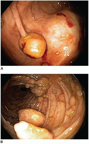
3 These polyps have a characteristic endoscopic appearance. They are multilobulated or rounded with a smooth surface that is similar in color to the background mucosa. Colorectal polyps may be pedunculated or sessile, and, when present, the stalk is usually thicker and longer than that of an adenoma (Figure 5.2). Patients with colorectal polyps almost always have small bowel polyps as well, the latter of which tend to produce clinical symptoms of recurrent abdominal pain secondary to intussusception. Individuals less than 30 years of age typically present with symptoms reflecting benign complications of disease, such as abdominal pain or bleeding, whereas malignant complications are more common presenting manifestations among older adults.


Pathologic Features

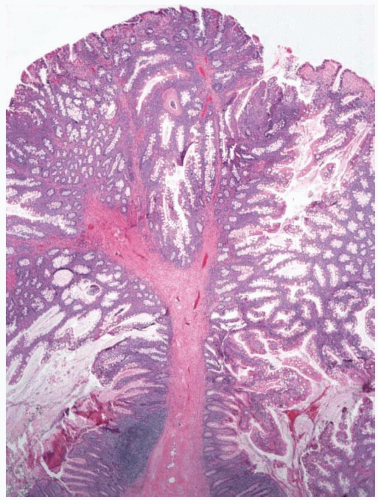
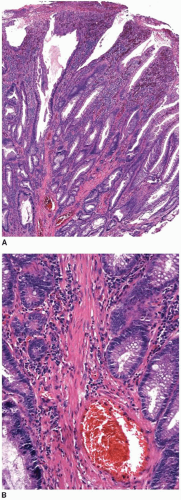
Peutz-Jeghers polyps are composed of nondysplastic mucosal aggregates and smooth muscle bundles that emanate in an arborizing fashion from the muscularis mucosae (
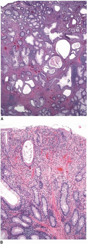
Figure 5.3). Polyps contain lobules of mature-appearing colonic epithelium supported by lamina propria (Figure 5.4). Crypts are normal or display mildly serrated epithelium and occasional cystic dilatation. Up to 10% of small-intestinal Peutz-Jeghers polyps are associated with displaced mucosa in the submucosa, muscularis propria, or subserosa of the subjacent bowel wall. This finding likely reflects a developmental phenomenon related to their hamartomatous nature, rather than trauma-related epithelial misplacement, and is usually lacking in colorectal hamartomatous polyps.

The differential diagnosis of Peutz-Jeghers polyps includes other types of hamartomatous polyps, mucosal prolapse polyps, adenoma with misplaced (displaced) epithelium, and invasive adenocarcinoma. The morphologic features of some Peutz-Jeghers polyps overlap with those of juvenile polyps, the latter of which may be seen in association with juvenile polyposis and PTEN hamartoma tumor syndrome. Overlapping features are particularly problematic among gastric and colonic Peutz-Jeghers polyps that do not show well-developed arborizing bundles of smooth muscle cells characteristic of small bowel polyps. Indeed, colonic and gastric Peutz-Jeghers hamartomas often display superimposed inflammatory changes more typically seen in juvenile polyps (Figure 5.5).8 Sporadic mucosal prolapse-type polyps contain abundant smooth muscle bundles extending into the lamina propria that simulate arborizing muscle bundles in Peutz-Jeghers polyps. However, prolapse-type polyps also show other features of luminal trauma, including erosions, hyperplasia and regeneration of crypts associated with ischemic-type surface epithelial changes, and inflammation (Figure 5.6). Mucosal prolapse polyps are most commonly observed in the distal colorectum and generally develop in older adults.


Table 5.1 Clinicopathologic and Molecular Features of Hamartomatous Polyposis Syndromes


Peutz-Jeghers Syndrome


Juvenile Polyposis Syndrome


PTEN Hamartoma Tumor Syndrome


Affected Gene


LKB1 (STK11)


SMAD4 and BMBR1A


PTEN


Gene Location


19p13.3


18q21.1 (SMAD4)


10q22.3 (BMBR1A)


10q22.3 (PTEN)


1p35-36 (SDHB)


11q23 (SDHD)


Gene Function


Serine threonine kinase


TGF-β-mediated signal transduction


Tumor suppressor gene


Prevalence in Affected Patients


70%


35%-60%


85%


Dermatologic Manifestations


Perioral, buccal, and conjunctival pigmentation


Oral papillomas, tricholemmomas, acral keratoses


Other Abnormalities


Cranial and cardiac abnormalities


Cleft palate


Polydactyly


Intestinal malrotation


Hereditary hemorrhagic telangiectasia


Autoimmune thyroiditis


Macrocephaly


Mental impairment


Fibrocystic breast disease


Glycogenic acanthosis


Distribution of Polyps


1. Small intestine


2. Colon


3. Stomach


1. Colon


2. Stomach


3. Small intestine


1. Colon


2. Stomach


3. Small intestine


Gastrointestinal Carcinomas


Carcinomas of colon, stomach, pancreas, small intestine


Carcinomas of colorectum, pancreas, stomach, duodenum


Carcinomas of colorectum and stomach


Other Malignancies


Ovarian sex cord-stromal tumor with annular tubules


Adenoma malignum of cervix


Testicular large cell calcifying


Sertoli cell tumor


Carcinoma of breast


Breast cancer


Thyroid cancer, follicular


FIGURE 5.2: Colonoscopic examination of a patient with Peutz-Jeghers syndrome demonstrates innumerable colonic polyps. One colonic polyp contains a long slender stalk composed of normal mucosal and submucosal elements (A), whereas others are sessile with a multinodular appearance (B). (Photographs courtesy of Dr. Randall Burt, University of Utah School of Medicine, Salt Lake City, UT.)


FIGURE 5.3: A cross-section through a Peutz-Jeghers polyp demonstrates a multinodular polyp head and dense stalk, reflecting smooth muscle cells emanating from the muscularis mucosae. The stalk shows some arborizing muscle bundles that extend into the head of the polyp (arrow).


FIGURE 5.4: Peutz-Jeghers polyps of the colorectum contain round aggregates of mucosal elements that are more numerous at the surface. Splayed bundles of smooth muscle cells fill the polyp stalk.

Peutz-Jeghers polyps that show epithelium in the subjacent bowel wall may simulate epithelial misplacement in adenomas as well as well-differentiated adenocarcinoma. Although both Peutz-Jeghers polyps and adenomas with epithelial misplacement contain lobules of epithelium surrounded by lamina propria, those of Peutz-Jeghers polyps are unassociated with features suggestive of polyp trauma, such as hemosiderin deposits, extruded mucin, hemorrhage, or scarring. A lack of these findings, in combination with the non-neoplastic nature of the epithelium, facilitates distinction of hamartomas from adenomas with epithelial misplacement. Importantly, mural mucosal aggregates occur almost exclusively in Peutz-Jeghers polyps of the small bowel where adenomas with misplaced epithelium are uncommon. Peutz-Jeghers polyps associated with mucosal elements in the subjacent wall may also simulate invasive adenocarcinoma, particularly with the former contain epithelium that is dysplastic.9 However, the Peutz-Jeghers polyps contain epithelium with a lobular architecture associated with lamina propria, rather than the desmoplastic stroma of an invasive carcinoma.


Molecular Alterations

More than half of patients with Peutz-Jeghers syndrome have detectable mutations in LKB1 (STK11), including approximately 70% of patients with inherited disease and 30% to 70% of those who are the first affected family members to be identified. This gene is located on


chromosome 19p13.3 and encodes a nuclear serine threonine kinase that normally inhibits cell growth by stimulating the promoter activity of p27.10, 11, 12 and 13 The disease phenotype tends to be more severe in patients with truncating mutations compared to missense mutations.11


FIGURE 5.5: A Peutz-Jeghers polyp of the colon is composed of numerous cystically dilated glands, some of which contain inspissated secretions. The lobular architecture of mucosal elements is lacking and smooth muscle bundles are not prominent (A). The lamina propria contains granulation tissue, eosinophils, and dilated crypts reminiscent of a juvenile polyp (B).


FIGURE 5.6: A mucosal prolapse polyp is eroded and contains cystically dilated crypts, some of which are serrated (A). Bundles of smooth muscle cells emanate from the muscularis mucosae into the polyp head, simulating the appearance of a Peutz-Jeghers polyp (B).


Juvenile Polyposis Syndrome


Clinical and Endoscopic Features

Unlike Peutz-Jeghers polyps, juvenile polyps may develop sporadically or in association with one of several polyposis syndromes, including juvenile polyposis syndrome, Cowden syndrome, and Bannayan-Riley-Ruvalcaba syndrome, as discussed in the next section. Isolated juvenile polyps of childhood show a predilection for the rectum and affect approximately 1% to 2% of children and adolescents.14 They are smooth, pedunculated, or sessile mucosal lesions that are somewhat friable and readily autoamputate in the fecal stream. Thus, juvenile polyps produce signs and symptoms related to gastrointestinal bleeding, including occult blood loss, hematochezia, anemia, passage of tissue per rectum, and mucosal prolapse.

Juvenile polyposis syndrome is an autosomal dominant hereditary polyposis syndrome affecting 1 in 100,000 persons in the United States. Phenotypic variants include infantile juvenile polyposis, juvenile polyposis coli, and generalized juvenile polyposis. Infantile juvenile polyposis is least common and unassociated with a family history of juvenile polyposis syndrome. It typically presents within the first 2 years of life with gastrointestinal polyposis, gastrointestinal bleeding, diarrhea, malnutrition, and protein-losing enteropathy and may result in early death.15 Patients with juvenile polyposis coli develop colorectal hamartomas, whereas those with generalized juvenile polyposis have numerous gastrointestinal polyps affecting the colon, stomach, and small intestine. Diagnostic criteria for juvenile polyposis include (1) three or more colorectal juvenile polyps, (2) any number of extracolonic juvenile polyps, or (3) any number of juvenile polyps in patients with a family history of the syndrome.16 However, only 20% to 50% of patients with syndromic disease have a family history of juvenile polyposis.

Approximately 15% of patients with juvenile polyposis syndrome also have congenital anomalies that include cranial and cardiac abnormalities, cleft palate, polydactyly, and intestinal malrotation (Table 5.1).17 The syndrome has also been reported in association with hereditary hemorrhagic telangiectasia characterized by vascular malformations affecting multiple organs, including those of the gastrointestinal tract.18

Only gold members can continue reading. Log In or Register to continue

Stay updated, free articles. Join our Telegram channel

May 22, 2016 | Posted by in ONCOLOGY | Comments Off on Hamartomatous Polyps and Polyposis Syndromes

Full access? Get Clinical Tree

Get Clinical Tree app for offline access