Fig. 1.1
The lactiferous ducts of a premenopausal breast lobe are filled with plastic. Blue colour indicates the ductal structure, while white colour indicates the TDLUs
The nipple-areola complex (NAC) is located on the most prominent part of the breast mound. Most textbooks state that there are 15–20 main ducts with 15–20 ductal orifices on the top of the nipple. According to classical anatomical descriptions, each lobe has its own main duct and orifice; however, some publications state less, 5–9 nipple ductal orifices. Possible explanations for the discrepancy may be the sebaceous glands that mimic the appearance of ducts but do not contribute to the ductal lobular infrastructure or that some ducts bifurcate shortly after emerging from the nipple [2]. The ducts have a zone which expands (lactiferous sinus) directly underneath the nipple [3]. They serve as a milk reservoir during lactation; however, recent studies doubt their existence [4]. The pigmented areola contains several prominences. They are modified accessory glands, so-called glands of Montgomery. Numerous sweat and sebaceous glands can be found among these, which keep the NAC lubricated and protected.
The breast can be divided practically into six portions. According to horizontal and vertical axes, four quadrants can be differentiated. The central substance is located behind the areola, while an extension of the breast extends into the axilla (Spence’s tail).
1.2 Fascial and Ligamentous Structure
The breast integrates into the superficial fascial system [5] of the trunk, which is located between the chest musculature and the skin. The superficial fascia of the breast encases the glandular tissue with its anterior and posterior lamella. The anterior lamella is located superficial to the gland at varying distances from the dermis. It is important to note that the superficial fascia of the breast is not a unified demarcation sheet, but a pitted cloak-like structure. The anterior lamella has clinical significance during skin-sparing mastectomies. The posterior lamella covers the undersurface of the gland and continues as Scarpa’s fascia [6] inferiorly. Between the posterior lamella and the pectoral fascia, the retromammary space (Chassaignac’s bursa) can be found [7]. This space contains fine areolar tissue, which can be dissected bluntly and serves as a pocket during sub-glandular breast augmentation.
The main suspensory ligaments of the breast course from the pectoral fascia through the glandular tissue to the skin. These are the so-called Cooper’s ligaments [8]. These ligaments have three distinct segments (deep, middle, superficial) created by the aforementioned anterior and posterior lamella of the superficial fascia of the breast. The deep segment is between the pectoral fascia and the posterior lamella, the middle segment is between the posterior and anterior lamella, and the superficial segment is between the anterior lamella and the skin (◘ Fig. 1.2). Cooper’s ligaments have significant oncologic importance. Their deep segment anchors the breast to the pectoral fascia, so total removal of the gland is facilitated by removal of the pectoral fascia. A tumour may also infiltrate these ligaments causing skin retraction, which is an important diagnostic sign of breast cancer. By contraction of the pectoralis major muscle, this skin sign can be provoked, which is an important clinical indicator of malignancy.
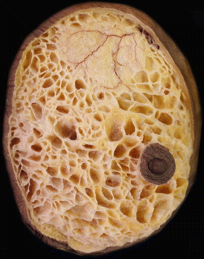

Fig. 1.2
Ligaments in between the skin and the anterior lamella of the superficial fascia of the breast. A superficial branch of the second internal mammary artery perforator is also seen
The posterior lamella of the superficial fascia continues as Scarpa’s fascia inferiorly. The anterior lamella also connects to the posterior lamella at the level of the fifth rib. This junction is fixed to the pectoral fascia, because the posterior lamella and the pectoral fascia get close to each other side to side. From this point several strong ligaments can be observed towards the dermis of the inferior pole and also the inframammary fold. They are considered the inframammary fold ligaments [9]. This anatomical feature may explain the fixed position of the inframammary fold during ageing.
There is a clinically important fibrous septum in the breast, called the septum fibrosum (◘ Fig. 1.3, Modified from Wuringer) [10].
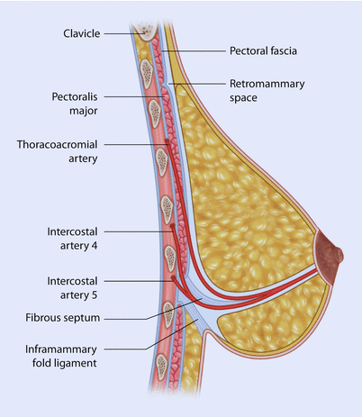

Fig. 1.3
Schematic representation of a lateral cross-sectional view of the fibrous septum of the breast
It is located horizontally at the level of the fifth rib towards the nipple. It divides the glandular tissue into an upper and lower part. It is reinforced medially and laterally by vertical horns. The medial horn of the septum fibrosum attaches parasternally between the second and fifth rib, while the lateral horn attaches to the lateral margin of the pectoralis major muscle. The septum fibrosum contains neurovascular structures supplying the NAC and gland. The thoracoacromial and deep branches of the lateral thoracic artery travel on the cranial side of this septum, while the fourth – rarely the fifth and sixth – intercostal artery perforator travels on the caudal side of it. The lateral cutaneous branches of the fourth intercostal nerve travel also along this septum. The breast can be dissected bluntly along this septum from the posterior side, so the blood supply can be preserved to the NAC during surgery.
1.3 Blood Supply
One can divide the arterial system of the breast into superficial and deep groups. Deep vessels penetrate the breast mainly along the septum fibrosum in the posterior to anterior direction, while the superficial ones travel in the subcutaneous layer towards the NAC (◘ Fig. 1.4). The superficial and deep vessels create an anastomosing subdermal net underneath the areola that supplies the NAC. One of the most important aspects of the vascular anatomy is the blood supply to the NAC. Blood supply of the NAC is beautifully described in relation to oncoplastic surgery in the work of van Deventer and colleagues (2004) [11]. Surgeons should be aware of the common patterns and how they may vary.
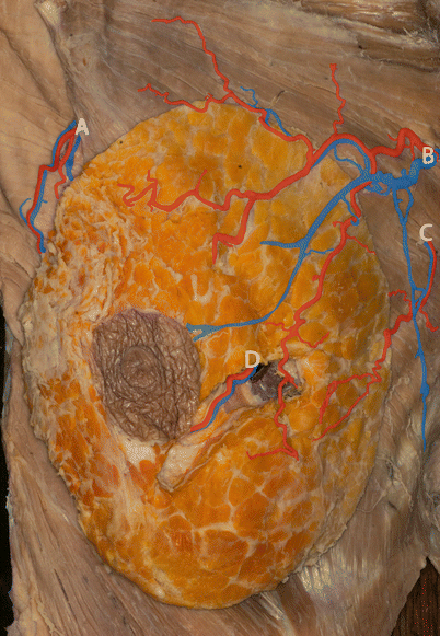

Fig. 1.4
Blood supply of the breast. Superficial and deep branches of the lateral thoracic vessels (A), second and third internal mammary vessel perforators (B, C), fourth intercostal vessel perforators (D)
The second to fourth internal mammary artery perforators belong to the superficial group. After piercing the chest wall, they get into the subcutaneous tissue, where they travel further towards the nipple. They are the dominant blood supply of the NAC [12], although there may be considerable anatomic variation in the number and direction of NAC feeding vessels which may account for some instances of nipple necrosis following surgery.
The breast receives its blood supply laterally from the lateral thoracic artery. It travels under the lateral margin of the pectoralis major muscle, then passes round the margin and gets into the substance of the breast. It has two main branches. One of them stays deep (deep group), the other becomes superficial (superficial group) and both travel towards the nipple.
The thoracoacromial artery (deep group) arises directly from the axillary artery and then branches underneath the pectoralis minor muscle. Its pectoral branches supply the upper pole of the breast.
The second to sixth intercostal artery perforators belong to the deep group of arteries. They are in general smaller, random vessels supplying the base of the breast. Sometimes some of these arteries can be more prominent, in particular the fourth intercostal artery perforator. It travels in the axis of the central parenchyma and supplies it. Sometimes this artery travels underneath the parenchyma and passes round the inferior pole of the breast. In particular, the fifth to sixth intercostal artery perforators are located in the area of the inframammary fold. Although they are smaller branches, their significance is enormous, because they serve as the blood supply to the inferior pole of the breast and hence are important in supporting inferior nipple pedicles during breast surgery.
The veins of the breast also have superficial and deep groups. The deep veins accompany the deep arteries. The superficial veins accompany the superficial arteries and are superficial to them. They create a superficial venous system. The venous blood flows towards the axillary, internal mammary and intercostal veins. The venous plexus underneath the areola is called the areolar venous plexus. These venous plexuses may become more apparent after augmentation surgery because of increased venous stasis.
1.4 Innervation
The sensory innervation of the breast is provided by the anterior and lateral cutaneous branches of the second to sixth intercostal nerves and the supraclavicular branches of the cervical plexus [8]. The anterior cutaneous branches of the intercostal nerves pierce the chest wall parasternally, and then they travel superficially to laterally and innervate the medial part of the breast.
Stay updated, free articles. Join our Telegram channel

Full access? Get Clinical Tree


