Gliomas, Ependymomas, and Other Nonembryonal Tumors of the Central Nervous System


Gliomas, Ependymomas, and Other Nonembryonal Tumors of the Central Nervous System


Williams D. Parsons

Ian F. Pollack

Daphne A. Hass-Kogan

Tina Y. Poussaint

Adekunle M. Adesina

Murali M. Chintagumpala


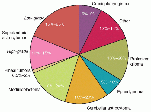
Tumors of the central nervous system (CNS) constitute the second most common pediatric cancer diagnosed in the United States each year, comprising approximately 25% of childhood cancers.1 Depending on the upper age chosen, the number of children, adolescents, and young adults who received diagnosis of a CNS tumor in 2013 was predicted to range between 3,160 (for ages 0 to 14 years) and 4,350 (for ages <20 years).2 The approximate incidence of the common pediatric CNS tumors is shown in Figure 26A.1.3

Although there has been a moderate increase in survival rates for children with CNS tumors over the last several decades such that the overall survival (OS) is now approximately 75%, CNS tumors remain the second leading cause of cancer death for children lesser than 20 years of age.1 In addition, the morbidity associated with CNS tumors and their therapy may be significant in terms of physical deficits as well as neuropsychological and neuroendocrine sequelae. Although not quantifiable, the long-term morbidity of childhood CNS tumors likely exceeds that associated with other pediatric malignancies. The optimal treatment of childhood CNS tumors, therefore, continues to pose a tremendous challenge that requires a multidisciplinary approach involving many pediatric specialists and subspecialists including neurosurgeons, neuropathologists, neurooncologists, neuroradiologists, radiation oncologists, neurologists, ophthalmologists, and physiatrists. In addition, the contributions of molecular biologists, pharmacologists, nurses, neuropsychologists, social workers, audiologists, nutritional experts, child-life specialists, and physical, occupational, and speech therapists are invaluable.

In the next two chapters, we review the current understanding of the biology of brain tumors and the principles associated with each of the diagnostic and treatment modalities. We then provide an overview of the clinical management and associated long-term sequelae of the more frequently encountered CNS tumors. This chapter includes information about gliomas, ependymomas, and other nonembryonal CNS tumors of childhood. Chapter 26B includes information about embryonal (e.g., medulloblastoma, supratentorial primitive neuroectodermal tumor [sPNET], atypical teratoid rhabdoid tumor) and pineal region tumors.


Figure 26A.1 Approximate incidence of common CNS tumors in children.


EPIDEMIOLOGY

The incidence of brain tumors peaks in the first decade of life, and then decreases until it peaks again, a second time, in older adulthood. The first peak is characterized by a predominance of males and by equal incidence rates for whites and blacks, except for the first 2 to 3 years of life, when a greater percentage of whites than nonwhites are affected.4 The male predominance is primarily explained by a disproportionate incidence of both medulloblastoma and ependymoma in males. For other tumor types, the genders are equally affected. During the first 2 years of life, supratentorial tumors predominate, whereas infratentorial lesions are more common through the rest of the first decade. Supratentorial tumors again predominate during late adolescence and through adulthood. Tumors of embryonal histology such as medulloblastoma, sPNETs, and pineoblastomas occur almost exclusively in children and young adults and primarily so during the first decade. High-grade gliomas, including glioblastoma multiforme, are much less common in children than in adults.


INHERITED SYNDROMES ASSOCIATED WITH CNS TUMORS

Less than 10% of children with brain tumors have a genetic disorder that places them at increased risk for developing a brain tumor. Although rare, these syndromes (Table 26A.1) place affected children at a markedly higher risk for developing non-CNS tumors as well.5

Turcot syndrome is an autosomal dominant disorder in which patients develop multiple adenomatous polyps and have an increased risk of primary brain tumors and colorectal cancer.6 Two subgroups of Turcot syndrome appear to exist. The first, which is due to mutation of the adenomatosis polyposis coli (APC) gene, is associated with an increased risk of medulloblastoma.6 Patients in the second subgroup, which is associated with mutations in DNA mismatch repair genes (PMS2, MLH1, MSH2, MSH6), develop gliomas.5,6

Children with Li-Fraumeni syndrome, which is caused by germline mutations in the TP53 gene, may develop multiple cancer types.7 The multifunctional protein encoded by this gene plays a role in cell cycle control, in ensuring DNA integrity and repair and, in some circumstances, in inducing apoptotic cell death. Children with inherited mutations of the TP53 gene most commonly develop low- or high-grade gliomas that may be multifocal.
They may also develop medulloblastomas, primitive neuroectodermal tumors (PNETs), or choroid plexus tumors.5


TABLE 26A.1 Inherited Disorders Associated with Brain Tumors


Syndrome


Gene(s)


CNS Tumor Type(s)


Cowden


PTEN


Dysplastic gangliocytoma of the cerebellum (Lhermitte-Duclos)


Hereditary retinoblastoma


RB1


Pineoblastoma, glioma, meningioma


Li-Fraumeni


TP53


Multiple brain tumor types, most commonly supratentorial PNET, medulloblastomas, and astrocytoma


Neurofibromatosis type 1


NF1


Neurofibroma, optic nerve glioma, astrocytoma


Neurofibromatosis type 2


NF2


Acoustic and peripheral schwannoma, meningioma, spinal ependymoma


Nevoid basal cell carcinoma (Gorlin syndrome)


PTCH1


Medulloblastoma, meningiomas


Rubenstein-Taybi


CREBBP


Medulloblastoma, oligodendroglioma, meningioma


Tuberous sclerosis


TSC1, TSC2


Subependymal giant-cell astrocytoma


Turcot


APC


Medulloblastoma (most common)


MLH1, PMS2, MSH2, MSH6


Astrocytoma and ependymoma (less common)


von Hippel-Lindau


VHL


Hemangioblastoma


Rhabdoid tumor predisposition syndrome


SMARCB1, SMARCA4


Atypical teratoid rhabdoid tumor


PNET, primitive neuroectodermal tumor.


Children with neurofibromatosis type 1 (NF1), due to mutation of the NF1 gene, are at risk for dermal and plexiform neurofibromas and have a markedly increased risk of astrocytoma.5 The astrocytomas are typically low-grade neoplasms that occur within the optic pathway and involve optic nerves, the chiasm, and the optic radiations. Low-grade gliomas, which can undergo malignant transformation, may also occur within the cerebral hemispheres, the brainstem, or the cerebellum.

Neurofibromatosis type 2, resulting from mutations in the NF2 gene, is associated with meningiomas within the brain and spinal cord and schwannomas of the cranial nerves, spinal nerves, and peripheral nervous system.5 Bilateral acoustic nerve schwannomas are highly associated with neurofibromatosis type 2. Gliomas and ependymomas also occur with increased frequency and tend to be located in the spinal cord.

Nevoid basal cell carcinoma syndrome (NBCCS, Gorlin syndrome) results from mutations in the PTCH1 gene, which is a cell surface receptor involved in the Sonic Hedgehog signaling pathway. In addition to multiple basal cell carcinomas, approximately 5% of children with NBCCS develop medulloblastoma, with a peak age of onset of 2 years.8

Children with rhabdoid tumor predisposition syndrome are at risk for atypical teratoid rhabdoid tumor of the CNS, in addition to extra-CNS rhabdoid tumors, due to germline deletion or mutation of the SWI/SNF chromatin remodeling complex gene SMARCB19 or less commonly SMARCA4.10 Germline alterations of SMARCB1 have also been reported in schwannomatosis.11

Finally, several rare tumor types occur most frequently in association with specific inherited disorders. Subependymal giant-cell astrocytomas, which occur in the anteromedial aspect of the brain near the foramina of Monro, are most frequent in children with tuberous sclerosis complex (TSC) due to mutations in TSC1 and TSC25 that lead to constitutive activation of the mammalian target of rapamycin (mTOR) signaling pathway.12 Inhibitors of the mTOR pathway have been found to decrease both tumor volume and seizure frequency in TSC patients with subependymal giant-cell astrocytomas, leading to FDA approval for the use of everolimus in this context.13,14

Cerebellar gangliocytoma (Lhermitte-Duclos) occurs in the context of Cowden syndrome, owing to mutation of the PTEN gene that encodes a dual-specificity phosphatase.15

von Hippel-Lindau syndrome, which arises from mutations of the VHL gene that appears to have a role in DNA replication, is associated with hemangioblastomas, most typically in the cerebellum, spinal cord, or retina.5

Data are inconclusive regarding less completely understood familial factors outside of known genetic syndromes associated with pediatric CNS tumors. Some studies show no influence of family history on the occurrence of brain tumors, whereas others report an increased risk of brain tumors with a family history of bone cancer, leukemia, and lymphoma. The children or siblings of persons with brain tumors may be at higher risk of developing brain tumors themselves.16,17,18 Reports of familial clustering of embryonal tumors, gliomas, and choroid plexus papillomas (CPPs) also exist.19,20,21


OTHER FACTORS ASSOCIATED WITH CNS TUMORS


Ionizing Radiation

Exposure to ionizing radiation is a well-documented cause of brain tumors. Children treated with radiation therapy for tinea capitis during the 1940s and 1950s were found to have increased risk for the development of meningiomas, gliomas, and nerve sheath tumors 22 to 34 years later.22 Brain tumors, most frequently meningiomas and high-grade astrocytomas, are also observed after cranial irradiation for non-CNS cancers such as acute lymphoblastic leukemia as well as for primary CNS tumors. The risk of secondary CNS malignancies is greater in childhood cancer patients who have inherited cancer syndromes associated with an increased genetic susceptibility to multiple primary malignancies that is enhanced by sensitivity to ionizing radiation. For example, patients with retinoblastoma, NF1, Li-Fraumeni syndrome, or NBCCS are
at substantial risk of developing radiation-related secondary CNS cancers as a result of the location of the radiation field for their primary tumors.23 The Childhood Cancer Survivor Study (CCSS) has provided precise estimates for radiation therapy and the development of subsequent malignant neoplasms of the CNS.24


Immunosuppression

CNS lymphomas occur with increased frequency in patients with a variety of underlying primary or secondary disorders of the immune system, including the Wiskott-Aldrich syndrome, ataxia-telangiectasia, acquired immunodeficiency syndrome, and after solid-organ transplantation.25


Environmental Exposures

The effect of environmental exposures, including diet, on the occurrence of childhood brain tumors has been studied by numerous investigators without conclusive evidence for an association.26 Studies of the effect of cellular telephone usage on the occurrence of childhood brain tumors have not been performed. An association of polyomavirus (e.g., JC and SV-40 viruses) with certain types of pediatric brain tumors such as medulloblastoma, ependymoma, and choroid plexus tumors has been reported27; however, there is no definitive evidence that such viruses are directly involved in the pathogenesis of these tumors.28


PATHOLOGIC CLASSIFICATION OF CNS TUMORS


Background

Classification of CNS tumors has historically been a challenge as several different histologic classifications have coexisted, reflecting a lack of consensus among neuropathologists. However, since the advent of the World Health Organization (WHO) classification of CNS tumors, there has been a significant improvement in consensus among neuropathologists. The prototype classification system proposed by Bailey in 1926, based on the cell-of-origin notion introduced by German pathologist Cushing, suggests that CNS tumors develop from cells arrested at various stages of development.


Methods of Classification


Morphologic and Histogenetic Classification

In addition to their cell-of-origin concept, Bailey and Cushing recognized that tumors were composed of heterogeneous cells. They decided to classify tumors on the basis of the morphology and presumed histogenesis of the predominant cell type. Hence, if the majority of cells resembled astrocytes, the tumor was called an astrocytoma, even though a small number of other cells (e.g., oligodendrocytes) were present. Most classifications in current use are based on this concept.

In 1949, Kernohan et al.29 introduced a grading system based on the concept that glial cells of all types become progressively more malignant over time. Criteria were advanced for grading glial tumors on a scale from 1 (most benign) to 4 (most malignant), and this scheme was to be applied to astrocytomas, oligodendrogliomas, and ependymomas. Based on histologic features, it has been readily adopted and is used commonly for astrocytomas for which clinical correlations exist. For oligodendrogliomas and ependymomas, the scheme has been an awkward fit, resulting in inconsistent use. Revision of the Kernohan grading system for astrocytomas was proposed by Daumas-Duport et al.30 An international panel of neuropathologists, working under the aegis of the WHO, expanded the concept of grading all CNS tumors, using the 1-to-4 scale to indicate biologic malignancy (Table 26A.2).31 Given the increasing knowledge of the genetic alterations underlying specific CNS tumor types (some of which demonstrate similar, if not identical, histologic features), it is likely that future classification systems will increasingly incorporate molecular features.


Phenotypic Classification

An alternative to classification is the phenotypic approach. Essentially, this involves evaluation of the tumor by identification of cell types comprising it.32 Beyond examination of sections stained by routine
hematoxylin and eosin (H&E), other special stainings, immunohistochemistry, ultrastructural study, and cytogenetics can now be used to determine the cell types comprising the tumor with greater precision than was possible a decade ago. Use of monoclonal antibodies to identify specific antigens, such as cytoskeletal and membrane proteins, hormonal polypeptides, and neurotransmitter substances, has been especially instrumental in classifying, on routine light microscopy, tumors with unusual morphologic features that previously were relegated to the “unknown” category. Table 26A.3 contains a listing of the widely available markers that are used most commonly and their utility in the differential diagnosis of tumors arising in the CNS. Use of this phenotypic approach, coupled with increasing information of the cytogenetics and microarray analyses of CNS tumors, has forced changes in the historic classification schemes.


TABLE 26A.2 World Health Organization Classification of Tumors of the Nervous System (Neuroepithelial Tumors Only)31


Tumors of neuroepithelial tissue


  1. Astrocytic tumors


    1. Pilocytic astrocytoma and pilomyxoid astrocytoma


    2. Subependymal giant cell astrocytoma


    3. Pleomorphic xanthoastrocytoma


    4. Diffuse astrocytoma (fibrillary, gemistocytic, protoplasmic)


    5. Anaplastic astrocytoma


    6. Glioblastoma (giant cell, gliosarcoma)


    7. Gliomatosis cerebri


  2. Oligodendroglial tumors


    1. Oligodendroglioma


    2. Anaplastic oligodendroglioma


  3. Oligoastrocytic tumors


    1. Oligoastrocytoma


    2. Anaplastic oligoastrocytoma


  4. Ependymal tumors


    1. Subependymoma


    2. Myxopapillary ependymoma


    3. Ependymoma (cellular, papillary, clear cell, tanycytic)


    4. Anaplastic ependymoma


  5. Choroid plexus tumors


    1. Choroid plexus papilloma


    2. Atypical chroroid plexus papilloma


    3. Choroid plexus carcinoma


  6. Other neuroepithelial tumors


    1. Astroblastoma


    2. Choroid glioma of the third ventricle


    3. Angiocentric glioma


  7. Neuronal and mixed neuronal-glial tumors


    1. Dysplastic gangliocytoma of cerebellum (Lhermitte-Duclos)


    2. Desmoplastic infantile astrocytoma/ganglioglioma


    3. Dysembryoblastic neuroepithelial tumor


    4. Ganglioglioma


    5. Anaplastic ganglioglioma


    6. Others (central neurocytoma, extraventricular neurocytoma, cerebellar liponeurocytoma, papillary glioneuronal tumor, rosette-forming glioneuronal tumor of the fourth ventricle, paraganglioma)


  8. Tumors of the pineal region


    1. Pineocytoma


    2. Pineal parenchymal tumor of intermediate differentiation


    3. Pineoblastoma


    4. Papillary tumor of the pineal region


  9. Embryonal tumors


    1. Medulloblastoma


      1. Desmoplastic/nodular medulloblastoma


      2. Medulloblastoma with extensive nodularity


      3. Anaplastic medulloblastoma


      4. Large cell medulloblastoma


    2. CNS primitive neuroectodermal tumor


      1. CNS neuroblastoma


      2. CNS ganglioneuroblastoma


      3. Medulloepithelioma


      4. Ependymoblastoma


    3. Atypical teratoid/rhabdoid tumor


TABLE 26A.3 Common Markers for Diagnosis of CNS Tumors


Marker


Tumor Types Containing Positive Cells


Glial fibrillary acidic protein


Astrocytoma, ependymoma, mixed glioma, gliosarcoma, ganglioglioma, glioblastoma multiforme, gliofibroma; positive cells occasionally may be found in oligodendroglioma, capillary hemangioblastoma, choroid plexus papilloma, PNET, AT/RT


Neurofilament


Ganglioglioma, gangliocytoma, PNET, neurocytoma, subependymal giant-cell tumor, AT/RT


Vimentin


Mesenchymal tumors, meningiomas, sarcoma, melanoma, lymphoma, ependymoma, astrocytoma, gliofibroma, chordoma, schwannoma, hemangioblastoma, carcinoma, PNETs, AT/RT


S100 and neuron-specific enolase


Positive in a variety of normal and neoplastic cells of neural and nonneural origin; of questionable utility for diagnostic purposes


Desmin


Tumors containing muscle (rhabdomyosarcoma, teratoma, etc.), PNET


Cytokeratin


Chordoma, choroid plexus tumors, meningioma, certain anaplastic gliomas, nongerminomatous germ-cell tumors, PNET, AT/RT


Epithelial membrane antigen


Meningioma, ependymoma, epithelial areas of teratomas, rhabdoid tumor (AT/RT)


Synaptophysin


PNET, ganglioglioma, gangliocytoma, central neurocytoma, neuroendocrine tumors


Smooth muscle actin


Tumors containing muscle, AT/RT


Retinal S-antigen


Pineal parenchymal tumors, PNETs, retinoblastoma


α-Fetoprotein


Embryonal carcinoma, endodermal sinus (yolk sac) tumor


Human chorionic gonadotrophin


Germinoma, choriocarcinoma


Placental alkaline phosphatase


Germ cell tumors


c-kit


Germinoma


AT/RT, atypical teratoid-rhabdoid tumor; PNET, primitive neuroectodermal tumor.


CLINICAL PRESENTATION

No single clinical finding is pathognomonic for the diagnosis of a childhood brain tumor. At the onset of illness, the nature of neurologic and systemic dysfunction is varied. Signs and symptoms may be a direct result of tumor infiltration into the adjacent brain and/or spinal cord, or a consequence of CSF flow obstruction with resultant increased intracranial pressure (ICP). The clinical presentation primarily reflects the site of tumor origin, the age and developmental level of the affected child, and, occasionally, the tumor type. Clinical prodromes may include features of increased ICP, symptoms and signs of a localizing nature, or symptoms and signs without a localizing quality.


Increased Intracranial Pressure

Brain tumors cause increased ICP directly by infiltrating or compressing normal CNS structures or indirectly by causing obstruction of the cerebrospinal fluid (CSF) pathways, resulting in noncommunicating hydrocephalus. Initial features of elevated ICP are typically insidious, nonspecific, and nonlocalizing. Among school-aged children, declining academic performance, fatigue, personality changes, and vague intermittent headaches are common. Over time, morning headaches, vomiting, and lethargy ensue. Papilledema may develop if the pressure is longstanding. Rapid progression of symptoms secondary to increased ICP is infrequent and is often associated with a quickly growing midline or posterior fossa tumor requiring immediate intervention.

Headaches resulting from brain tumors may have ominous features distinct from tension headaches or migraines. When children with a tumor are recumbent, increased ICP may worsen, resulting in a headache that wakens them at night or is present on waking in the morning. On arising, vomiting may occur along with some relief of pain. Once such patients are upright, the headache may diminish over the course of the day. Over time, headaches gradually increase in severity and frequency and clearly differ from any previous pain. The pain, which usually is frontal or occipital rather than temporal, may be further exacerbated with Valsalva maneuvers. The clinical suspicion for tumor should be the greatest in those children with recent and continuing complaints of headache, and should prompt a careful history and evaluation for related symptoms and signs. In fact, by 6 months from headache onset, nearly 100% of children have associated neurologic signs, such as papilledema, strabismus, ataxia, or weakness.33

Signs and symptoms of elevated ICP in infants and young children, whose skulls may more easily accommodate the growth of a mass lesion, may be quite different and may include irritability, anorexia, failure to thrive, and developmental delay or regression. Chronically increased pressure may lead to macrocephaly and separation of the cranial sutures. Infants may develop a tense or bulging anterior fontanelle associated with a shrill, neurogenic cry. Funduscopic evaluation of these patients may reveal only optic pallor but no evidence of papilledema. The setting-sun sign, a seemingly forced downward deviation of the eyes and part of Parinaud syndrome, may also be seen.

Parinaud syndrome is a collection of ophthalmologic findings stemming from increased ICP at the dorsal midbrain. In addition to the impaired upward gaze seen in infants, older children also display large pupils with impaired reflex constriction to light but
not with accommodation. Convergence of gaze may evoke repetitive, bilateral, adducting nystagmus with retraction of the globes in the orbit. Cranial nerve IV palsy, with the affected eye deviated upward and laterally, may also occur. Affected children often compensate for the trochlear nerve palsy by tilting their heads toward the shoulder of the unaffected eye.

A head tilt may occur also with increased ICP because of a stiff neck and cervical root irritation from incipient cerebellar herniation of a posterior fossa mass. Other signs of increased ICP include listlessness and horizontal diplopia from pressure on the long, free intracranial course of the abducens nerve.


Localizing Symptoms and Signs

Children with supratentorial tumors (i.e., of the cerebrum, basal ganglia, thalamus, hypothalamus, and optic chiasm) may demonstrate various localizing symptoms and signs that precede those of increased ICP. The most common of these signs and symptoms include hemiparesis, hemisensory loss, hyperreflexia, seizures, and visual complaints.

Vision loss may localize to any location in the optic pathway. Complaints occasionally start insidiously with such events as a failed school eye examination or a need for eyeglasses. Tumors confined to the optic nerve produce monocular vision loss. Chiasmatic tumors often present with a complex visual field loss and decrement in acuity, whereas lesions located more posteriorly, in the optic tract, lateral geniculate nucleus, optic radiations, or the occipital cortex, demonstrate some aspect of hemianopsia. A paradoxical increase in pupillary size to light when moving the source from one eye to the other indicates a relative afferent pupillary defect (the Marcus-Gunn pupil), a potential sign of tumor at the optic nerve or chiasm. Among infants, chiasmatic tumors may result in unilateral or bilateral pendular nystagmus, with head nodding and head tilt, a triad known as spasmus nutans.

In contrast to the experience in adults, seizures are seldom the sine qua non of a supratentorial mass in children. Nevertheless, all simple and complex partial (i.e., focal) seizures and most unexplained generalized (grand mal) seizures mandate computed tomography (CT) or MRI of the brain. After a first seizure and subsequent neuroimaging, less than 1% of patients are found to have a tumor.34 Examples of seizure features that are associated with an increased risk of a tumor include a change in the character of preexisting seizures, status epilepticus as the first seizure, prolonged postictal paralysis, resistance to medical control, and the presence of focal symptoms or deficits. An initially normal CT scan in patients with any of these seizure characteristics or with persistent epilepsy does not rule out the possibility of a tumor, and repeat imaging with MRI may be indicated.

Other localizing signs of a supratentorial tumor may be more subtle. For example, children with frontal lobe tumors may have a long history of behavioral problems. Likewise, hypothalamic tumors in infants may cause failure to thrive and emaciation with a paradoxical euphoric mood and increased appetite, the so-called diencephalic syndrome, rather than motor or visual symptoms.

For infratentorial tumors—those arising from the cerebellum and brainstem—localizing features may include ataxia, long-tract signs, or cranial neuropathies. Initial cerebellar dysfunction may be insidious, with clumsiness, worsening handwriting, difficulty with hopping or running, or slow or halting speech. Tumors arising in the cerebellar hemispheres more commonly cause lateralizing signs, such as appendicular dysmetria and nystagmus, whereas midline cerebellar masses lead to truncal unsteadiness or increased ICP.

Cranial neuropathies often suggest brainstem pathology. Diplopia, with images seen side by side, is common from invasion of the abducens nerve within the pons. Inability to abduct one or both eyes (abducens palsy), however, can be a false localizing sign, because it may result also from increased ICP trapping the abducens nerve against the edge of the tentorium. Inability to deviate both eyes conjugately (gaze palsy) or the inability to adduct one eye properly on attempted lateral gaze implies an intrinsic brainstem disorder. These latter findings alone or, more likely, in combination with deficits of the trigeminal, facial, or auditory nerve strongly suggest tumor involving the brainstem. Masses involving the cerebellopontine angle may result in facial weakness, absent corneal reflex, and hearing loss. Weakness of an entire half of the face (peripheral seventh nerve palsy) suggests a posterior fossa tumor; weakness of the lower face on one side, with spared eyelid closure and forehead movement (central seventh nerve palsy), suggests involvement anywhere superior to the pons. Drooling and swallowing difficulties may arise from involvement of the medulla. A partial Horner syndrome (ipsilateral ptosis, miosis, and anhidrosis) may also be present in some patients with hypothalamic, brainstem, or upper cervical cord disease as a result of compromise of the descending sympathetic tracts.


Nonlocalizing Symptoms and Signs

Some symptoms are characteristic of a brain tumor but not specifically localizing. Affected children may display changes in affect, energy level, motivation, or behavior. They may exhibit weight gain or loss with anorexia. Sexual precocity or delayed puberty, growth failure, somnolence, or symptoms of an autonomic nature may suggest hypothalamic or pituitary dysfunction or may be nonspecific. Vomiting can occur with irritation of the area postrema in the floor of the fourth ventricle from a generalized increase in ICP or as a result of direct irritation by a mass.

As many as 15% of primary CNS tumors, particularly medulloblastoma, germ cell tumors, ependymoma, and high-grade gliomas, have disseminated to other CNS sites by the time of diagnosis.35 Although such dissemination usually is asymptomatic, neurologic dysfunction from such lesions sometimes overshadows the symptoms of the primary tumor, confusing the localization of tumor origin. For example, spinal cord and cauda equina involvement may cause back or radicular pain, bowel or bladder dysfunction, or long-tract symptoms. Thus, examination at the time of diagnosis should include a search for local tenderness of the spine, focal extremity weakness, or sensory loss.


Syndromes Specific to Tumor Types

Although a pathologic brain tumor diagnosis requires tissue biopsy, certain patterns of symptoms and signs are particularly suggestive of specific tumor histologies. In the suprasellar region, pilocytic astrocytomas of the optic pathway and hypothalamus may cause visual field loss, nystagmus, and diencephalic syndrome. Craniopharyngiomas also occur in the suprasellar and sellar regions, and are more often associated with visual deficits and endocrinopathies, particularly short stature and diabetes insipidus.

Germ cell tumors may occur in the anterior hypothalamus or the pineal region (See Chapter 26B). Hypothalamic tumors frequently cause endocrinologic abnormalities such as growth failure and diabetes insipidus that precede the diagnosis by several years. Emotional and behavioral disturbances also can occur.

Pineal region tumors, including germ cell tumors, pineal parenchymal tumors, pineoblastoma or pineocytoma, are apt to be associated with Parinaud syndrome. Focal motor deficits appear more commonly with infiltrating glial tumors in the pineal region (see Chapter 26B).36

In the posterior fossa, brainstem glioma, medulloblastoma, ependymoma, and pilocytic astrocytoma comprise the oncologic differential diagnosis. Medulloblastoma and ependymoma often compress the fourth ventricle, leading to signs and symptoms of increased ICP. Vomiting may be extreme with ependymoma because of invasion of the area postrema, an emetic chemoreceptor on the dorsal medulla that protrudes into the fourth ventricle.

The classic brainstem glioma, a diffusely infiltrative pontine glioma, presents with a prodrome of less than 6 months consisting
of a triad of long-tract signs, ataxia, and cranial neuropathies, particularly abducens palsy. The atypical, focal brainstem glioma presents with a longer prodrome, often without abducens palsy.

Cerebellar pilocytic astrocytomas frequently present first with vague symptoms and then with ataxia of long duration, usually a period of 18 months. In the rare cerebellar hemangioblastoma, an elevated hemoglobin level may be noted, secondary to extramedullary hematopoiesis.37

Although a single seizure seldom is the presenting symptom for histologically malignant cerebral tumors, long-standing epilepsy may be associated with low-grade neoplasms. In children with long-standing epilepsy found to harbor a tumor, the most common diagnoses are ganglioglioma, dysembryoplastic neuroepithelial tumor (DNET), oligodendroglioma, and low-grade gliomas.30,38 Tumors are found in 12% to 33% of children who undergo surgery for intractable seizures.39

Among infants with brain tumors, seizures may occur in conjunction with macrocephaly as the harbinger of desmoplastic infantile ganglioglioma (DIG), a massive, cystic, and malignant-appearing tumor with a favorable prognosis.40 CPP presents during infancy with hydrocephalus in nearly all cases. In congenital brain tumors, the most common diagnoses are malignant astrocytoma, teratoma, embryonal tumors, and CPP.41 For those tumors diagnosed within 2 months of birth, the mass occupies more than one-third of the intracranial volume in 75% of patients.


NEUROIMAGING IN PEDIATRIC CNS TUMORS: CURRENT STATUS AND FUTURE DIRECTIONS


Magnetic Resonance Imaging

Preoperative assessment of tumor type and extent, by imaging, is based on the combination of anatomic location, tissue characterization, and enhancement pattern coupled with the clinical history. Since its introduction into clinical practice, MRI has superseded CT as the diagnostic tool of choice for pediatric brain and spinal cord tumors. Advantages of MRI include the ease of imaging in multiple planes without the need to move the patient, imaging without the use of irradiation, and improved anatomic detail as well as superior resolution. Nevertheless, the clinical presentation of children with brain tumors most frequently leads to initial evaluation by unenhanced CT. Whenever MRI is readily available in a timely fashion, CT with iodinated contrast is not recommended because of its inferiority in delineating tumor extent when compared with gadolinium-enhanced MRI.

Routine MRI sequences include T1-weighted imaging (T1WI) before and after gadolinium, T2-weighted, and FLAIR (fluid-attenuated inversion recovery) imaging. Postgadolinium imaging may be performed with magnetization transfer suppression, which amplifies contrast enhancement by suppressing the signal intensity of normal background brain tissue. As a result, the detection of contrast enhancement is increased by a factor of two to three.42 This can be useful in demonstrating enhancement within a tumor, extension of the tumor along white matter pathways, and the subarachnoid spread of tumor. Notable is that contrast enhancement is a reflection not of vascularity but of breakdown of the blood-brain barrier (BBB) and, given this factor, neither CT nor MRI defines the true extent of tumor spread or tumor grade.

MRI offers other advantages over CT scanning such as FLAIR sequences and fast-echo planar imaging. In addition, the availability of higher field strengths allows for faster scan acquisition and improved resolution. The penumbra of edema surrounding a tumor, which may contain metastatic foci, can be delineated with a FLAIR sequence. This sequence may be useful to the radiation oncologist for targeting focal therapy, although it tends to overestimate the extent of tumor. Fast-echo planar imaging has enabled the development of diffusion, perfusion, and gradient echo (GE) techniques, which are discussed later in this section. Finally, with the advent of frameless stereotaxy, MRI has superseded CT for preoperative planning since the resultant three-dimensional (3D) volumetric data can be reformatted in any plane in the operating room, allowing for tumor localization in relation to markers placed on the skin. Intraoperative MR magnets are being used to guide conventional tumor resection as well as stereotactic and minimally invasive tumor resection.43

Because it has been replaced by magnetic resonance (MR) angiography, conventional angiography is rarely performed in pediatric CNS tumors. Digital subtraction angiography may still be indicated, however, in those cases displaying a mass with blood and a differential diagnosis of vascular malformation versus hemorrhagic tumor. In addition, if a highly vascular tumor is suspected, diagnostic angiography may be performed as part of a neurointerventional procedure before resection to minimize blood loss.

Assessment of pediatric brain tumors has historically focused on morphology. However, with the introduction of higher field strengths, faster gradients, parallel imaging, and new sequence design, together with new contrast agents, the ability to combine parameters of function with anatomy may provide meaningful insights into tumor physiology.44 For example, T1WI as well as T2*-weighted (T2*-W) dynamic gadolinium-enhanced perfusion imaging can be used to assess vascularity, permeability, and microcirculation of brain neoplasms. Arterial spin labeling (ASL) perfusion techniques that use endogenous blood as a tracer and do not require exogenous contrast have been used in pediatric brain tumors providing measures of cerebral hemodynamics.45 Functional MR is used for localizing the eloquent areas in the brain controlling language, motor skills, and memory. Relative increased blood flow and increased oxygen utilization in activated portions of the brain are used for localization. Diffusion-weighted imaging can be used to better delineate and even differentiate tumors. Spectroscopic chemical shift imaging allows for metabolic mapping both within and around tumors and helps to differentiate tumor recurrence from radiation necrosis and can potentially be used to assess response to therapy. The combined use of these new techniques enables us to learn more about the pathophysiology of CNS lesions in vivo and may ultimately lead to improvements in the planning of therapy as well as prognostication.

It has been shown that repeated follow-up at appropriate time intervals is the best method for detecting early recurrence. To this end, techniques for relocalizing whole-brain acquisitions, so that they are more strictly comparable over time for the radiologist to read and compare, may improve the accuracy of interpretation and allow for earlier intervention, when required. Perhaps, the single most important and highest impact factor will be the establishment and implementation of rigorous imaging protocols that will result in strictly comparable images with the exact same order and timing of image acquisition.


Magnetic Resonance Spectroscopy

Magnetic resonance spectroscopy (MRS) is a noninvasive in vivo technique that provides measurement of metabolites within the tissue under investigation. For example, proton MRS (HMRS) determines in both a qualitative and a quantitative fashion the chemical environment of the hydrogen nuclei within the tissues targeted. Frequency-domain spectra, which reflect the distribution of resonance frequency of the particular nuclei in the sample, form the data for analysis. Spectra are represented by a series of peaks with positions expressed in parts per million (ppm); the result can be considered a histogram of nuclei with different precession frequencies.

Spectra can be acquired using a single or multivoxel techniques, with short (10 to 30 ms) or long (135 to 280 ms) echo times. In using a short echo time, more peaks are captured, but the spectrum is superimposed by a complicated baseline, and its
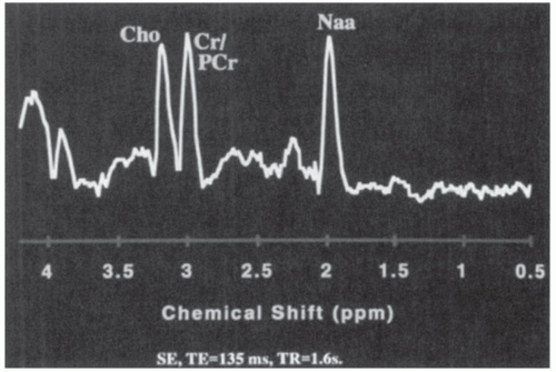
analysis is more difficult. With longer echo times, fewer peaks are captured, but the measurement precision is improved. In pediatric brain tumors, the three most important metabolic peaks (reading from right to left) are N-acetyl aspartate (NAA), 2.02 ppm; creatine-phosphocreatine (Cr/PCr), 3.02 ppm; and choline (Cho), 3.22 ppm (Fig. 26A.2).


Figure 26A.2 Normal long-echo single-voxel spectrum from the cerebellum of an age-matched control for a patient with a posterior fossa brain tumor. Reading from right to left, note normal peaks of N-acetyl aspartate (NAA), creatine-phosphocreatine (CR/PCr), and choline (Cho).

NAA is a marker of neuronal and axonal integrity. Cr is a marker for energy metabolism. Cho is a marker for cell membrane turnover and, as such, is elevated in tumors. The relative ratios of different metabolites vary, depending on the location of the voxel in the brain and on the age of the child. Normal age-matched control data from the same brain region are useful for interpreting the spectra from young children. As a general rule, the NAA increases over time, especially during the first 18 months of life, whereas the Cho slightly decreases over the same period. Cr/PCr tends to remain rather stable over time; for this reason, it has historically been used as an internal control when metabolic data are expressed as ratios.

The finding of a lipid-lactate peak usually indicates the presence of ischemia or necrosis. Lactate is a marker of anaerobic metabolism in hypoxic regions. Lactate peaks have been found to be more prominent in malignant gliomas than in low-grade gliomas.46

Single-voxel MRS can be used to interrogate new tumors having a volume greater than 1 mL3. However, a multivoxel technique, such as two-dimensional chemical shift imaging, in which several subcentimeter voxels can be examined simultaneously, can be very helpful in detecting and quantifying abnormal brain metabolites, identifying tumor tissue, differentiating tumor types, and distinguishing recurrent tumor from radiation necrosis.47

With higher-field-strength magnets that provide improved spectral resolution with shorter acquisition times, 3D spectroscopic evaluation of tumors can readily be performed.48,49

Single-voxel long-TE MRS data obtained by plotting Cr:Cho against NAA:Cho ratios have been used with benefit in separating posterior fossa medulloblastoma from pilocytic astrocytoma and ependymoma and in predicting disease progression.50 MRS also appears to be useful in monitoring the response of histologically proven pediatric glioma to adjuvant chemotherapy or radiation therapy as demonstrated by correlations between the ratio of tumor Cho to brain Cho versus tumor volume or clinical response.51 In patients with recurrent brain tumors, the Cho:NAA ratio also appears to have prognostic significance; children with a maximum Cho:NAA ratio of 4.5 or less had a projected survival of more than 50% at 63 weeks.52 Increasing Cho:NAA ratio has been associated with tumor progression, and nonprogressing or stable tumors exhibited a decrease in the Cho:NAA ratio.53 Single and multivoxel spectroscopy has been useful in the assessment of pediatric patients with diffuse intrinsic glioma.54


Gradient Echo Imaging

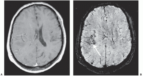
T2* gradient echo (GE) imaging has been used with good effect in detecting the presence of altered blood and blood products within tumors. There is some evidence for its lack of sensitivity and it has been replaced by susceptibility-weighted imaging (SWI), which is a high-spatial-resolution 3D GE MR imaging technique that uses phase processing to accentuate the paramagnetic properties of blood products.55 SWI has already proven itself superior to conventional GE imaging in the detection of hemorrhage in traumatic brain injury, coagulopathic or other hemorrhagic disorders, in vascular malformations, in the demonstration of venous thrombosis, and in delineating neoplasms that have hemorrhage, increased vascularity, or calcification.56 An example of a hemorrhage within a tumor demonstrated on SWI is shown in Figure 26A.3.


Diffusion-Weighted Imaging

MR diffusion images reflect the molecular translational motion (Brownian motion) of water within the section of the brain studied. Restriction of the water molecule motion can be observed in normal white matter tracts. This is called anisotropy; water molecules are restricted orthogonal to the white matter tracts. The motion of the molecules between the applications of the diffusion gradients leads to attenuation of the signal within each image voxel. From these images, a diffusion coefficient termed the apparent diffusion coefficient (ADC) can be calculated. MR diffusion using predominately echoplanar techniques has been useful in the characterization of tissue, tumor cellularity, tumor grading, tumor response to treatment, and distinction of tissue types. Numerous studies have confirmed that diffusion MRI is a biomarker for treatment response.57,58

Diffusion tensor imaging (DTI) provides visualization of fiber bundle integrity and direction with in vivo characterization of the rate and direction of white matter diffusion. This imaging is based on the diffusion tensor, which is a mathematical model of the 3D pattern of diffusion anisotropy of white matter tracts.59 It is used for presurgical planning or coregistration of tractography data with radiosurgical planning and functional MR data.60 DTI has also been used to assess the white matter tracts before and during therapy in pediatric brainstem glioma61 and as a biomarker to monitor effects of radiotherapy in children with medulloblastoma.62

Newer diffusion analyses such as functional MR diffusion maps are being applied to predict the treatment response in pediatric brain tumors.63 ADC histogram techniques are also being used to improve the identification of different types of brain tumors.64


Magnetic Resonance Perfusion Imaging

MR perfusion imaging is used to evaluate cerebral perfusion dynamics by analysis of the hemodynamic parameters of relative cerebral blood volume, relative cerebral blood flow, and mean transit time. The techniques used to perform perfusion imaging include T2*W dynamic susceptibility techniques, ASL techniques, and T1WI dynamic contrast-enhanced perfusion techniques. These techniques use either exogenous tracer agents, such as paramagnetic contrast material, or endogenous tracer agents, such as magnetically labeled blood (arterial water).65

The development of fast-echoplanar imaging has made possible an assessment of the vascularity of tumors using a gadolinium first-pass bolus technique. Imaging is performed dynamically (rapid imaging over time during a bolus injection), using echoplanar imaging-based spin echo or GE sequences. This relies on changes in the T2 signal of intravenous paramagnetic contrast agent administration (gadolinium) as it passes
through the region of interest.66 Resulting data, reflected in maps of relative cerebral blood volume, provide some semiquantitative analysis of the blood flow to a particular region. Early work has suggested a positive correlation between relative cerebral blood volume and tumor grade.67 Perfusion imaging may be helpful in targeting a lesion for biopsy. Application of perfusion imaging may be particularly useful for the study of neovascularization and angiogenesis inhibition particularly using the T1WI technique. A 3D dynamic sequence is employed. Kinetic modeling of the dynamic signal changes can yield estimates of regional fractional blood volume and microvascular permeability (Kps), which is an indicator of BBB disruption and correlates with angiogenesis.68


Figure 26A.3 A: T1-weighted MRI with contrast. B: Susceptibility-weighted image (SWI) showing hemorrhage in a glioma (arrow). (Courtesy of Mark Haacke, Ph.D., The Magnetic Resonance Imaging Institute for Biomedical Research, Detroit, Michigan.)

Arterial spin-labeling (ASL) is an MR perfusion technique that does not use an intravenous contrast agent and has been used in pediatric brain tumors to provide measures of cerebral hemodynamics. The major techniques used include pulsed and continuous ASL and a third type pseudocontinuous ASL.45


Activation Functional Magnetic Resonance Imaging

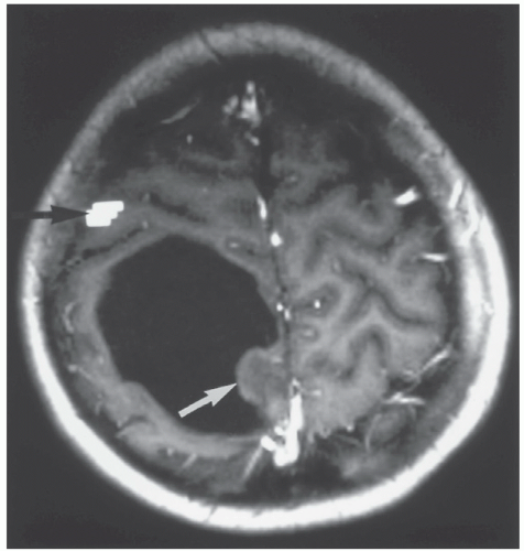
After the use of a stimulation paradigm or task designed to activate a specific functional area of the brain, the target can be located anatomically by an increase in blood flow to that area. Functional MRI (fMRI) uses the BOLD (blood oxygen level dependent) technique to generate differential signals based on the relative blood flow within the activated portions of the brain during an appropriate stimulus. Repetition of the task improves the robustness of the data, and subtraction of rest from activity reduces background signal. Data are presented on maps that outline the activated area of interest in relation to the lesion, and these may be useful in preoperative planning. An example involving finger-thumb opposition and the motor cortex of the hand is shown in Figure 26A.4. Different activation tasks can be designed to stimulate other eloquent areas of the brain for vision, hearing, and language.


Positron Emission Tomography

PET can serve as an imaging biomarker that enables early detection of response and can be correlated with outcome and/or progression-free survival (PFS) in children with brain tumors. The most commonly used tracer in the evaluation of brain tumors has been fluorodeoxyglucose. However, molecular imaging researchers are evaluating new markers of tumor physiology through novel
18F-labeled agents such as fluorothymidine (FLT) for the assessment of cell proliferation and fluoro-DOPA and fluoromisonidazole for the assessment of protein synthesis and hypoxia within the tumor, respectively. Recent work in children with brainstem gliomas demonstrated that FDG-PET parameters correlated with survival.69


Figure 26A.4 Preoperative functional magnetic resonance imaging in an 11-year-old boy presenting with left-sided focal motor seizures, performed for surgical planning. The bright pixels (black arrow) represent the statistical parametric map produced by using a left-sided finger-thumb opposition paradigm. The bright pixels reflect the increase in oxygenated blood flowing away from the right-sided sensorimotor cortex. The white arrow indicates the solid tumor component within a cystic mass displacing the sensorimotor cortex anteriorly. Histopathology demonstrated a cystic dysplastic ganglioglioma that later was removed successfully via a posterior approach without damage to the child.


Future Developments

Technological advances in imaging with higher-field-strength scanners, sophisticated imaging-processing techniques, and radiopharmaceuticals will serve to characterize direct or surrogate imaging markers of response to new therapies aimed at improving our understanding of the physiologic and metabolic profiles of pediatric brain tumors.


NEUROSURGERY: DIAGNOSIS AND TREATMENT

For most CNS tumors, surgical intervention forms the initial step in the treatment plan by providing tissue to establish the histologic diagnosis and, when possible, reducing the tumor burden. Notable exceptions to this rule are certain unresectable tumor types, such as diffuse infiltrative brainstem gliomas and globular chiasmatic gliomas in children with NF1. Each of these tumors has an almost diagnostic MRI appearance in association with consistent histologic features. As surgery has been shown not to improve prognosis in patients with these tumors and the histologic diagnosis rarely is in question, operative intervention is not required, although with the increasing adoption of molecularly targeted therapeutic strategies, stereotactic needle biopsy may in the future play a role in guiding protocol-directed therapy, particularly in brainstem gliomas, based on recent evidence that it can be accomplished with low morbidity.70 Similarly, for certain deep-seated parenchymal tumors, the risks imposed by a conventional surgical approach to them may be high, and for establishing a histologic diagnosis, stereotactic biopsy techniques provide a reasonable alternative to open tumor debulking. CT- or MRI-guided stereotactic procedures are highly accurate in targeting such deep-seated lesions and are associated with a morbidity of less than 5% and a mortality of less than 1% of patients.71,72

For the majority of pediatric brain tumors, however, open operations are preferred; the goal in such procedures is to remove as much tumor as is safely possible. Although a truly complete tumor resection is feasible only for well-circumscribed benign tumors, such as pilocytic astrocytomas and some craniopharyngiomas, an extensive, near-total resection can be achieved with many parenchymal tumors. The limitation to complete resection in such tumors is the imperceptible blending of the neoplasm into the surrounding brain; scattered tumor cells may infiltrate past the margins of the resection into the normal parenchyma. Unlike the situation with solid, non-CNS tumors, it is rarely feasible to resect the tumor with surrounding margins of normal tissue because of the unacceptable risks of producing irreversible neurologic deficits. An outline of the neurosurgical approach and considerations for gliomas, ependymomas, and other nonembryonal tumors of the CNS are provided in Table 26A.4.


Preoperative and Perioperative Considerations

In occasional affected children who present with obtundation from a large mass lesion, resection is performed urgently. In children who are awake and alert but nonetheless harbor a large lesion that is producing substantial mass effect, the tumor resection is performed on the next operating day. Smaller lesions without significant mass effect can be managed more selectively.

Because peritumoral edema commonly contributes to the neurologic impairment produced by the tumor, moderate doses of corticosteroids generally are administered preoperatively. For example, 0.1- to 0.5-mg/kg dexamethasone may be given every 6 hours. This dosing often will lead to a dramatic improvement in the patient’s symptoms and signs, avoiding the need for emergency surgery in the vast majority of cases. Corticosteroids are typically continued intraoperatively and during the early postoperative period. If a significant reduction in tumor volume has been achieved at the time of operation, corticosteroid therapy is tapered and then discontinued within several days of surgery.

Another factor that commonly contributes to increased ICP in children with brain tumors is the presence of obstructive hydrocephalus, observed most commonly in tumors arising near the aqueduct, such as with pineal region tumors, or the fourth ventricle, as seen with cerebellar vermian lesions. Although resection of the tumor often opens the CSF pathways and leads to resolution of the hydrocephalus, the resection frequently is rendered safer if the elevated pressure is relieved as an initial step. The use of preoperative shunting carries a small risk of upward herniation through the tentorial hiatus. In some institutions, preoperative relief of hydrocephalus is accomplished by endoscopic third ventriculostomy; however, a more common approach is to place an external ventricular drain immediately before the craniotomy for tumor resection, which has the advantage of allowing drainage of bloody spinal fluid and debris in the early postoperative period. If the operative procedure opens the CSF pathways and the patient’s absorptive pathways remain patent, the external ventricular drain often can be removed within several days of surgery.73 If the hydrocephalus persists, a third ventriculostomy can be performed or, if the hydrocephalus appears to be of a communicating type, a ventriculoperitoneal shunt can be inserted. Although in the past, a great concern was that shunting would provide a route for systemic dissemination of tumor, an increased risk of tumor spread via the shunt has not been demonstrated.73

The endocrinopathies commonly manifested by hypothalamic tumors can be exacerbated by tumor resection. Patients undergoing removal of tumors in this location typically require stress doses of hydroxycorticosteroids before, during, and after surgical intervention. Although thyroid hormone replacement is occasionally instituted preoperatively, it is most commonly initiated postoperatively. Patients in whom the posterior pituitary stalk is sectioned or injured during surgery often manifest a triphasic response of impaired fluid regulation, characterized by an initial period of transient diabetes insipidus lasting 1 to 2 days, a subsequent period of inappropriate antidiuretic hormone release lasting several days, and a final phase of persistent diabetes insipidus. In view of the rapid changes in vasopressin levels during the first several days postoperatively, careful attention to fluid replacement and cautious administration of synthetic vasopressin, where indicated, are essential to avoid potentially deleterious swings in electrolyte levels and fluid balance.

Children with cerebral cortical tumors and those in whom cortical retraction is required in the approach to a deep-seated lesion may be at risk for seizures during the perioperative period. Preoperatively, such patients often are started on an anticonvulsant medication (e.g., phenytoin) that is continued during the postoperative period, even if they have not experienced previous seizures. Patients generally are maintained on anticonvulsants for at least 1 week postoperatively; the decision to continue such therapy in patients without documented seizures is of uncertain benefit. In patients who experience a preoperative seizure disorder from the tumor and have been rendered seizure-free by tumor resection, anticonvulsants often can be stopped within several months after surgery.


Intraoperative Considerations and Surgical Technique

Multiple studies have demonstrated that the extent of surgical resection has a major impact on the likelihood of long-term survival
for children with many types of pediatric brain tumors, particularly ependymomas,74 high-grade gliomas,75,76 medulloblastomas (see Chapter 26B), low-grade gliomas,77 and choroid plexus tumors.78 Accordingly, with the exceptions noted earlier, in which surgical resection is not indicated or in which stereotactic biopsy may be a preferable initial step, extensive resection is the goal for many types of pediatric brain tumors.


TABLE 26A.4 Management Schema for Pediatric Brain Tumorsa


Tumor Type


Surgical Interventionb


Common Morbidity


Comments


Supratentorial hemispheric


Low-grade glioma


GTR if possible


Varies with site (e.g., hemiparesis, hemisensory deficits, hemianopsia, seizures)


Outcome improved by extensive resection


Pilocytic


Fibrillary


High-grade glioma


GTR if possible


Varies with site


Outcome improved by extensive resection


Mixed neuronal-glial neoplasms


GTR if possible


Varies with site


Outcome improved by extensive resection


Ganglioglioma


DIG


DNET


Ependymoma


GTR if possible


Varies with site


Outcome improved by GTR


Choroid plexus tumors


GTR if possible


Hemiparesis, subdural hygromas, life-threatening blood loss in infants


Outcome improved by GTR


Supratentorial midline


Chiasmatichypothalamic glioma


Resection reserved for exophytic lesions with substantial mass effect; biopsy used if the diagnosis is uncertain; in NF-1, the diagnosis is often made by imaging


Vision loss common with biopsy; hypothalamic dysfunction and hemiparesis with extensive resection


In infants, possible to delay need for adjuvant therapy by extensive resection; remains controversial whether this improves results over those obtained with biopsy and chemotherapy


Craniopharyngioma


Depending on lesion type, GTR, subtotal resection, or stereotactic approaches potentially optimal


Increased neuroendocrine deficits common after extensive resection; vision loss, personality changes, and neurologic impairments also common


Progression-free survival possibly improved by extensive resection, but potential for morbidity also increased


Infratentorial


Cerebellar astrocytoma


GTR if possible


Dysmetria, ataxia


GTR usually curative


Ependymoma


GTR if possible


Dysmetria, ataxia, cranial nerve palsy, paresis, transient mutism


Outcome improved by GTR


Diffuse (malignant) brainstem glioma


Generally an imaging diagnosis, except in rare instances; biopsy may have a future role in directing molecularly targeted therapy


Cranial nerve dysfunction, paresis


Benefits unproven


Benign (focal) brainstem glioma


Craniotomy, near-total resection feasible in selected cases


Cranial nerve dysfunction, paresis


Excellent outcome for dorsally exophytic and cervicomedullary tumors after near-total resection; for other focal tumors, remains uncertain whether results after resection are superior to those after biopsy and irradiation


aAlthough this schema summarizes general management approaches, a more extensive presentation of treatment caveats for individual tumor types is provided within the text. Because the details of treatment for many tumor types have evolved over time and will likely continue to evolve, treatment decisions for individual patients are best made in the context of a multidisciplinary approach.

b Perioperative measures, such as the use of corticosteroids to reduce mass effect and the use of anticonvulsants for lesions at high risk for seizures, are provided in the text.


GTR, gross-total resection; DIG, desmoplastic ganglioglioma; DNET, dysembryoplastic neuroepithelial tumor.


A major limitation to the widespread incorporation of extensive surgical resections in the management of childhood brain tumors has been the fact that aggressive resections may increase the risk of immediate- and long-term morbidity, particularly for tumors in functionally critical locations. Although morbidity generally is less than 10% for polar supratentorial gliomas and less than 20% for cerebellar astrocytomas, more deep-seated lesions,
such as ependymomas and craniopharyngiomas, carry morbidity rates in excess of 40% with extensive resection.79 Although some studies have observed that morbidity is lower if operations are performed by neurosurgeons who do such operations frequently,80 other studies show that pediatric neurosurgeons are more likely to attempt extensive removals, on the basis of their recognition that this influences prognosis, and therefore may have overall rates of management of morbidity comparable with those of general neurosurgeons, albeit with a higher frequency of complete or nearly complete tumor resections.81

During the last 10 to 20 years, a number of intraoperative modalities have been developed or refined to allow tumor resection to be performed more safely and efficiently. Foremost among these are the progressive improvements in operative microscopy, which facilitates illumination and visualization of the interface between the neoplasm and the normal brain. Localization techniques, such as frame-based and frameless stereotactic guidance systems, allow preoperative targeting of the tumor so that the surgical approach can be tailored precisely to minimize manipulation of normal brain structures and to maximize the extent of resection of deep-seated subcortical lesions.82

Ultrasonographic guidance also is useful in this regard. Intraoperative MRI units have become available in an increasing number of centers and may help to refine further the accuracy of intraoperative decision making,83 provided that issues of cost and the difficulties involved in operating in or adjacent to a high-field-strength magnet can be resolved effectively.

In children whose tumors are in and around functionally critical brain regions, intraoperative monitoring of visual, auditory, and somatosensory pathways and direct assessment of motor and speech pathways often are used in an attempt to improve the safety of the tumor resection. In addition, areas of essential cortex overlying a deep-seated tumor may be delineated using cortical stimulation techniques to plan an approach to the tumor that avoids traversing important structures. Functional MRI and diffusion tensor imaging also provide a noninvasive way of delineating localizing important cortical and subcortical areas84 to identify a safe trajectory to an underlying lesion. Finally, in children with intractable seizures from cerebral neoplasms, intraoperative or extraoperative electrocorticography (ECOG) may be used to define areas of epileptogenic cortex in and around the tumor to increase the likelihood that seizure control will be obtained postoperatively.85

For supratentorial craniotomies, children generally are placed in the supine or lateral position or prone for occipital lesions. For infratentorial craniotomies, the prone or lateral position is used more often than the “sitting” position because of concern over potential venous air embolism. Intraoperatively, the head of an infant often is positioned on a soft headrest, rather than being held by pins that can perforate the skull or cause a depressed fracture. For cortical and many subcortical tumors, the surgical approach follows the most direct trajectory to the lesion. However, for deep-seated lesions that are subjacent to functionally critical regions of the brain, alternate approaches often are required. Details of the operative approaches for specific tumor types are provided in subsequent tumor-specific sections of this chapter and Chapter 26B.

The actual tumor resection often is aided by the use of ultrasonic aspiration, which provides a relatively atraumatic way to debulk many pediatric brain tumors. The surgical laser also may be used, depending on the consistency and location of the tumor. In general, tumors are resected “from the inside out.” With many extra-axial tumors and a small percentage of intraparenchymal lesions, a clearly defined peritumoral plane is encountered through which the tumor may be dissected carefully from the surrounding brain, cranial nerves, and vessels after the central portion of the mass has been debulked. However, for most intraparenchymal tumors, a well-defined tumor capsule is not present, and the resection must proceed via gradual internal debulking until the boundary between the tumor and the normal brain is reached.

Because the extent of resection is so important in defining prognosis and choice of subsequent therapy for many tumor types, objective confirmation of the volume of residual tumor, if any, is essential before embarking on further therapy. Because a surgeon’s impression of the extent of tumor resection is subject to error, postoperative confirmation of the extent of resection generally is established by CT or, preferably, by MRI. This imaging typically is performed within the first 24 to 72 hours postoperatively to minimize the impact of postoperative inflammation on the delineation of areas of residual tumor.

A trend over the last decade in the surgical management of selected types of brain tumors has been the concept of second-look surgery. For large, relatively vascular tumors in which an initial complete resection cannot be obtained, the patient is treated with several courses of postoperative chemotherapy in the hope of making the tumor amenable to complete resection at a second procedure. This approach has been applied anecdotally in ependymomas, a tumor type in which the extent of residual disease before initiation of radiation therapy has a substantial impact on long-term outcome.86 What remains to be determined is whether patients who undergo a second-stage complete resection have as good a prognosis as those who were amenable to complete resection initially; this issue is being examined systematically in studies of the Children’s Oncology Group (COG).

Surgical resection has been used increasingly as a component of the management of recurrent disease, particularly in children without evidence of tumor dissemination. For children with malignant lesions, this relieves mass effect in preparation for additional investigational chemotherapy on phase I or II clinical trials. Some recurrent tumors, such as JPAs and craniopharyngiomas, can be treated with reoperation alone, without the need for additional adjuvant therapy, if a gross-total resection (GTR) can be achieved. For children with recurrence of other, more malignant tumors that may also be subject to dissemination, data are lacking for a survival benefit from re-resection.


Postoperative Considerations

The postoperative recovery of patients who undergo a complete or partial resection of a posterior fossa tumor, particularly a tumor in or around the cerebellar vermis, may be complicated by posterior fossa syndrome. Posterior fossa syndrome, also known as “cerebellar mutism,” may range from a mild to severe disorder that is characterized by mutism, ataxia, hemiparesis, cognitive impairment, behavioral changes, cranial nerve palsies, bulbar palsy, and tremor.87 The onset is typically within the first week after surgery and appears to occur more commonly with medulloblastoma than with other posterior fossa tumors.88

The etiology of the posterior fossa syndrome is unknown but appears to be associated with edema in the brachium pontis or with decreased blood flow during surgery. Previously reported as a rare complication of posterior fossa surgery, for unclear reasons, this syndrome is now observed in 10% to 25% of patients following tumor resection in the posterior fossa.88 Although recovery may be complete, particularly for the mutism, there are patients in whom it is incomplete with long-term neurologic sequelae.88,89


RADIATION THERAPY

Radiation therapy is a central component of curative as well as palliative therapy for a majority of children with CNS tumors. The potential efficacy of radiation in pediatric CNS tumors has been apparent since the middle of the 20th century. During the last 25 years, the potential detrimental effects of radiation in the developing and mature CNS have also been quantified. Recognition of the unique CNS vulnerabilities in children and the concurrent demonstration of brain tumor responsiveness to chemotherapy introduced a paradigm of delaying or avoiding
irradiation in children. With the introduction of sophisticated, 3D-image-guided radiation techniques capable of relative sparing of the normal brain structures, the focus of pediatric brain tumor trials has shifted to investigating broader indications for radiation as a key element in achieving disease control. Current studies are addressing ways to optimize the risk-to-benefit ratio of accurate, limited-volume radiation delivery, sometimes in the setting of reduced radiation dose as well.

The rational application of radiation therapy in pediatric brain tumors requires an understanding of brain development, biologic effects of ionizing radiation on the brain of a child, the behavior and natural history of various brain tumors, radiobiology and physics, techniques and technology of radiation therapy, and the interactions of radiation with other treatment modalities, such as chemotherapy.


Indications for Radiation Therapy

The indications for radiation therapy depend on tumor histology. Pretreatment histologic diagnosis is required except in cases of diffusely infiltrating pontine gliomas and visual pathway gliomas, both diagnosed on the basis of neuroimaging and neurologic findings. For ependymomas and many of the astroglial tumors, the use and timing of radiation also depend on the anatomic site of involvement and the degree of resection. Specific indications for radiation therapy and the controversies concerning its use are discussed later under the individual tumor-type headings.


Radiation Volume

The radiation target volume is determined by tumor histiotype, anatomic extent, and known patterns of spread and failure. Advances in neuroimaging (particularly MRI, with current treatment planning often based on fusion of MRI and CT imaging and the use of fMRI and PET imaging currently under exploration)90 have made tumor localization more accurate.

Local target volumes are used for tumors that are typically confined to a single anatomic location (e.g., ependymomas, craniopharyngiomas, and most astroglial tumors). Determining the target volume requires a complex integration of preoperative and postoperative/preirradiation imaging (accounting for reconfiguration of the brain following surgery and, in clearly defined settings, response to chemotherapy) to identify the “tumor” or “tumor bed” as the gross target volume (GTV). Depending on the tumor type, a margin for potential microscopic infiltration (the clinical target volume or CTV) is identified by a 3D expansion of the GTV by approximately 1 cm (e.g., discrete pilocytic astrocytomas, craniopharyngioma, ependymomas), 1 to 2 cm (e.g., medulloblastomas when boosting only the tumor bed region [see Chapter 26B]), or 2 cm (e.g., high-grade gliomas, diffuse-infiltrating brainstem gliomas, or infiltrating WHO grade II astrocytomas). Finally, a volumetric expansion of the CTV (typically by 0.3 to 0.5 cm) defines the planning target volume (PTV), recognizing some variability in the daily setup or patient positioning despite considerable attention to immobilization and reproducibility.

Craniospinal irradiation (CSI) is a technically demanding technique that provides homogeneous irradiation to the cranium and spine, targeting the entire subarachnoid space in tumors with known potential for (or established) intracranial and/or spinal leptomeningeal metastasis (e.g., medulloblastoma or disseminated ependymoma).


Radiobiologic Considerations

The biology of radiation cell lethality for tumors and normal tissues is outlined in Chapter 13. There are differences in inherent tumor cell radiosensitivity; for instance, medulloblastoma cell lines show significantly greater cell death after 2-Gy exposure than glioblastoma cell lines.91 Fractionation (the principle of multiple, relatively small doses of radiation protracted over time) is particularly important in the normal tissue tolerances seen in the brain, spinal cord, and several other critical organs. Most data have been derived from radiation effects following conventional fractionation (i.e., the use of a single daily fraction of 1.8 to 2.0 Gy, typically on a 5-days-per-week schedule). Hyperfractionated delivery (delivering two daily fractions of 1.0 to 1.2 Gy each to total doses up to 20% to 30% higher than “tolerance levels” defined following conventional fractionation) was purported to show a significant benefit in childhood brainstem gliomas.92 The therapeutic ratio (i.e., risk-to-benefit ratio) was felt to be improved after high-dose hyperfractionated irradiation. The primary theoretical benefit of hyperfractionated radiation is the opportunity to escalate the total radiation dose without increasing the damage to normal structures. In theory, benefit from hyperfractionation should result because the antitumor effects from irradiation are primarily related to the total dose rather than the dose per fraction, whereas the side effects of irradiation correlate more often with dose per fraction.93

Radiation effects on brain generally are characterized as delayed reactions, reflecting slow turnover times of normal brain parenchymal cells or indirect effects on cerebrovascular structures. Prospective data from a number of cooperative-group and single-institution experiences show no clear benefit in tumor response or outcome following hyperfractionated irradiation in brainstem gliomas.94 Details about hyperfractionation in medulloblastoma are outlined in Chapter 26B.


Techniques in Radiation Therapy


Conventional External-Beam Radiation Therapy

Conventional radiotherapeutic techniques are appropriate for pediatric brain tumors only when large volumes are treated (e.g., craniospinal or full-cranial irradiation). Simple geometric field arrangements (typically two or three per radiation volume) with customized blocking provide relatively homogeneous irradiation within the defined target volume.


Three-Dimensional Conformal Radiation Therapy

Advances in neuroimaging and sophisticated 3D computerized treatment planning systems have greatly improved the ability to target the tumor while significantly sparing the surrounding normal tissues. Three-dimensional conformal radiation therapy (3D-CRT) typically involves multiple, individually shaped (or collimated) fields, arranged in coplanar, nonaxial, and noncoplanar orientation delivered in either static or dynamic mode. Compared with conventional radiation therapy, 3D-CRT more accurately targets the CTV while simultaneously reducing the volume of normal brain exposed to high-dose radiation.


Intensity-Modulated Radiation Therapy

Intensity-modulated radiation therapy (IMRT) is a more complex form of 3D-CRT, combining two advanced concepts with 3D-CRT: (a) inverse treatment planning (in which both the target volume and the adjacent or subtended normal tissues are assigned specific dose levels), allowing optimization of beam trajectories and weights in an overall plan, and (b) computer-controlled intensity modulation of the radiation beam during treatment. IMRT allows a high degree of flexibility in reducing the dose to the surrounding normal tissues by the creation of so-called avoidance areas during the treatment planning process. Most IMRT programs prioritize dose avoidance over target volume homogeneity; the latter is a potential consideration in intrinsic brain tumors.95

Some have hypothesized that IMRT may increase the risk of second malignancies because of the use of multiple fields and higher leakage doses, a hypothesis that rests on the assumption that second malignancy risk is heavily influenced by low-dose exposure. However, a comprehensive review of 22 large cohort and case-control studies with IMRT led to the conclusion that IMRT
and multi-beam plans do not significantly increase the risk of second malignancy and that the risk increases with dose.96


Stereotactic Radiosurgery

Radiosurgery is generally administered by a Gamma-Knife unit (based on a highly collimated dose array using 192-201 fixed cobalt-60 sources), CyberKnife, or modified linear accelerator unit. Radiosurgery systems are designed to deliver a high-radiation dose to a small intracranial target in one fraction by focusing multiple small-radiation beams from different directions to the target, resulting in a steep dose gradient just outside the edge of the target. The application of radiosurgery usually is limited to tumors measuring 3 cm or less in maximum diameter, to minimize the risk of toxicity to normal tissue surrounding the target. Lesions within or immediately adjacent to critical structures (optic nerve/chiasm, brainstem) may be treated, though the size limitation may be stricter. Radiosurgery commonly is used for the management of brain metastases, acoustic neuromas, arteriovenous malformations (AVMs), vestibular schwannomas, meningiomas, and a variety of other tumors. In children and adolescents, radiosurgery has been used quite selectively in well-circumscribed, intrinsic low-grade gliomas (e.g., JPAs in the midbrain) not amenable to complete resection and in focal areas of residual craniopharyngiomas.97 Note that subacute reactions (intralesional necrosis, often associated with transient expansion of a space-occupying lesion) limit enthusiasm for this approach in treating lesions intrinsic to the brainstem.98


Particle Beam Irradiation

There is considerable current interest in the use of particle beams (most often protons) in radiation therapy for children with CNS tumors. Proton beam radiation provides an advantage over photon radiation, in that the proton energy can be modulated to provide radiation to a selected depth, nearly obviating the exposure of underlying tissue (compared with photon beams that pass through the target region with gradually diminishing dose intensity). This can result in an improvement of the dose distribution and administration of lower doses to the surrounding normal tissues. However, many questions remain regarding not only the clinical benefit but also the precise dose distribution of protons and this has resulted in continued controversy. Nowhere is this controversy more evident than in the treatment of children requiring CSI. Some have argued that the incontrovertible superiority of proton radiotherapy over photons for treatment plans such as CSI mandates the offer of referral of children requiring such therapy to centers capable of proton radiotherapy;99 however, such arguments reference published reports of lower radiation-related second neoplasms, reports that if examined carefully fail to document any advantages in second malignancy rates.100,101 Published literature has failed to demonstrate that protons can mitigate the major late morbidities following CSI in children, consisting of growth retardation, neurocognitive decline, ototoxicity, and endocrinopathies.102 For example, in examining ototoxicity, the dosimetry of protons appears favorable, but clinical data fail to show a translation into clinical benefit compared to IMRT with photons.103,104 In studies by experienced pediatric centers, the incidence of high-grade hearing loss with protons versus IMRT photons was nearly identical.103,104 Furthermore, studies of patterns of relapse after proton treatment for medulloblastoma have raised concern for higher-than-expected “spine-only” recurrences105 and toxicity studies have highlighted significant rates of brainstem toxicity following proton radiotherapy for brain tumors.106

The physical advantages of proton beam radiotherapy must be weighed against proton beam contamination by neutrons that may theoretically increase the risk of second malignancies, particularly in children. Such increased risk may be mitigated, as passive scatter techniques are replaced by active scanning approaches.107 In passive scanning, protons are scattered by a foil to cover the target, which leads to production of neutrons and, in turn, increases the total-body dose up to 10-fold compared with IMRT. Conversely, active scanning techniques offer better conformity of highest doses, potentially lower doses to normal structures, and reduced beam contamination by neutrons. Investigations into the clinical efficacy and long-term toxicities of proton radiotherapy are ongoing and of critical importance in the pediatric population.


Brachytherapy

Brachytherapy, or interstitial irradiation, involves the implantation of radioactive sources directly into brain tumors. Iodine-125 and iridium-192 are radioisotopes that have been used for either permanent or temporary implants. Brachytherapy has been used for patients with glioblastoma as an additional boost after external-beam radiotherapy and for selected patients with recurrent high-grade glioma. Anecdotal favorable results have also been reported in patients with low-grade gliomas. Dose homogeneity and ready availability of 3D-CRT or IMRT have supplanted much of the enthusiasm for technically demanding implant procedures in CNS tumors. Intralesional brachytherapy (typically phosphorus-32) has been utilized with some success in cystic neoplasms, most often craniopharyngioma cysts.108


Radiation and the Developing Brain

Brain development is most rapid during the first 3 years of life. Axonal growth and synaptogenesis are most active during the growth phase. The rate of growth and development decrease after 6 years of age. However, maturation of the brain, judged by the degree of myelinization, is not complete until puberty.109 For infants and young children, white matter alterations after radiation appear to primarily mediate the functional and neurocognitive changes that are observed in these patients.110 Younger children are more vulnerable to white matter changes that may be localized within or adjacent to the high-dose regions or may extend, presumably by axonal degeneration, along white matter tracts remote from the primary tumor.111


Radiation Effects on the Brain

CNS responses following irradiation are classically divided temporally as (a) acute reactions, occurring during treatment; (b) subacute or early delayed reactions, occurring a few weeks to 2 months after irradiation; and (c) late reactions, occurring several months to years after treatment.112 The pathogenesis of early and, more often, subacute radiation-induced brain injury includes inflammatory-like changes with associated intra- or perilesional edema; direct damage to oligodendrogliocytes resulting in inhibition of myelin synthesis and consequent white matter degeneration/loss; and damage to the vascular endothelium, resulting in areas of hypoxia or release of necrosis factors with attendant white matter necrosis.113,114,115 Immediate peritumoral edema or intralesional necrosis is uncommon with conventional fraction sizes (i.e., 1 to 3 Gy per fraction).

Subacute reactions include imaging and clinical findings that may mimic the primary tumor or that result in constitutional symptoms (e.g., lassitude, low-grade fever, less often alterations in recent memory; diffuse white matter changes identified as leukoencephalopathy); these reactions are typically time-limited, resolving within several weeks to a few months.

Late reactions are more clearly dose- and volume-dependent, occurring beyond 6 to 12 months after radiation; these effects are typically permanent. Late reactions include focal radiation necrosis, a more diffuse pattern of radiation- and/or chemotherapy-induced leukoencephalopathy, neuropsychological effects, cerebrovascular effects, and secondary neoplasms (both benign and malignant). Late effects can be progressive, irreversible, and sometimes fatal. Late neuropsychological effects are particularly of concern for younger children and include intellectual impairment, memory deficits, and limited ability to acquire new knowledge.115 Impairment in cognition is most pronounced in children younger
than 4 to 7 years.116 Deterioration in IQ is more prevalent in children following whole-brain or “focal” supratentorial radiation than after treatment confined to the posterior fossa.115

The presence and severity of radiation reactions depend on (a) radiation treatment factors, including total dose, fraction size, interfractional interval, and treatment volume; (b) patient factors, such as age, presence of preexisting brain injury by tumor or surgery, infection, and vascular diseases; and (c) other treatment modalities, most commonly surgery and chemotherapy. The influence of certain factors, such as fraction size, treatment volume, and dose homogeneity, can be modified or optimized to limit the incidence and severity of brain injury.112


Radiosensitivity of Specific Structures in the CNS


Brainstem

In the modern radiotherapy era, incidental brainstem necrosis is quite rare; there are no data suggesting the brainstem is more sensitive to irradiation than other normal brain structures. Subacute radiation effects, such as those that occur in the management of diffuse intrinsic pontine gliomas (DIPG) or large, focal intrinsic JPAs, can be problematic. For example, imaging changes can be difficult to differentiate from tumor progression and clinical signs may be quite pronounced, particularly focal intrinsic brainstem reactions. With 3D-CRT to “tolerance” levels of 54 to 60 Gy, one can see subacute white matter changes on MRI several months after radiation; changes are often asymptomatic and transitory but may progress to frank necrosis.117


Spinal Cord

In view of its location, the tolerance of spinal cord to radiation is a major dose-limiting factor in delivering high-dose radiation to tumors of the head and neck region, the thorax, and the upper abdomen; cord tolerance is more problematic in general adult radiation oncology than in pediatrics, in which tolerance levels are often approached only in intrinsic spinal cord tumors or tumors with sizable metastatic subarachnoid foci. In pediatric radiation oncology, the spinal cord is more often the targeted tumor volume than an unintended critical structure. CSI is common although dose levels to the entire spine rarely approach or exceed the 44-Gy level identified as “safe” in the United States using conventional fractionation.112,118 More limited spinal volumes are often treated to dose levels approximately 45 to 50 Gy or, occasionally, 54 Gy.112,119 With contemporary 3D, image-guided radiation for low-lying posterior fossa lesions (e.g., fourth ventricular ependymomas) or white matter lesions, as described previously for the brainstem, can occur in the cervicomedullary or upper cervical cord regions.117 Although most subacute effects are transitory (white matter on MRI, Lhermitte syndrome of a shock-like sensation radiating down the extremities associated with neck flexion), such changes can be associated with significant neurologic signs and symptoms.

Frank postirradiation myelopathy can occur from 1 year to several years after treatment.119 The traditional dogma concerning the pathogenesis of radiation myelopathy rests on postmitotic cell death in the endothelial cells or oligodendrocytes (or both).112 The current view is that radiation produces cell death that induces a complex pathophysiologic reaction in which the response of surviving cells may contribute to the impact of radiation on tissue integrity and functions. Cytokines, such as tumor necrosis factor and interleukin-6 (IL-6), appear to play important roles.120 Also, some researchers suggest that the tolerance of the spinal cord is 5% to 10% lower in children than in adults.120 Large fraction sizes (≥25 Gy) are disproportionately associated with untoward biologic effect on the spinal cord. An apparent volume relationship suggests that the dose to the spinal cord should be reduced when the irradiated length is large.112 Results of primate studies suggest that an increase in treatment volume reduces the threshold and steepens the slope of the sigmoid dose-response curve for myelopathy.120


Cranial Nerves

Most cranial nerves are relatively resistant to radiation-induced damage. Two cranial nerves, the second (optic) and eighth (vestibulocochlear), are particularly worth mentioning in the radiation treatment of pediatric cancers. The optic nerve and visual pathway can be damaged during the delivery of therapeutic radiation to periorbital tumors (e.g., orbital rhabdomyosarcoma, optic glioma, paranasal, and nasopharyngeal tumors) and suprasellar tumors (e.g., craniopharyngioma, pituitary adenoma, germ cell tumor, hypothalamicchiasmatic glioma).121 The risk of radiation-induced optic neuropathy is related to total radiation dose, fraction size, and irradiated volume. For example, no injuries were observed in 106 optic nerves that received a total dose of less than 59 Gy. However, the 15-year actuarial risk of optic neuropathy after a dose >60 Gy was 11% when treatment was administered in fraction sizes of <1.9 Gy, as compared with 47% when given in fraction sizes of >1.9 Gy.122

The vestibular cochlear nerve and auditory apparatus must be considered in the delivery of high-dose radiation to posterior fossa tumors, such as medulloblastoma, ependymoma, and astrocytoma. Although the incidence of early ototoxicity is typically related to cisplatin delivery (and potentially enhanced when administered with or after irradiation), radiation exposure to more than 40 to 50 Gy is associated with a small but finite incidence of late radiation-induced ototoxicity, presumably as a late effect on vestibular nerves.123 The combination of radiation and cis-platinum may be associated with a greater incidence of ototoxicity, highlighting the importance of prospective audiologic studies in children with CNS tumors receiving both therapies.124 To minimize the risk of hearing loss, some recommend a cumulative cochlear dose of less than 35 Gy for patients planned to receive 54 to 59.4 Gy in 30 to 33 treatment fractions.123


Retina

The retina, a specialized neural end organ supplied by an endarterial system, is sensitive to vascular injury and has little ability for repair.118 It is sensitive to radiation as well. Deterioration of vision, resulting from radiation-induced progressive obliteration of small retinal vessels, can occur 1.5 to 6.0 years after irradiation. The dose-response curve is steep (with increasing incidence at dose levels between 50 and 60 Gy), and 45 Gy produces a 5% risk of visual injury within 5 years.125 Again, as the fraction size increases up to 2.5 Gy or more, the frequency of injury increases.125


Lens

The lens is one of the most radiosensitive organs, even to very low doses of radiation. For example, 1 Gy can lead to cataract formation. From total-body irradiation data, the risk of developing a cataract requiring surgery was 20% for fractionated doses of 12 to 16 Gy.126 The dose that produces a 5% risk of damage to the lens within 5 years is 10 Gy.


Hypothalamic-Pituitary Axis

Irradiation of the region of the hypothalamus and pituitary gland can result in significant neuroendocrine abnormalities and longterm sequelae. The hormones affected include growth hormone (GH), thyroid-stimulating hormone, adrenocorticotropic hormone, and follicle-stimulating hormone and luteinizing hormone. The largest volume of data concerns the effect of cranial irradiation on GH production and release. Impaired serum GH response with provocative testing is apparent in 60% to 80% of children who have survived brain tumors.93,127 A dose-response relationship is seen with a threshold of 18 to 25 Gy. The higher the dose of radiation, the earlier the GH deficiency occurs.

Deficiencies of other hypothalamic-pituitary hormones have also been described. Constine et al.128 have described non-GH abnormalities (thyroidal, gonadal, prolactin, and adrenal) in 20
children with brain tumors not involving the hypothalamic-pituitary region and treated with either cranial or craniospinal irradiation. In patients receiving only cranial irradiation, the hypothalamic-pituitary region is estimated to receive a mean dose of 53.6 Gy (40 to 70 Gy).128


PRINCIPLES OF CHEMOTHERAPY

The role of chemotherapy in the treatment of childhood brain tumors has become increasingly important over the past several decades particularly for some of the embryonal (Chapter 26B) and low-grade glial neoplasms. The specific indications for chemotherapy in childhood CNS tumors are described in the respective tumor-specific sections.


Factors Influencing Drug Exposure in the CNS

The BBB and blood-CSF barrier are natural membrane barriers in the CNS that profoundly influence the penetration of most substances into the CNS. A detailed description of these barriers, the factors that influence CNS tumor penetration of an agent across them, and mechanisms to circumvent them (including disruption of the BBB, high-dose systemic chemotherapy, and intrathecal chemotherapy) are described in Chapter 10 and several review articles.129,130


Novel Agents and Approaches


Differentiating Agents

The potential utility of differentiating agents, such as retinoic acid and inhibitors of histone deacetylase (phenylbutyrate, valproic acid, depsipeptide, and suberoylanilide hydroxamic acid [SAHA]), in the treatment of pediatric brain tumors, has been demonstrated preclinically.131,132 In vitro studies suggested, in addition to inducing differentiation and suppressing tumor growth,131,133,134 that these agents may also cause a direct apoptotic effect.135 Current or recently completed investigations with histone deacetylase inhibitors include a phase I studies of valproic acid, which has also been shown to enhance nuclear receptor activity through mitogen-activated protein kinase (MAPK) activation,136,137 and phase I and II trials of newer generation histone deacetylase inhibitors such as depsipeptide138 and SAHA with and without cis-retinoic acid.139 Phase II trials that incorporate valproic acid or SAHA either alone or in combination with retinoic acid are in progress for children with newly diagnosed high-grade gliomas, including brainstem gliomas.


Antiangiogenic Agents

Abnormal angiogenesis has been implicated in the development of many tumor types including brain tumors and presents an attractive strategy for the targeted development of new anticancer agents. Antiangiogenic agents have been evaluated in adults with high-grade glioma. Bevacizumab is the most promising to date with demonstrated antitumor activity in adults with recurrent high-grade gliomas when administered with or without irinotecan.140,141 The U.S. Food and Drug Administration granted accelerative approval for bevacizumab as a single agent in adults with progressive glioblastoma multiforme in 2009. Most recent presentations at the American Society of Clinical Oncology annual meeting showed that OS was not improved by adding bevacizumab to temozolomide and radiotherapy.142 Studies evaluating the antitumor activity of bevacizumab in children with recurrent143 or newly diagnosed high-grade gliomas and other CNS tumors are ongoing.


Molecularly Targeted Therapies

There is increasing knowledge about the underlying molecular biology of pediatric CNS tumors, particularly with respect to aberrant signal transduction pathways. A number of small molecule inhibitors for abnormal signaling pathways are currently in various stages of preclinical and clinical development, particularly agents that inhibit receptor tyrosine kinase pathways. Some of the agents that have recently been evaluated in cooperative groups are briefly discussed.

Activation of the MAPK signaling pathway appears to be a common feature of pilocytic astrocytomas as well as other low-grade astrocytic, neuronal, and glioneuronal tumors (in addition to a variety of other cancers). This finding has resulted in significant interest in the development of agents targeting this pathway. A significant number of these tumors have a specific mutation in the BRAF gene (V600E) for which agents (e.g., vemurafanib, dabrafenib) are currently in clinical trials. Other MAPK pathway members are also being targeted, such as MEK by the inhibitor AZD6244, which is currently in clinical trials for low-grade glioma through the Pediatric Brain Tumor Consortium. Similar strategies are being used to target specific molecular subgroups of other CNS tumor types. For example, approximately 30% of patients with medulloblastoma have evidence of Sonic hedgehog (SHH) pathway activation, prompting clinical trials evaluating a selective SHH antagonist, GDC0449.144 Future protocols are likely to address the SHH subgroup of medulloblastoma patients with more specific agents directed against these mutations in combination with cytotoxic chemotherapy, in an attempt to improve outcomes while lessening long-term side effects (by decreasing the doses of cytotoxic agents used).


Immunotherapy

The CNS is a relatively immunologically privileged site. The brain lacks defined lymphatic drainage,145 the expression of major histocompatibility complex antigens is low,146 and the BBB limits the interaction of the peripheral host immune system and the brain.147 Nevertheless, the privilege is not absolute. For example, patterns of allogeneic and xenogeneic tissue transplant rejection from immunologically naive148 and non-naive brains149 suggest that peripheral T-cell activity may be carried into the CNS.150

Immunotherapy of CNS tumors is based on the hypothesis that stimulation of the immune system, or blocking of the immunosuppressive effects of tumors, might enhance an antitumor response. Immunotherapy has been studied primarily in the preclinical setting and some preliminary adult phase I studies of patients with malignant gliomas have been performed.151 Pediatric data are in their very early stages. Strategies of immunotherapy are based on eliciting systemic antitumor immune responses that are carried into the CNS and on inducing a primary immune response in the brain itself. Adults with malignant gliomas are known to have, to some degree, altered immunity, owing to effects on T-cell proliferation, natural killer-cell activity, and immunoglobulin production. The current thought is that these effects most likely are due to production of transforming growth factor β. These observations form the basis for another immunotherapy strategy that of decreasing tumorigenicity of malignant gliomas by blocking the immunosuppressive effects of transforming growth factor β. A variety of approaches have been studied: administration of cytokines, such as interleukins and interferons; delivery of monoclonal antibodies; and the use of cancer vaccines.

The majority of CNS tumor vaccines have been examined in high-grade glial tumors. Strategies for vaccination include the use of cytokine-transfected tumor cells, adoptive transfer of tumor-activated T-cells, and antigen-pulsed dendritic cell vaccines. A randomized trial that incorporates a vaccine against the EGFR variant III (EGFRIII) mutation commonly found in adult glioblastomas is ongoing in adults152; however, this is not a widely identified mutation in high-grade gliomas of childhood. An approach that utilizes adoptive transfer of HER2-specific T cells has shown marked activity in preclinical studies of both gliomas and medulloblastomas and is currently being studied in clinical trials.153 More recently, studies of vaccination with tumor associated antigens presented by dendritic cells or admixed with an immunostimulatory vehicle
have been studied in adults154 and children155,156 with evidence of intriguing preliminary activity.

The role of CMV in the pathogenesis and spread of GBM is the subject of ongoing research. Specifically, Cobbs and others have shown that CMV gene products can corrupt multiple cellular pathways in GBM including mutagenesis, apoptosis avoidance, angiogenesis, and microscopic invasion.157 CMV proteins are being pursued as targets for cellular therapies.158 Recent clinical trials show that blockade of the B7-H1(programmed death ligand 1 [PD-L1])/PD-1 pathway with anti-PD-1 or anti-PD-L1 is active in several malignancies and produces durable responses in a subset of patients.159


EPENDYMOMA


Demography

Ependymomas, which constitute approximately 10% of all primary CNS tumors in children, usually arise within or adjacent to the ependymal lining of the ventricular system or the central canal of the spinal cord. Ninety percent of the tumors are intracranial, and up to two-thirds of these occur in the posterior fossa. In children younger than 3 years, more than 85% of tumors may occur in the posterior fossa.160 The highest incidence of ependymoma in children occurs in the first 7 years of life, with a second peak in the third to fifth decades of life.161 Although in the past, the ratio of male to female patients was reported to be near unity, contemporary series report a male-to-female ratio of between 1.3 and 2.0.162


Imaging

Ependymomas may be supratentorial or infratentorial in location. Within the posterior fossa, they are the fourth most common posterior fossa tumor in children, following medulloblastoma, cerebellar astrocytoma, and brainstem glioma. Ependymomas arise from ependymal cells lining the ventricles; they grow out of the fourth ventricle via the foramina of Luschka and Magendie into the cisterna magna, basilar cisterns, cerebellopontine angles, and through the foramen magnum into the upper cervical canal around the spinal cord. On CT scans, the tumor has a mixed density, with punctate calcification in 50% of cases, and variable enhancement. These tumors are heterogeneous on MR reflecting a combination of solid component, cyst, calcification, necrosis, edema, or hemorrhage. On T1-weighted images, ependymomas are usually hypointense, and on T2-weighted images, the mass is often isointense to gray matter with foci of dark T2 signal related to calcification or blood and foci of bright T2 signal related to cyst or necrosis within the tumor. Following contrast administration, there is heterogeneous enhancement in the tumor.

CT scans of supratentorial ependymomas tumors reveal a heterogeneous mass with calcification and cyst formation. On MR imaging, these tumors are heterogeneous containing cysts, calcification, and occasional hemorrhage as well as irregular, heterogeneous enhancement with gadolinium. In the posterior fossa, ependymomas demonstrate significantly higher ADC values than seen in medulloblastoma and lower ADC values than are noted in astrocytomas.163 Recent work with support vector machine-based classifiers using ADC histogram features has been used to discriminate among pediatric posterior fossa tumor types and ADC textural features.164 MR spectroscopy of ependymoma has demonstrated considerable heterogeneity.165


Pathology and Patterns of Spread

Ependymomas are typically soft, tan masses with well-demarcated borders and may have areas of calcification, hemorrhage, and cysts. Microscopically, the classical pattern is a mono-morphic nuclear morphology with round to oval nuclei having multiple chromocenters and indistinct nucleoli (a so-called salt-and-pepper pattern). A key histologic feature is the perivascular pseudorosette, which is characterized by tumor cell processes converging on vessels, creating a perivascular fibrillary zone (Fig. 26A.5). Less commonly, the true ependymal rosette, composed of radially aligned columnar cells about a central lumen, is present. The following histopathologic variants of ependymoma can be distinguished: cellular, papillary, clear cell, and tanycytic. There is also a myxopapillary variant, which is a slowly growing tumor almost exclusively located in the region of the conus medullaris and filum terminale of the spinal cord.166


Figure 26A.5 Section from a fourth ventricular ependymoma displaying typical perivascular pseudorosettes (H&E, ×200).

Ependymomas vary from well-differentiated tumors with no anaplasia, rare or absent mitoses and mild pleomorphism to highly cellular lesions with brisk mitotic activity, anaplasia, microvascular proliferation, and pseudopalisading necrosis. The former are low-grade tumors (WHO grade II) and the latter are high-grade, anaplastic tumors (WHO grade III).31 Poorly differentiated tumors lacking distinct formation of classic ependymal rosettes or perivascular pseudorosettes may demonstrate ultrastructural features of ependymal differentiation including intracellular intermediate filaments, junctional complexes, such as desmosomes, cilia, surface microvilli or microvilli within microlumina. Although the impact of histology on disease behavior and outcome has been debated for two decades, some contemporary reviews suggest a significant correlation between anaplastic histology and a higher rate of disease recurrence.167

Ependymomas are locally invasive tumors that spread contiguously into the adjacent brain. Tumors arising in the posterior fossa frequently infiltrate the brainstem. In as many as one-third of these cases, tumor may project through the foramina of Luschka and/or Magendie to involve the cerebellopontine angle and upper spinal canal.168 The incidence of spinal subarachnoid dissemination has been estimated to be 7% to 12%, most commonly occurring in high-grade and posterior fossa tumors.161,168 Systemic metastases are rare and, when present, show a predilection for the liver, lung, and bone.


Biology

Recent studies have provided significant insight into both the developmental origins of ependymomas and the genetic alterations found in these tumors, demonstrating that ependymomas occurring in specific anatomic locations (supratentorial, posterior fossa, spine) include specific molecular subtypes with characteristic biologic and clinical characteristics.169 In 2010, cross-species genomic studies revealed that ependymomas arising in different CNS sites share gene expression profiles with neural stem cells
in the corresponding anatomic regions.170 More recent genome-scale and profiling analyses, including whole genome sequencing, have shown a relative paucity of focal copy number alterations and somatic mutations in ependymomas.171,172 but did identify a recurrent fusion (C11orf95RELA) involving RELA, a key gene in the NF-κB signaling pathway, in approximately 70% of supratentorial ependymomas.171 These genomic data have provided evidence that ependymoma, like other pediatric CNS tumors, comprises a number of distinct molecular subtypes (C11orf95-RELA-positive and -negative supratentorial tumors, CPG island methylator phenotype [CIMP]-positive and -negative posterior fossa tumors, and spinal cord tumors).171,172,173 As a result of these studies, investigational treatment strategies targeting epigenetic mechanisms are of particular interest for children with ependymomas.


Prognostic Considerations

The single most important prognostic factor that emerges from review of single- and multi-institutional experience with ependymoma is the extent of tumor resection. Whether gauged by the surgeon’s estimate or measured by postoperative MRI, the survival rate is higher following a gross-total (66% to 75%) versus a less-complete resection (0% to 11%). Perhaps related to degree of resection has been the finding in some series that location of primary intracranial tumor and patient age are prognostic.174,175 However, these data are inconsistent with the exception of ependymomas of the spinal cord, which are associated with the best outcome. Younger children are more likely to have tumors arising from the posterior fossa, and in this location, tumors tend to be more invasive, making a GTR more difficult. Delay or postponing the use of radiation therapy to the time of recurrence especially in the younger child (<2 to 4 years) has a negative impact on PFS.176 Finally, histologic subtype, particularly anaplastic ependymoma, has inconsistently been associated with a worse prognosis versus that for patients with a well-differentiated ependymoma. Because this factor has seemed to be an important prognostic variable in larger studies, especially those with consistent central neuropathology review, it was examined prospectively as a therapeutic stratification variable in the recently completed COG ACNS0121 study and is also a variable in the ongoing COG ACNS0831 study. The clinical impact of integration of molecular ependymoma subtype with these known prognostic factors for children with ependymoma will require further prospective study.


LOW-GRADE GLIOMAS

Low-grade glial neoplasms are a diverse group of tumors that include pilocytic astrocytoma, diffuse (also called protoplasmic or fibrillary) astrocytoma, oligodendroglioma, ganglioglioma, and such mixed tumors as oligoastrocytoma. Their unifying features are their generally slowly evolving, clinical behavior, and relatively benign histologic appearance. In general, high rates of long-term survival are characteristic as well, despite low but steady rates of disease progression even 10 years from diagnosis.189 Optic pathway tumors (OPTs) are generally low-grade glial neoplasms that are not routinely biopsied. As a result of their unique location and association with NF1, these tumors will be discussed in a separate section.


Demography

Astrocytomas of the cerebellum are the most prevalent, representing 15% to 25% of all CNS tumors; followed in prevalence by cerebral hemispheric and tumors of deep midline structures, each representing 10% to 15% of all CNS tumors; and then tumors of the optic pathway, which account for approximately 5% of all CNS tumors.190 About 70% to 75% of cerebellar astrocytomas occur in childhood.191,192 The average age at diagnosis ranges from 6.5 to 9 years.193 Boys are affected more commonly than are girls.194 Neuraxis dissemination of low-grade gliomas from any location in the brain is distinctly uncommon, occurring in approximately 5% of cases.195 Tumors arising from the hypothalamus and periventricular areas may be more likely to disseminate.


Imaging


Pilocytic Astrocytoma

Pilocytic astrocytomas in the infratentorium can occur in the midline or in the cerebellar hemispheres. These tumors may be associated with hydrocephalus due to compression of the aqueduct or fourth ventricle. They classically appear as a cerebellar mass consisting of a large cyst with a solid tumor nodule. However, on imaging, they may present with a wide spectrum of appearances including cystic, solid, or a mix of cystic and solid. The solid, enhancing component of cerebellar pilocytic astrocytomas has greater ADC values than other pediatric cerebellar tumors such as ependymoma, rhabdoid tumor, and medulloblastoma.196

Despite the benign clinical course of pilocytic astrocytomas, MRS shows high lactate concentrations, consistently high Cho content, and high Cho/NAA and Cho/Cr ratios.197 In addition, these tumors may have increased perfusion despite the low-grade pathology.

Supratentorial pilocytic astrocytomas within the cerebral hemispheres are typically well demarcated with the T2 signal abnormality matching the amount of gadolinium enhancement, occasionally presenting with an associated cyst; however, solid enhancement can occur.


Ganglioglioma

The temporal lobe is by the far the most common location of gangliogliomas, followed by the parietal lobe, frontal lobe, occipital lobe, third ventricle, and hypothalamus. The cerebellum, brainstem, and spinal cord can also be affected.

On standard CT scans, these tumors tend have low attenuation (38%), followed in decreasing order of frequency attenuations that are mixed (32%), isodense (15%), or hyperdense (15%).198 Calcification is seen approximately 35% to 50% of the time. Contrast enhancement can be seen in the solid component of the lesion. When located peripherally, erosion of the adjacent inner table of calvarium may be present. On standard MR examinations, these tumors tend to have a variable and nonspecific appearance. In general, they typically appear hypointense to isointense relative to gray matter on short TR images and hyperintense to gray matter on long TR images. They also tend to be solid or mixed solid and cystic in nature; the solid elements usually, but not always, enhance. It is interesting to also note that gangliogliomas demonstrate high cerebral blood volume, which helps to differentiate them from other low-grade gliomas.199


Oligodendroglioma

Oligodendrogliomas account for approximately 2% to 5% of all brain neoplasms and less than 1% of all pediatric CNS neoplasms. These lesions are primarily supratentorial in the frontal or temporal lobes. On standard CT scans, they typically appear as either hypodense or isodense masses with the majority containing coarse calcifications. Cystic degeneration, hemorrhage, and remodeling of the adjacent calvarium may all be seen. Enhancement is variable, with up to 50% showing enhancement. On standard MRI examinations, oligodendrogliomas typically are heterogenous in nature and are mostly hypointense on T1-weighted images and hyperintense on T2-weighted images compared with gray matter. As on CT, heterogenous enhancement may be seen. The presence of enhancement tends to be seen in more aggressive oligodendrogliomas, but this is not significantly sensitive. Studies have shown that increased cerebral blood volume seen on perfusion imaging in combination with elevated Cho/Cr ratios on MR spectroscopy yields a higher accuracy in differentiating high-grade and lowgrade oligodendrogliomas.200

Only gold members can continue reading. Log In or Register to continue

Stay updated, free articles. Join our Telegram channel

Aug 25, 2016 | Posted by in ONCOLOGY | Comments Off on Gliomas, Ependymomas, and Other Nonembryonal Tumors of the Central Nervous System

Full access? Get Clinical Tree

Get Clinical Tree app for offline access