GESTATIONAL TROPHOBLASTIC DISEASE
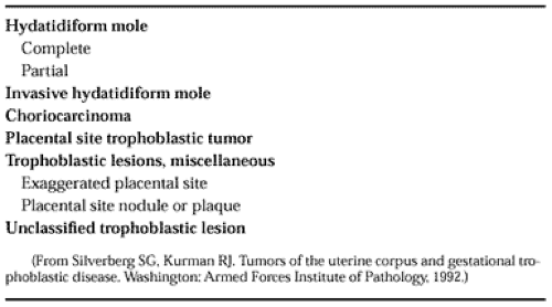
Gestational trophoblastic disease is an abnormal proliferation of trophoblastic tissue, reflecting its inherent capacity for invasiveness. Some lesions contain villus structures, whereas others are avillus. Historically, gestational trophoblastic disease was classified into three well-defined types: hydatidiform mole, invasive mole, and choriocarcinoma. Today, additional categories are recognized, some of which show overlap with spontaneous abortions and implantation site reactions. The World Health Organization classification of trophoblastic disease is presented in Table 111-1.4
HYDATIDIFORM MOLE
The classic complete hydatidiform mole is readily recognized. It is composed of free vesicles (“bunches of grapes”) without recognizable placenta or fetus (Fig. 111-6). All villi are dilated to varying degrees, ranging from a few millimeters to several centimeters. Histologically, the villi are largely avascular, and larger ones show central cisternae. No blood is present in the occasional vessel. Proliferation of cytotrophoblast and syncytiotrophoblast is present on the surface of the villi. This shows a haphazard arrangement without the typical cap of syncytiotrophoblast on cytotrophoblast noted on the young avascular villi of an early pregnancy. Free aggregates of trophoblast are also noted in the intervillus space. An exaggerated placental site reaction with numerous giant cells in the myometrium is often present.4,5,6 and 7
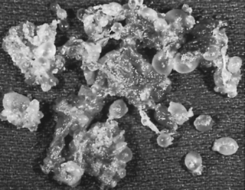 FIGURE 111-6. Gross appearance of complete hydatidiform mole. Villi are dilated as individual free, grape-like vesicles. |
A syndrome of partial hydatidiform mole has been distinguished from complete mole. In these, the villus tissue is organized into a placenta, and a fetus or embryo is often found. The villi display a variable pattern. Some show hydatidiform change, whereas others are small. Capillaries are frequently present and may contain blood. The surface trophoblast, predominantly syncytiotrophoblast, is somewhat hyperplastic. The outlines of the villi tend to be irregular and map-like, leading to the frequent presence of stromal trophoblastic inclusions on section (Fig. 111-7).
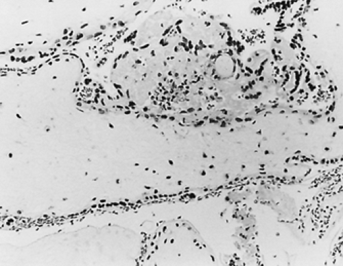 FIGURE 111-7. Section of hydropic villus from a partial hydatidiform mole. Macerated embryo was present, and tissue culture showed the karyotype to be 69,XXY. Villus outline is irregular, and mildly proliferated syncytiotrophoblast is present on the surface. Adjacent small villi were present. (Hematoxylin and eosin; ×400)
Stay updated, free articles. Join our Telegram channel
Full access? Get Clinical Tree
 Get Clinical Tree app for offline access

|
