General Principles of Chemotherapy


General Principles of Chemotherapy


Peter C. Adamson

Susan M. Blaney

Rochelle Bagatell

Jeffrey M. Skolnik

Frank M. Balis


Since the introduction of chemotherapy for the treatment of childhood leukemia more than 60 years ago,1 the prognosis of childhood cancer has improved dramatically (Fig. 10.1). The 5-year survival rate for this group of diseases, many of which were uniformly fatal in the prechemotherapy era, was 83% for all forms of childhood cancer diagnosed between 2002 and 2008.2 This striking improvement in survival is a direct result of the incorporation of anticancer drugs into treatment regimens that previously relied only on surgery or radiotherapy for the primary tumor. The multimodality approach, which integrates surgery and radiotherapy to control local disease with chemotherapy to eradicate systemic (metastatic) disease, has become the standard approach to treating most childhood cancers.


PRINCIPLES OF CANCER CHEMOTHERAPY

The ultimate goal of the multimodality treatment approach, in which anticancer drugs play a critical role, is to cure the patient of his or her cancer. The feasibility of achieving cures by the addition of anticancer drugs to surgery or radiation was first demonstrated in chemosensitive childhood cancers, such as Wilms tumor. However, curing the underlying disease is not the goal of most pharmacological interventions. With the exception of antimicrobial and anticancer chemotherapy, the common classes of drugs (e.g., antihypertensives) are administered with the intent of controlling the disease or the symptoms caused by the disease, rather than curing the underlying disease. The model for curing cancer is based on the successful model of curing bacterial infections. This strategy attempts to exploit differences between cancer and normal host cells and eradicate or kill all cancer cells in the body. This “killing paradigm”3 has had a profound impact on our approach to anticancer drug discovery, drug development, and the design of treatment regimens that incorporate anticancer drugs.

The predominant strategy for anticancer drug discovery has historically been high-throughput in vitro screening to evaluate the antiproliferative or cancer cell-killing effects of candidate drugs in tumor cell lines.4 The precise mechanism of action of the candidate drugs was not critical to the selection process, and for many agents (e.g., doxorubicin), the mechanism of action was not defined until after the drugs were in widespread clinical use. This non-mechanistically-based screening method identified drugs that are cytotoxic and nonselective. As a result, most conventional anticancer drugs produce substantial toxicity.

For classic cytotoxic drug development, the initial dose-finding (phase I) clinical trials define the maximum tolerated dose (MTD), which is based on the severity of toxicity, as the optimal dose, rather than using a therapeutic end point to establish the optimal dose. This design is based on the premise that the highest tolerable dose will produce the maximum achievable cancer cell kill. In the subsequent (phase II) trials, the MTD is evaluated in small cohorts of patients with different types of cancer to establish whether the drug has activity, which is defined as a ≥50% decrease in the size of measurable tumors in at least 20% to 30% of patients with a specific type of cancer.

Conventional frontline treatment regimens for most types of childhood cancer are composed of multiple anticancer drugs that are administered at their MTD intensity, even though these regimens typically produce substantial toxicity. Methods of rescuing or circumventing anticancer drug toxicity, such as the administration of hematopoietic growth factors and bone marrow or stem cell transplant to alleviate hematological toxicity, have been incorporated into treatment regimens to allow for administration of higher doses of anticancer drugs.

The basic principles that guide our current use of cancer chemotherapy are based on the goal of curing patients by eradicating all cancer cells and on empiric observations made in early clinical trials involving children with drug-sensitive cancers, such as acute lymphoblastic leukemia (ALL), Burkitt lymphoma, and Wilms tumor. These principles include the use of multidrug combination regimens (i.e., combination chemotherapy), the administration of chemotherapy before the development of clinically evident metastatic disease (i.e., adjuvant chemotherapy), and the administration of drugs at the maximally tolerated dose rate (i.e., dose intensity).


Combination Chemotherapy

The importance of administering anticancer drugs in combination regimens was first appreciated in the treatment of ALL. Compared with single-agent therapy, the use of drug combinations significantly increased the percentage of patients achieving complete remission and prolonged the duration of their remissions.5 At best, only 60% of patients treated with a single agent achieved complete remissions, but standard three- and four-drug combination induction regimens achieve complete remission rates that exceed 95%. Almost all patients on single-agent therapy experienced a relapse within 6 to 9 months, despite continuation of therapy with the same drug. Long-term remissions and cures were attained only after the institution of combination chemotherapy that incorporated the most active single agents.

The primary scientific rationale for the use of combination chemotherapy is to overcome drug resistance to individual agents, the incidence of which can often exceed 50% even in newly diagnosed
cancers.6 Because it is not feasible to accurately predict whether a particular patient’s tumor will respond to a given drug, administering anticancer drugs in combination ensures a greater chance of achieving a response (i.e., exposing the tumor to at least one active agent). In addition to providing a broader range of coverage against naturally resistant tumor cells, combination chemotherapy may also prevent or delay the development of acquired resistance in initially responsive tumors and provide additive or synergistic cytotoxic effects if agents with different mechanisms of action are selected.

A thorough knowledge of the clinical pharmacology of individual anticancer drugs is required to design effective combination chemotherapy regimens. Traditionally, combination chemotherapy regimens contain drugs with demonstrated single-agent activity against the type of tumor being treated, with a preference for agents that produced complete responses in patients with advanced or recurrent disease; drugs that are non-cross-resistant to overlap against drug-resistant subpopulations of tumor cells; drugs with nonantagonistic (i.e., additive or synergistic) mechanisms of action; and drugs with nonoverlapping toxicity profiles, allowing each agent to be administered at its optimal dose and schedule.


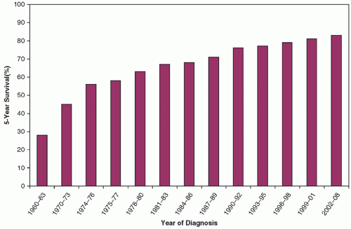
Figure 10.1 Five-year survival rate for all childhood cancers diagnosed between 1960 and 2004.(From Silverberg E, Boring CC, Squires TS. Cancer statistics, 1990. CA Cancer J Clin 1990;40(1):9-26; Jemal A, Murray T, Ward E, et al. Cancer statistics, 2005. CA Cancer J Clin 2005;55:10-30; Jemal A, Siegel R, Ward E, et al. Cancer Statistics, 2009. 2009;59(4):225-249.)


Adjuvant Chemotherapy

Anticancer drugs are most effective when administered in the adjuvant setting to patients who are without overt evidence of residual disease after local therapy with surgery or radiation but who are at high risk for relapse at metastatic sites. Before the routine use of adjuvant chemotherapy, relapse at metastatic sites occurred in 60% to 95% of children with localized solid tumors after local therapy. The aim of adjuvant chemotherapy is to prevent metastatic recurrence by eliminating micrometastatic tumor deposits that are present at the time of diagnosis in the lungs, bone, bone marrow, lymph nodes, or other sites.7 Adjuvant chemotherapy is efficacious for most of the common pediatric cancers, including Wilms tumor, Ewing sarcoma, lymphoma, rhabdomyosarcoma, astrocytoma, and osteosarcoma.8,9,10,11,12

Theoretical considerations and experimental evidence support the use of adjuvant chemotherapy.13,14 Microscopic foci of tumor should be more chemosensitive on a cell-kinetic basis, because a larger fraction of the cells are actively proliferating and potentially susceptible to the cytotoxic effects of the drugs. The smaller burden of tumor cells also implies a lower probability that drug-resistant cells are present. The mathematical modeling experiments of Goldie and Coldman, which assume that a curable tumor is one with no drug-resistant tumor cells and that the development of drug resistance is the result of a random genetic event, predict that the chance for cure is maximized if all available active drugs are given simultaneously in the adjuvant setting, when there is minimal residual disease and the probability that drug-resistant cells are present is low.6,15

Clinical experience has demonstrated a correlation between low tumor burden and the efficacy of chemotherapy.14,15,16 Children presenting with extensive or disseminated tumors are less likely to be cured than children with the identical type of cancer but with a low tumor burden. For example, the 5-year event-free survival for patients with metastatic Ewing sarcoma treated on the Children’s Cancer Group-Pediatric Oncology Group Ewing sarcoma trial (INT-0991) was 22% compared with 69% for children presenting with localized disease.17

The selection of appropriate drugs and the optimal timing of drug therapy relative to the definitive local therapy are important considerations in the design of successful adjuvant chemotherapy regimens. Traditionally, drugs have been selected on the basis of their activity in advanced disease. Animal models and clinical experience have shown that regimens producing the most dramatic responses in metastatic or recurrent disease have the greatest likelihood of being curative in the adjuvant setting.16

Adjuvant chemotherapy should begin as soon as possible after definitive local therapy. A delay to allow for recovery from surgery or radiation therapy may compromise the chance of curing the patient. One strategy to avoid delays caused by potential adverse interactions between chemotherapy and surgery or irradiation is the administration of the drug therapy before definitive local therapy.
This approach, called primary or neoadjuvant chemotherapy, may also improve local control of the primary tumor by shrinking the primary and making it more amenable to surgical resection, in addition to providing earlier therapy for micrometastases.18


Dose Intensity

Most cytotoxic anticancer drugs have a steep dose-response curve, and a small increment in the dose can significantly enhance the therapeutic effect of a drug in preclinical studies. In animal tumor models, a twofold increase in the dose of cyclophosphamide can result in a tenfold increase in tumor-cell killing.19,20 Retrospective clinical studies have also demonstrated a relationship between dose intensity of anticancer drugs and disease outcome, but this relationship has not been consistently confirmed in randomized prospective trials.

In a meta-analysis of chemotherapeutic regimens containing cyclophosphamide, methotrexate, and fluorouracil for metastatic breast cancer, Hryniuk and Bush observed a strong correlation between response rate and the relative dose intensity of the various regimens.21 The relative dose intensity is calculated by normalizing the dose rate (mg/m2/week) for each agent to the dose rate in an arbitrarily selected standard regimen and then averaging the relative dose intensities for all agents in the regimen to derive the relative dose intensity for the regimen. Over a threefold range in relative dose intensity, the response rate in metastatic breast cancer ranged from 12% to 84%. Retrospective meta-analyses in stage II breast cancer, ovarian cancer, colorectal cancer, and lymphoma have also demonstrated a correlation between the dose intensity of the drug regimen and disease outcome.22 However, prospective randomized trials have failed to demonstrate a survival advantage for more dose-intensive regimens, including high-dose chemotherapy with bone marrow or stem cell rescue, compared with standard dose regimens in breast, ovarian, and small cell lung cancers23,24,25 and for more dose-intensive cisplatin in germ cell tumors.26

For children with ALL and osteosarcoma, relapse rates are significantly lower in patients receiving more dose-intense chemotherapy.27,28,29,30,31 In a randomized trial, patients with ALL receiving standard doses of methotrexate and mercaptopurine had a median survival of 15 months compared with 6 months for the group randomized to a half-dose maintenance regimen.27 In children with high-risk ALL, those who received less than 94% of the protocol-prescribed dose of vincristine, anthracycline, and L-asparaginase during intensification therapy were 5.5 times more likely to experience a subsequent adverse event than patients who received at least 99% of the prescribed dose of these agents.28 Oral mercaptopurine dose intensity during maintenance therapy is also predictive of event-free survival in ALL.29 However, in the latter study, lower mercaptopurine dose intensity was primarily the result of missed doses rather than reductions of the daily dose, leading the authors to conclude that prescribing higher doses of mercaptopurine could be counterproductive if greater hematological toxicity resulted in treatment delays.

Retrospective analyses of osteosarcoma trials demonstrated a twofold higher relapse rate in patients receiving less than 75% of their recommended dose of chemotherapy compared with patients receiving 75% or more in one study30 and a threefold higher relapse rate in a second study using 80% of the protocol-prescribed dose as a cutoff.31

Meta-analyses, such as those performed by Hryniuk and Bush, have also been performed for several pediatric tumors.32,33 Analysis of 44 clinical trials involving 1,592 patients older than 1 year with stage IV neuroblastoma revealed a fivefold to tenfold range in the dose intensity of the individual agents studied.32 The dose intensity of four drugs (i.e., teniposide, cisplatin, cyclophosphamide, doxorubicin) significantly correlated with response and survival. Similarly, examination of the relation between individual drug dose intensities and disease outcome in osteosarcoma and Ewing sarcoma suggests that doxorubicin dose intensity is an important determinant of response in osteosarcoma and disease-free survival of patients with Ewing sarcoma.33

Prospective randomized trials to assess the importance of dose intensity in childhood cancers have been performed for a few tumor types. The administered dose intensity of dactinomycin and doxorubicin in pulse-intensive regimens for Wilms tumor was significantly higher than for the standard treatment regimens, but there was no survival advantage associated with the enhanced dose intensity.34,35 In a randomized trial of Filgrastim in children with high-risk ALL, the treatment interval was shorter with Filgrastim, resulting in a slight increase in dose intensity but no impact on event-free survival.36 A 33% increase in dose intensity was achieved in a five-drug chemotherapy regimen for Ewing sarcoma by shortening the dosing interval (interval compression) from 21 to 14 days. Compared with the standard every 21-day regimen, event-free survival was higher (76% vs. 65%) on the compressed regimen.37

Methods for maximizing dose intensity include greater patient and physician willingness to tolerate drug toxicities; more aggressive supportive care of patients experiencing these side effects; selective rescue of the patient from toxicity, such as with bone marrow or peripheral stem cell transplantation or the administration of Filgrastim; the use of regional chemotherapy (e.g., intra-arterial, intrathecal delivery) to achieve high drug concentrations at local tumor sites while minimizing systemic drug exposure; and the development of new treatment schedules, such as long-term continuous infusions, which may allow more drug to be administered over a given period.


CLINICAL PHARMACOLOGY OF ANTICANCER DRUGS

The primary role of the pediatric oncologist is to coordinate the administration of complex combination chemotherapy regimens to children in the setting of multimodal (i.e., surgery, radiotherapy, and chemotherapy) therapy. Special care must be taken because the anticancer drugs used in these regimens have the lowest therapeutic index of any class of drugs and predictably produce significant, even life-threatening, toxicity at therapeutic doses (Fig. 10.2).38 However, implementing significant dose reductions or delays in therapy to attenuate these toxicities may compromise the therapeutic effect and place the patient at an increased risk for disease recurrence, a uniformly fatal event with most childhood cancers. The cancer chemotherapist must carefully balance the risks of toxicities from therapy against the risk of tumor recurrence from inadequate treatment. Unfortunately, the crucial adjustments in the dose and schedule of chemotherapy needed to achieve this balance often must be made empirically, because therapeutic drug monitoring for most agents is not available.

To ensure that these drugs are used safely and effectively, the pediatric oncologist must have an in-depth knowledge of the clinical pharmacology of these agents, including the mechanisms of drug action, pharmacokinetics, pharmacogenetics, spectrum of toxicities, potential drug interactions, and mechanisms of drug resistance.


Mechanism of Action

Although recent advances in cancer cell genomics and cell biology have provided critical insights into the pathogenesis of many forms of childhood cancer and offer hope for the development of targeted, more selective new cancer treatments, most current conventional anticancer drugs used in the frontline treatment of childhood cancers are cytotoxic agents that nonselectively and irreversibly damage vital macromolecules (e.g., DNA) or metabolic pathways that are also critical to normal cells. As a result, they cause many undesirable and potentially severe toxic effects.


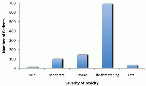
Figure 10.2 The worst degree of any toxicity experienced by patients (n = 1,062) treated on one of the eight treatment arms of the Intergroup Rhabdomyosarcoma Study III. Seventy-eight percent of patients had at least one severe or life-threatening toxicity, and there were thirty-two toxicity-related deaths. (Adapted from Table 6 in Crist W, Gehan EA, Raghab AH, et al. The third Intergroup Rhabdomyosarcoma Study. J Clin Oncol 1995;13:610.)

Most anticancer drugs produce their cytotoxic effects by interfering with the synthesis or function of DNA and RNA (Fig. 10.3). For example, the alkylating agents are chemically reactive compounds that damage DNA by covalently bonding to and cross-linking nucleobases within the DNA, and the antimetabolites block the synthesis of nucleotide precursors or are directly incorporated into DNA as fraudulent bases. The topoisomerases are also important targets of anticancer drugs. These nuclear enzymes maintain the three-dimensional structure of DNA and are critical for DNA replication, transcription, repair, and recombination. The topoisomerases work by cleaving and religating DNA, and agents such as the anthracyclines, epipodophyllotoxins, and camptothecins interfere with religation, resulting in protein-associated DNA strand breaks.39


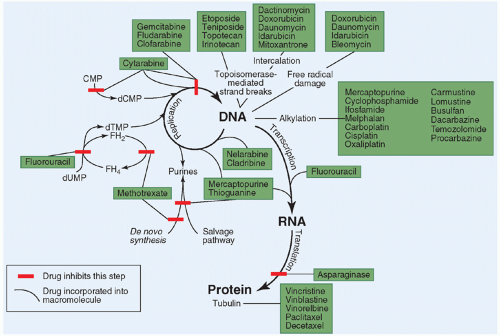
Figure 10.3 Site of action of the commonly used cytotoxic anticancer drugs.

Dysregulation of the cell cycle, of signal transduction pathways that regulate cell proliferation, and of programmed cell death (apoptosis) are common to most forms of cancer.40,41,42 Mutations to genes involved in these highly complex, highly regulated and
interrelated pathways can result in loss of control of DNA replication and cell division and suppression of the apoptotic response to receptor-linked or DNA-damage-induced signals. In addition to their role in tumorigenesis, mutations in cell cycle regulatory genes and genes involved in apoptosis may modulate the sensitivity of cancer cells to conventional anticancer drugs.40,42 The cellular damage produced by most anticancer drugs appears to induce apoptosis in chemosensitive cancer cells. Overexpression of oncogenes that promote apoptosis, such as MYCC and MYCN, can enhance the chemosensitivity of tumor cells, whereas overexpression of bcl-2, which blocks the apoptotic pathway, can attenuate drug-induced apoptosis and convey pleiotropic resistance to anticancer drugs.

The cell cycle is regulated by negative feedback controls or checkpoints that block the cell from proceeding to the next cell cycle event until the prior event is completed and until needed repairs to DNA are performed.43,44,45,46 Normal cells are arrested in G1 phase in response to DNA damage caused by cytotoxic drugs, allowing for DNA repair.47,48 If this DNA damage is not repaired, the complex process of DNA replication and cell division is disrupted. Mutations in cell cycle regulatory genes that have been implicated in tumorigenesis most frequently involve genes controlling the transition from the G1 to S phases of the cell cycle,40 and loss of checkpoint function (e.g., p21) can enhance chemo- and radiosensitivity.40,49 The same mechanisms that play a role in the pathogenesis of cancer could also sensitize the cancer cell to DNA-damaging anticancer drugs. Somatic mutations to the tumor-suppressor gene TP53 and loss of p53 function occur in half of all human cancers. Clinical studies correlating TP53 mutational status to disease outcomes have found shorter survival and poor response to treatment, especially for mutations within the DNA-binding motifs.50

A number of new anticancer drugs block the activation of cellular signal transduction pathways by inhibiting protein kinase activity of critical cell membrane receptors and downstream effector proteins.51,52,53 Activation of these pathways occurs through sequential phosphorylation of pathway proteins usually on a tyrosine, serine, or threonine moiety. Activating mutations in receptors and effector proteins, such as the BCR-ABL fusion protein created by the t(9;22) translocation in chronic myelogenous leukemia, play a critical role in the pathogenesis of many cancers. Targeted drugs, such as imatinib, that inhibit the protein kinase activity of these constitutively activated signaling proteins block the transduction of the aberrant signal and, thereby, control cellular proliferation. Cellular receptors and effectors that are targeted by this new class of molecularly targeted anticancer drugs include EGFR, VEGFR, ERBB2, FGFR, KIT, RET, ALK, BRAF, MET, JAK, and PDGFR.53


TABLE 10.1 Pharmacokinetic Terms


Term


Common Abbreviation


Units


Definition


Clearance


Cl


Vol/time (mL/min)


Used to quantify the rate of drug elimination; expressed in terms of volume of plasma cleared of drug per unit of time. Total clearance is the sum of renal, metabolic, spontaneous chemical degradation, and biliary (fecal) elimination. When the true bioavailability of a drug is not known (e.g., drugs with only an oral formulation), the term “apparent” clearance is used and is abbreviated Cl/F.


Half-life


t1/2


Time (h)


Time required to reduce the drug concentration by 50%. Plasma drug disappearance frequently has multiple phases with differing rates of disappearance (e.g., rapid distribution phase, terminal or elimination phase). Half-lives listed for drugs in this chapter are the postdistributive (terminal, elimination) half-lives, unless otherwise noted.


Area under the curve


AUC


Conc. × time (µM • h)


Quantitates total drug exposure; integral of drug concentration over time or the area under the plasma concentration-time curve; used in calculation of clearance and bioavailability


Volume of distribution


Vd; Vdss


Volume (L)


Relates plasma concentration to total amount of drug in the body (i.e., volume required to dissolve the total amount of drug to give the final concentration found in plasma); a property of the drug rather than a real volume or physiologic compartment


Bioavailability


F


Fraction (%)


Rate and extent of absorption of a drug, frequently synonymous with the fraction of a dose absorbed when administered by some route other than intravenous.


Biotransformation


Enzymatic metabolism of a drug; may result in the activation of a prodrug, conversion to other biologically active intermediates, or inactivation of a drug


Conc., concentration.


An understanding of the mechanism of drug action is useful in predicting which cancers may respond to the drug based on their biochemical, cytokinetic, and genomic profiles, and which drug combinations may produce additive or synergistic antitumor effects. Combining agents that together could enhance the inhibition of vital intracellular processes through sequential or concurrent blockade or lead to complementary inhibition of specific metabolic pathways has been a traditional strategy for the design of combination regimens.54 Devising approaches to combine molecularly targeted drugs with conventional cytotoxic chemotherapy is the next challenge for pediatric oncologists. The role of tumor genetic profiling for selection of molecularly targeted drugs is under study.

A drug’s schedule of administration may also be influenced by its mechanism of action. For example, the antimetabolites, which are inhibitory only during S phase in the cell cycle, tend to be more cytotoxic if administered by prolonged infusion. This approach ensures that a greater number of tumor cells are exposed to the drug as they pass through S phase.


Pharmacokinetics

The discipline of pharmacokinetics deals with quantitative aspects of drug disposition in the body, including drug absorption, distribution, metabolism, and excretion, referred to as ADME (Table 10.1). Although the pharmacokinetic behavior of most of the commonly used anticancer drugs has been studied in adults, many of these agents have not been as well studied in children.
As the technology to measure the concentration of these drugs and their metabolites in biologic fluids has improved, a greater emphasis has been placed on studying anticancer drug pharmacokinetics in children with cancer.

Pharmacokinetic studies have revealed substantial interpatient variability in drug disposition and systemic drug exposure with most anticancer drugs.55,56 Administering a standard dose of etoposide, doxorubicin, or cyclophosphamide to a group of children results in a twofold to tenfold range in systemic drug exposure, as measured by the area under the plasma drug concentration-time curve (area under the curve; AUC),57 and substantial variability in systemic drug exposure is also observed with orally administered agents such as methotrexate and mercaptopurine.58 Assuming that drug effect is more closely related to systemic drug exposure than dose, these differences in drug disposition could account for the variability in toxicities and responses observed with most combination chemotherapy regimens employing standardized doses of individual agents.59 Variability in anticancer drug disposition in children may result from age-related developmental changes in body composition and excretory organ function, variation in rate of metabolism and excretion of drug by the kidneys or liver, variation in the extent of drug-protein binding, drug interactions, and pharmacogenetics.60,61,62,63

The most important determinant of variability in anticancer drug pharmacokinetics is the rate of drug metabolism. Drug-metabolizing enzymes are divided into two groups based on the type of reaction that they catalyze. Phase I reactions (e.g., oxidation, hydrolysis, reduction, and demethylation) introduce or expose a functional group (e.g., hydroxyl group) on the drug. Phase I reactions usually diminish the drug’s pharmacological activity, but some prodrugs, such as cyclophosphamide, are converted to active metabolites by these enzymes. Phase II conjugation reactions covalently link a highly polar conjugate (e.g., glucuronic acid, sulfate, glutathione, amino acids, or acetate) to the functional group created by the phase I reaction. The conjugated drugs are highly polar, usually devoid of pharmacological activity, and rapidly excreted.

The significant interpatient variation in systemic drug exposure with current dosing methods, the toxic nature of these agents, and the potential importance of dose intensity in cancer chemotherapy point to the need for more precise, individualized dosing methods for anticancer drugs,56,60,64,65,66 such as the adaptive dosing techniques that have been successfully applied to individualize carboplatin dose67 and therapeutic drug monitoring of methotrexate that plays a critical role in determining the duration of leucovorin rescue following high-dose methotrexate therapy.56,66,68 A prerequisite for these individualized dosing methods is the establishment of the relation between a drug’s pharmacokinetics and pharmacodynamics (toxicity or therapeutic effect). Systemic drug exposure (AUC) of anticancer drugs is usually the best correlate of the drug’s toxic or therapeutic effects. However, this usually requires plasma sampling at multiple times over a prolonged period, which may not be practical for monitoring large numbers of patients. Through pharmacokinetic modeling, a limited number of sampling times that can reliably estimate the AUC can often be identified, providing a more practical pharmacokinetic monitoring schedule.56,69,70,71 Parameters other than AUC, such as peak or trough concentration or average steady-state concentration, can also be evaluated for clinical correlations.

Even though therapeutic drug monitoring has yet to play a significant role in the day-to-day management of the patient with cancer, the pharmacokinetic parameters are important for determining the optimal dose, schedule, and route of administration of the drug. Knowledge of the route of elimination of a drug is also helpful in adjusting the dosage for patients with hepatic or renal dysfunction.72,73

Physiologic differences between children and adults can affect drug disposition and must be considered in determining the appropriate dose and schedule of the drug for children. Developmental differences in drug absorption, plasma protein or tissue binding, functional maturation of excretory organs, and distribution of drug in the various tissues of the body (Table 10.2) can result in differences in systemic drug exposure for children compared with adults treated with the same dose. The most dramatic changes in excretory organ function and body composition occur during the first few days to months of life, but there are very limited data on the disposition of anticancer drugs in infants.


Pharmacogenetics

In addition to the effects of ontogeny, disease, organ dysfunction, and drug interactions on the interpatient variability in response to drugs, genetic factors can influence both the efficacy of a drug and the likelihood of toxicity.74 The field of pharmacogenetics began with the study of drug-metabolizing enzymes, but it now encompasses the study of the influence of genetic variation (polymorphisms) on the entire spectrum of drug action, including drug disposition (absorption, distribution, metabolism, and excretion), drug targets, and treatment-modifying genes.75

Pharmacogenetically based variability in response to drugs is more apparent for drugs that have a narrow therapeutic index, such as anticancer drugs. The study of mercaptopurine methylation in large measure ushered in the modern era of pharmacogenetics.76 There are more than 57 active CYP drug-metabolizing enzymes in humans,77 and the majority have genetic variation (polymorphisms) that, in certain cases, translates into functional changes in the encoded enzyme.75,78 Phase I enzymes include the cytochrome p450 (CYP) superfamily of enzymes that catalyze oxidation and demethylation reactions. The CYPs are responsible for 70% to 80% of all phase I drug metabolism (Fig. 10.4) and are categorized into families and subfamilies according to their amino acid sequence similarity. Sequences that are >40% identical belong to the same family designated by an initial number (e.g., CYP1); sequences that are >55% identical are in the same subfamily designated by a letter suffix (e.g., CYP1A). Subfamilies may contain multiple isoforms (e.g., CYP1A2). The CYP1, CYP2, and CYP3 families are primarily responsible for hepatic drug and xenobiotic metabolism in humans, with CYP3A being the most important subfamily, accounting for the metabolism of nearly half of all drugs (Fig. 10.4). A number of CYP genes are known to have functionally relevant polymorphisms,79,80 and CYP2A6, CYP2B6, CYP2C9, CYP2C19, and CYP2D6 have the greatest variability. CYP3A4 does not have critical functional polymorphisms.77

Phase II enzymes also have functionally relevant polymorphisms (Fig. 10.4). Thiopurine-S-methyl transferase (TPMT), which is the enzyme that methylates the thiol group on mercaptopurine and thioguanine, is the classic example for anticancer drugs. Since the original description of enhanced drug toxicity associated with this genetic variation in TPMT, the identification of the gene encoding the enzyme and the DNA sequence variations associated with this inherited trait have been identified81,82,83,84,85 and studied in diverse populations.86,87,88,89 Another example is irinotecan, the toxicity of which is a function of the pharmacogenetic variation observed in the phase II enzyme UDP-glucuronosyltransferase.90,91,92

Lastly, genetic differences may also have indirect effects on drug response, as has been observed with methylation of the methylguanine methyltransferase (MGMT) gene promoter. The expression of the DNA repair protein, MGMT, in tumor modulates the response of gliomas to carmustine.93


Toxicity

In therapeutic doses, actively dividing normal host cells, such as those in the bone marrow or the mucosal epithelium, are sensitive to the cytotoxic effects of anticancer drugs.94 The nonselective mechanisms of action and resulting low therapeutic indices of these agents mean that a high incidence of potentially severe
toxicities must be tolerated to administer effective doses.95 Acute toxicities common to many of the anticancer drugs include myelosuppression, nausea and vomiting, alopecia, orointestinal mucositis, liver function abnormalities, allergic or cutaneous reactions, and local ulceration from subcutaneous drug extravasation. These acute toxicities occur over hours to weeks after a dose and are usually reversible. Many drugs also have unique toxicities affecting specific organs or tissues, such as cardiotoxicity associated with the anthracyclines, hemorrhagic cystitis associated with cyclophosphamide and ifosfamide, peripheral neuropathy from vincristine, cisplatin, and paclitaxel, nephrotoxicity from cisplatin and ifosfamide, ototoxicity from cisplatin, and coagulopathy from L-asparaginase. Many of these latter toxicities are cumulative (i.e., occur after multiple doses), and in some cases they are not completely reversible (e.g., anthracycline cardiotoxicity).


TABLE 10.2 Physiologic Differences in Children that May Influence Drug Disposition


Organ or Compartment


Value at Birtha


Age Adult Values Are Reachedb


Effect on Drug Dispositionc


Kidney


Size


Renal blood flow


1 y


↓Renal excretion


Glomerular filtration


6 mo-1 y


↓Renal excretion


Tubular function


1 y


↓Tubular secretion


Liver


Size


Phase I drug-metabolizing enzymesd


Variable (oxidative enzymes increase rapidly after birth)


↑ activity in young children


↓ Metabolic clearance


↑ Metabolic clearance


Phase II drug-metabolizing enzymese


↑ Sulfation


↓ Other enzymes


Variable (6 mo for glucuronidation)


↓ Metabolic clearance


Biliary excretion


6 mo


↓ Biliary excretion


Gastrointestinal


Acid secretion


3 mo


Altered drug absorption and stability


Motility


6-8 mo


↑ Transit time in young children


Delayed absorption


More rapid absorption


Body Composition


Blood volume


Adolescence


Extracellular fluid


48 mo


↑ Distribution volume


Total body water


4 mo


↓ Distribution volume


Fat


Adolescence


↑ from 4-12 mo of age


↓ Distribution volume of lipophilic drugs


↑ Distribution volume of lipophilic drugs


Cerebrospinal fluid volume


3 y


↑ Distribution volume of intrathecal drugs


Protein binding


1 y


↑ Free drug levels


a ↓, decreased; ↑, increased (compared with adult values and relative to body surface area or weight).

b Relative to body surface area or weight.

c Refer to Table 10.4 to determine which drugs may be affected by alteration of renal, biliary, or metabolic function.

d Oxidation, hydrolysis, reduction, and demethylation.

e Conjugation, acetylation, and methylation.


The toxicity profile of molecularly targeted tyrosine kinase inhibitors (TKIs) differs from conventional cytotoxic anticancer drugs. TKIs are usually not myelosuppressive, and common toxicities include fatigue, anorexia, nausea, vomiting, diarrhea, abdominal pain, edema, hypertension, and skin rashes including hand-foot skin reactions. Many TKIs have also been associated with thyroid dysfunction96 and cardiotoxicity.97

A significant portion of an oncologist’s time is spent in providing supportive care for patients experiencing acute and long-term drug toxicities. A number of therapeutic approaches have evolved to attenuate these toxicities, to make the therapy more tolerable, and to safely increase the dose intensity of regimens by circumventing dose-limiting toxicities.94,98,99 Bone marrow or peripheral stem cell transplantation to rescue patients from myeloablative doses of anticancer drugs is an example of this rescue approach. Other widely used forms of rescue include the administration of leucovorin or glucarpidase100 to counteract the toxicities of high-dose methotrexate, the use of antiemetics to block nausea and vomiting,101 the use of mesna to prevent the hemorrhagic cystitis caused by the oxazaphosphorines,102 the use of colony-stimulating factors (e.g., filgrastim, pegfilgrastim) to alleviate myelosuppression,103,104 and the use of dexrazoxane to prevent anthracycline cardiotoxicity.105,106

The toxicity of anticancer drugs has a major impact on the dosing of these agents. The end point of the phase I dose-finding studies for most cytotoxic anticancer drugs is the identification of the MTD, which is considered the optimal dose. The dosing interval (every 21 to 28 days) for cytotoxic anticancer drugs is determined by the duration of acute toxicities, and dose modifications are usually based on the severity or duration of toxicities on the prior treatment cycle. The lifetime cumulative dose of the anthracyclines and bleomycin is limited to prevent cardiotoxicity and pulmonary toxicity. This toxicity-based dosing approach for anticancer drugs reflects the lack of data on the relationship between dose and anticancer effect.

The severity, incidence, and time course of toxicities are important factors in designing optimal drug combinations or adjusting
doses to avoid overlapping toxicities. For example, nonmyelosuppressive agents such as vincristine, prednisone, L-asparaginase, and high-dose methotrexate with leucovorin rescue can often be administered with traditional myelosuppressive drugs without compromising the dose of myelosuppressive agents. Some regimens administer nonmyelosuppressive agents during the period of marrow suppression from myelotoxic drugs to ensure continuous exposure of the tumor to cytotoxic therapy.107


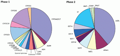
Figure 10.4 Phase I and II enzymes involved in drug metabolism. Almost all of the major human enzymes responsible for modification of functional groups or conjugation with endogenous substituents exhibit common polymorphisms at the genomic level. The percentage of phase I and phase II metabolism of drugs that each enzyme contributes is estimated by the relative size of each section of the corresponding chart. ADH, alcohol dehydrogenase; ALDH, aldehyde dehydrogenase; CYP, cytochrome P450; DPD, dihydropyrimidine dehydrogenase; NQO1, NADPH:quinone oxidoreductase or DT diaphorase; COMT, catechol-O-methyltransferase; GST, glutathione S-transferase; HMT, histamine methyltransferase; NAT, N-acetyltransferase; STs, sulfotransferases; TPMT, thiopurine methyltransferase; UGTs, uridine 59-triphosphate glucuronosyltransferases. (Adapted with permission from Evans WE, Relling MV. Pharmacogenomics: translating functional genomics into rational therapeutics. Science 1999;286(5439):487-491. Copyright 1999 AAAS)

The long-term side effects of cancer chemotherapy are also of particular concern to the pediatric oncologist because of the high cure rates and the long life spans of successfully treated patients. The adverse late effects of chemotherapy on growth, development, and reproductive function; possible permanent cardiac, pulmonary, or renal damage; and possible carcinogenic and teratogenic effects are discussed in Chapter 47.


Drug Resistance

Although toxic effects of anticancer drugs are usually predictable, the response of any given tumor to individual agents is not. Clinical resistance to anticancer drugs is the primary reason for treatment failure in childhood cancers. Drug resistance can be present at the outset of treatment or can become clinically apparent under the selective pressure of drug exposure. The magnitude of the problem of drug resistance was appreciated early in cancer chemotherapy for childhood cancers. Less than one-half of children with ALL who were treated with single-agent therapy achieved a complete remission, and almost all of the patients who did respond eventually relapsed despite continuation of the drug that produced the remission.113

The development of most forms of drug resistance has a genetic or epigenetic basis.6,114 The inherent genetic instability of tumor cells results in the spontaneous generation of drug-resistant clones as a consequence of a mutation, deletion, gene amplification, translocation, chromosomal rearrangement, or alterations in gene expression.6,114 These alterations are presumed to be random events, which may account for the variability in response observed in most clinical trials. This genetic and epigenetic basis for drug resistance means that resistance can be inherited by subsequent generations of tumor cells, and under the selective pressure of drug exposure, drug-resistant cancer cells become the predominant subpopulation. At a biochemical level, there are a variety of mechanisms by which tumors become drug resistant. In most cases, these alterations in cellular metabolism can be related to an increase, a decrease, or an alteration in some gene product, such
as the gene amplification identified in methotrexate-resistant cells that results in overproduction of dihydrofolate reductase (DHFR), the target enzyme of methotrexate.115 Mechanisms of resistance to molecularly targeted receptor and nonreceptor TKIs include point mutations in the kinase domain that lower drug affinity or activation of downstream effectors or alternative signaling pathways through epigenetic alterations.116,117

Genetically based molecular or biochemical alterations in cancer cells can produce anticancer drug resistance that is specific to a single agent or a class of agents or provides protection from a broad range of anticancer drugs. In the latter form of resistance, termed multidrug resistance, a single cellular alteration conveys resistance simultaneously to multiple unrelated drugs, including drugs to which the cancer has not been exposed.118 The best-studied multidrug-resistant phenotypes are associated with decreased intracellular drug accumulation and an increase in plasma membrane, ATP-dependent drug efflux pumps.118,119,120,121,122

ATP-dependent drug efflux pumps are part of a family of transporters, the adenosine triphosphate-binding cassette (ABC) transporters.123 To date, 48 human ABC genes have been identified and classified into 7 subfamilies (ABCA to ABCG).124 The functional protein is usually comprised of two nucleotide-binding folds and two transmembrane domains. Most of our knowledge of ABC transporters and their role in drug resistance stems from studies of P-glycoprotein, the product of the ABCB1 (MDR1) gene. P-glycoprotein (P-gp) is expressed in human tumor specimens, including ALL, neuroblastoma, rhabdomyosarcoma, neuroepithelioma, Ewing sarcoma, retinoblastoma, and osteosarcoma,125,126,127,128,129,130 and in a variety of normal human tissues, such as the biliary canaliculi in the liver, the proximal tubules in the kidney, the mucosal lining of the jejunum and colon, the adrenal gland, hematopoietic progenitor cells, and the endothelial cells of blood vessels within the central nervous system (CNS) and testis. P-glycoprotein appears to be responsible for excretion of toxic compounds from these normal cells.125,131 Although the presence of P-glycoprotein in tumor specimens has been associated with a worse prognosis and a poor response to therapy in some studies, the clinical significance of P-glycoprotein expression in childhood cancers remains controversial, in part because of the lack of a universal standard for quantifying expression at an RNA or protein level.118,127,132

The lack of expression of P-gp in some multidrug-resistant cells led to the discovery of ABCC1 (MRP1), initially cloned from a human lung carcinoma cell line.133 The structural similarity between MRP1 and P-gp underlie the significant overlap in substrate specificity. Notable exceptions to this are the taxanes, which are poor substrates for MRP1. The second member of the MRP (ABCC) family, MRP2, transports a number of MRP1 substrates as well as cisplatin.134 MRP4 and MRP5 transport nucleoside analogs.123,135

Other mechanisms for multidrug resistance include an enhanced capacity to repair DNA damage produced by alkylating agents,136 the detoxification of chemically reactive forms of alkylating agents and anthracyclines by glutathione, decreased levels of topoisomerase II, the target enzyme of the anthracyclines, epipodophyllotoxins, and dactinomycin, and suppression of apoptotic pathways.41 The loss of DNA mismatch repair activity results in multidrug resistance by impairing the cancer cell’s ability to detect DNA damage and activate apoptosis.114,137

The mechanism of drug resistance is an important consideration in selecting agents to be included in combination regimens or as second-line therapy in relapsed patients. Ideally, drug combinations should be composed of non-cross-resistant agents, and relapse treatment regimens should avoid the use of drugs that are cross-resistant with drugs used in the frontline regimen. With advances in our understanding of the mechanisms of drug resistance, specific treatment approaches may be devised to prevent the development of, or overcome, drug resistance in tumor cells.

In the remainder of this chapter, the pharmacological characteristics of the anticancer drugs used to treat pediatric cancers are reviewed. Tables summarize the general pharmacological properties (Table 10.3) and pharmacokinetic parameters ( Table 10.4) of the commonly used anticancer drugs.


ALKYLATING AGENTS

The alkylating agents have a broad range of clinical activity in childhood cancers. These drugs are chemically reactive compounds that exert their cytotoxic effect through the covalent bonding of an alkyl group to important cellular macromolecules (Fig. 10.5). Although a number of nucleophilic macromolecules and their precursors are potential targets for alkylation intracellularly, damage to the DNA template and the resulting induction of apoptosis appears to be the major determinant of cytotoxicity.42,138 With the bifunctional alkylating agents that have two alkylating groups, this damage appears to result primarily from interstrand and intrastrand DNA-DNA and DNA-protein cross-links.138

Alkylating agents have steep dose-response curves in experimental model systems.139 A log-linear relationship exists between tumor-cell killing and the concentration of the alkylating agent, and this correlation is maintained through 4 to 5 orders of magnitude of cell killing. This steep dose-response relationship for alkylating agents provides a strong rationale for their use in high-dose therapy regimens. Because of the significant myelosuppressive effects of these drugs, high-dose alkylator therapy is generally administered in conjunction with bone marrow or peripheral stem cell transplantation to prevent permanent bone marrow aplasia. The use of melphalan and busulfan in childhood cancers is limited almost exclusively to high-dose transplantation preparative regimens, and other alkylating agents, such as cyclophosphamide and thiotepa, are also frequently incorporated into these regimens.140,141

Myelosuppression is the major dose-limiting toxicity for most of the commonly used alkylating agents. Other common acute toxic effects include nausea and vomiting, alopecia, allergic and cutaneous reactions, and gastrointestinal and neurologic toxicity at high doses. Of particular concern to the pediatric oncologist are the potential long-term effects of alkylator therapy. Alkylating agents can produce gonadal atrophy, permanently affecting reproductive function. The nitrogen mustards and the nitrosoureas have been linked to pulmonary fibrosis, and nephrotoxicity of the nitrosoureas, cisplatin, and ifosfamide can permanently impair renal function.142,143 These agents are also highly carcinogenic, mutagenic, and teratogenic.144

The pharmacokinetics of the alkylating agents has been difficult to study because the chemical reactivity and inherent chemical instability of the active alkylating species make their measurement in biologic fluids difficult. Spontaneous hydrolysis of alkylating agents or their active metabolites in solution can be a major route of drug elimination. Most alkylating agents also undergo some degree of enzymatic metabolism, which can produce active and inactive metabolites.145

Several mechanisms for the development of resistance to alkylating agents have been described, including a decrease in drug uptake or transport by the cell; an increase in intracellular thiol compounds (glutathione) that are capable of detoxifying active alkylating species; enhancement of intracellular enzymatic catabolism to inactive metabolites; and an increase in the capability for repair of DNA damage produced by alkylation.136,146,147,148 Loss of DNA mismatch repair capacity induces resistance to the methylating agents procarbazine and temozolomide, busulfan, and the platinum analogs.137 In vitro studies indicate that resistance to alkylating agents is difficult to induce despite protracted exposure of cells to the drugs and that, after resistance has been induced, it is often not stable without the drug in the medium to create continuous selection pressure. Cross-resistance to these drugs is not common in preclinical models.149,150,151

Of the various classes of alkylating agents, the nitrogen mustards and the nitrosoureas are most frequently used in the treatment of


the childhood cancers. The chemical structures of these agents and several nonclassical alkylators are shown in Figures 10.6, 10.7, 10.8, and 10.9.


TABLE 10.3 Pharmacologic Properties of the Commonly Used Anticancer Drugs


Drug


Synonyms


Routea


Dose/m2


Scheduleb


Mechanism of Action


Toxicitiesc


Antitumor Spectrum


Mechanisms of Resistanced


Alkylating agents


Mechlorethamine


Mustargen, HN2, nitrogen mustard


IV


6 mg


Weekly × 2, q 28 d


Alkylation; cross-linking


M, N&V, A, phlebitis, vesicant, mucositis


Hodgkin disease


↓ Transport, ↑ DNA repair, ↑ GT


Cyclophosphamide


Cytoxan, CTX


IV


250-1800 mg


Daily × 1-4 d, q 21-28 d


(Prodrug) alkylation; cross-linking


M, N&V, A, cystitis, water retention; cardiac (HD)


Lymphomas, leukemias, sarcomas, neuroblastoma


↑ IC catabolism, ↑ DNA repair, ↑ GT


PO


100-300 mg


Daily


Ifosfamide


IFOS, IFEX


IV


1600-2400 mg


Daily × 5, q 21-28 d


(Prodrug) alkylation; cross-linking


M, N&V, A, cystitis, NT, renal; cardiac (HD)


Sarcomas, germ cell


↑IC catabolism, ↑ DNA repair, ↑ GT


Melphalan


Alkeran, L-PAM


IV


10-35 mg


q 21-28 d


Alkylation; cross-linking


M, N&V; mucositis & diarrhea (HD)


Rhabdomyosarcoma; sarcomas, neuroblastoma, & leukemias (HD)


↓ Transport, ↑ DNA repair, ↑ GT


PO


4-20 mg


Daily for 1-21 d


IV


140-220 mg


Single dose (BMT)


Lomustine


CeeNU, CCNU


PO


100-150 mg


Single dose, q 4-6 wk


Alkylation; cross-linking; carbamylation


M, N&V, renal & pulmonary


Brain tumors, lymphoma, Hodgkin disease


↓ Uptake, ↑ IC catabolism, ↑ DNA repair


Carmustine


BiCNU, BCNU


IV


200-250 mg


Single dose, q 4-6 wk


Alkylation; cross-linking; carbamylation


M, N&V, renal & pulmonary


Brain tumors, lymphoma, Hodgkin disease


↓ Uptake, ↑ IC catabolism, ↑ DNA repair


Busulfan


Myleran


PO


1.8 mg


Daily


Alkylation; cross-linking


M, A, pulmonary; N&V, mucositis, NT, hepatic (HD)


CML; leukemias (BMT)


↑ DNA repair, ↑ GT


PO


37.5 mg


q 6 h for 4 d (BMT)


Cisplatin


Platinol, CDDP


IV


50-200 mg


Over 4-6 h, q 21-28 d


Platination; cross-linking


M (mild), N&V, A, renal, NT, ototoxicity, HSR


Testicular and other germ cell, brain tumors, osteosarcoma, neuroblastoma


↓ Uptake, ↑ DNA repair, ↑ GT


IV


20-40 mg


Daily × 5, q 21-28 d


Carboplatin Oxaliplatin


CBDCA Eloxatin


IV


400-600 mg


Single dose or daily × 2, q 28 d


Platination; cross-linking


Platination; cross-linking


M (Plt), N&V, A, hepatic (mild), HSR


NT


Brain tumors, germ cell, neuroblastoma, sarcomas


Colorectal cancer


↓ Uptake, ↑ DNA repair, ↑ GT


↓ Uptake, ↑ DNA repair


IV


IV


100-175 mg


85-130 mg


Weekly × 4, q 6 wk


Single dose q 21 d


Dacarbazine


DTIC


IV


250 mg


Daily × 5, q 21-28 d


(Prodrug) methylation


M (mild), N&V, flulike syndrome, hepatic


Neuroblastoma, sarcomas, Hodgkin disease


↑ DNA repair


Temozolomide


TMZ


PO


200 mg


Daily × 5, q28 d


(Prodrug) methylation


M, N&V


Brain tumors


↑ DNA repair


Procarbazine


Matulan, PCZ


PO


100 mg


Daily for 10-14 d


(Prodrug) methylation; free-radical formation


M, N&V, NT, rash, mucositis


Hodgkin disease, brain tumors


↑ DNA repair


Antimetabolites


Methotrexate


MTX


PO, IM, SC


7.5-30 mg


Weekly or biweekly


Interferes with folate metabolism


M (mild), mucositis, rash, hepatic; renal, NT (HD)


Leukemia, lymphoma, osteosarcoma


↓ Transport, ↑ target enzyme, ↓ polyglutamation


IV


10-33,000 mg


Bolus or CI (6-42 h)


Mercaptopurine


Purinethol, 6-MP


PO


75-100 mg


Daily


(Prodrug) incorporated into DNA & RNA; blocks purine synthesis, interconversion


M, hepatic, mucositis


Leukemia (ALL, CML)


↓ Activation, ↑ IC catabolism


Thioguanine


6-TG


PO


75-100 mg


Daily × 5-7


(Prodrug) incorporated into DNA & RNA; blocks purine synthesis, interconversion,


M, N&V, mucositis, hepatic (VOD)


Leukemia (ALL, AML)


↓ Activation, ↑ IC catabolism


PO


40-60 mg


Daily


Fludarabine Phosphate


F-ara-AMP


IV


25 mg


Daily × 5


(Prodrug) incorporated into DNA; inhibits DNA polymerase, ribonucleotide reductase


M, opportunistic infectious, neurotoxicity (high dose)


Leukemia (AML, CLL), indolent lymphomas


↓ Membrane transport, ↓ IC activation, ↑ IC catabolism,


Clofarabine


Clolar


IV


52 mg


Daily × 5


(Prodrug) incorporated into DNA; inhibits DNA polymerase, ribonucleotide reductase


M, hepatic, hypokalemia, Systemic inflammatory response syndrome


Leukemia


Cladribine


2-CdA


IV


8.9 mg


Daily × 5


(Prodrug) incorporated into DNA; inhibits DNA polymerase, ribonucleotide reductase


M, opportunistic infectious


Leukemia (AML, CLL), indolent lymphomas


↓ Membrane transport, ↓ IC activation, ↑ IC catabolism,


Nelarabine


Arranon


IV


400-650 mg


Daily × 5


(Prodrug) incorporated into DNA;


Somnolence, peripheral neuropathy, Guillan-Barre


T-cell leukemia


Cytarabine


Ara-C, Cytosine arabinoside, Cytosar


IV, SC


100-200 mg


q 12 h or CI for 5-7 d


(Prodrug) incorporated into DNA; inhibits DNA polymerase


M, N&V, mucositis, GI, flulike syndrome; NT, ocular, skin (HD)


Leukemia, lymphoma


↓ Activation, ↓ transport, ↑ dCTP, ↑ IC catabolism,


IV


3,000 mg


q 12 h for 4 to 8 doses


Gemcitabine


Gemzar, dFdC


IV


1,000 mg


Weekly × 3


(Prodrug) incorporated into DNA; inhibits DNA polymerase, ribonucleotide reductase


M, N&V, hepatic, mucositis, flulike syndrome, edema, rash


Hodgkin; possibly sarcomas


Fluorouracil


5-FU


IV


500 mg


Single or daily × 5


(Prodrug) inhibits thymidine synthesis; incorporated into RNA, DNA


M (bolus), mucositis, N&V, diarrhea, skin, NT, ocular, cardiac


Carcinomas, hepatic tumors


↑ IC catabolism, ↓ Activation, ↑ target enzyme, altered target enzyme


IV


800-1,200 mg


CI (24-120 h)


Topoisomerase Inhibitors


Doxorubicin


Adriamycin, ADR


IV


45-75 mg


Single, q 21 d


Intercalation; DNA strand breaks (Topo II); free radical formation


M, mucositis, N&V, A, diarrhea, vesicant, cardiac (acute, chronic)


Leukemia (ALL, ANL), lymphomas, most solid tumors


Multidrug resistance, ↓ Topo II


IV


20-30 mg


Weekly


IV


45-90 mg


CI (24-96 h)


Daunomycin


Daunorubicin, DNR


IV


30-45 mg


Daily × 3 or weekly


Intercalation; DNA strand breaks (Topo II); free radical formation


M, mucositis, N&V, diarrhea, A, vesicant, cardiac (acute, chronic)


Leukemia (ALL, ANL), lymphomas


Multidrug resistance, ↓ Topo II


Idarubicin


IDA


IV


10-15 mg


Daily or weekly × 3


Intercalation; DNA strand breaks (Topo II); free radical formation


M, mucositis, N&V, diarrhea, A, vesicant, cardiac (acute, chronic)


Leukemia (ALL, ANL), lymphomas


Multidrug resistance, ↓ Topo II


PO


30-40 mg


Daily × 3


Mitoxantrone


Novantrone, MITO


IV


8-12 mg


Daily × 3-5 d


Intercalation; DNA strand breaks (Topo II);


M, mucositis, N&V, A, bluish color to urine, veins, sclerae, nails


Leukemia (ALL, ANLL), lymphomas


Multidrug resistance, ↓ Topo II


Dactinomycin


Cosmegen, ACT-D, actinomycin D


IV


0.45 mg (15 µg/kg)


Daily × 5, q 3-6 wk


Intercalation; DNA strand breaks (Topo II)


M, N&V, A, mucositis, vesicant, hepatic (VOD)


Wilms, sarcomas


Multidrug resistance, ↓ Topo II


IV


1.35-1.8 mg (45-60 µg/kg)


Single dose q 3-6 wk


Etoposide


VePesid, VP-16


IV


60-120 mg


Daily × 3-5, q 3-6 wk


DNA strand breaks (Topo II)


M, A, N&V, mucositis, mild NT, hypotension, HSR, secondary leukemia; diarrhea (PO)


Leukemias (ALL, ANL), lymphomas, neuroblastoma, sarcomas, brain tumors


Multidrug resistance, ↓ or altered Topo II, ↑ DNA repair


PO


50 mg


Daily × 21 d q 4 wk


Topotecan


Hycamptin


IV


1.4-4.5 mg


Daily × 5, q 3 wk


DNA strand breaks (Topo I)


M, diarrhea, mucositis, N & V, A, rash, hepatic


Neuroblastoma, rhabdomyosarcoma


↓ or altered Topo I, multidrug resistance


Irinotecan


CPT-11, Camptosar


IV


50 mg


Daily × 5, q 3 wk


(Prodrug) DNA strand breaks (Topo I)


M, diarrhea, N &V, A, hepatic, dehydration, ileus,


Rhabdomyosarcoma


↓ or altered Topo I, multidrug resistance


Tubulin Inhibitors


Vincristine


Oncovin, VCR


IV


1-1.5 mg (max, 2 mg)


Weekly × 3-6


Mitotic inhibitor; blocks microtubule polymerization


NT, A, SIADH, hypotension, vesicant


Leukemia (ALL), lymphomas, most solid tumors


Multidrug resistance, altered tubulin subunit


Vinblastine


Velban, VLB


IV


3.5-6 mg


Weekly × 3-6


Mitotic inhibitor; blocks microtubule polymerization


M, A, mucositis, mild NT, vesicant


Histiocytosis, Hodgkin, testicular


Multidrug resistance, altered tubulin subunit


Vinorelbine


Navelbine


IV


30 mg


Weekly


Mitotic inhibitor; blocks microtubule polymerization


M, mild NT, A, vesicant


?


Multidrug resistance, altered tubulin subunit


Paclitaxel


Taxol


IV


135-250 mg


CI for 3 or 24 h, q 3 wk


Mitotic inhibitor; blocks microtubule depolymerization


M, HSR, A, NT, mucositis, cardiac, EtOH poisoning


?


Multidrug resistance, altered tubulin subunits, ↑ Raf kinase


Docetaxel


Taxotere


IV


100-125 mg


q 3 wk


Mitotic inhibitor; blocks microtubule depolymerization


M, HSR, A, NT, rash, edema, mucositis,


?


Multidrug resistance, altered tubulin subunits


125 mg


Weekly × 4, q 6 wk


Small Molecule Pathway Inhibitors


Imatinib mesylate


Gleevec, STI-571


PO


340 mg


Daily


Inhibits BCR-ABL, VEGF, c-Kit kinases


N & V, Fatigue, M, headache, GI,


Ph+ CML


Mutations in BCR-ABL Multidrug resistance


Dasatanib


Sprycel


PO


85


Daily


Inhibits BCR-ABL, c-KIT, PDGFb receptor, EPHA2, SRC family kinases


Fluid retention events, rash, nausea, bleeding, diarrhea


CML, Ph+ ALL


Sorafenib


Nexavar


PO


200


Twice daily


Inhibits VEGFR-2, PDGFR-β, FLT-3, c-KIT, RAF


Rash, hypertension, diarrhea, N&V, bleeding


Renal cell carcinoma, hepatocellular carcinoma


Sunitinib


Sutent


PO


Adults: 50 mg (flat dosing)


Daily × 4 weeks followed by 2 weeks rest


Inhibits c-KIT, FLT-3, VEGFR2, PDGFRβ


Cardiac, hypertension, diarrhea, N&V, GI, mucositis, bleeding, rash


GIST, renal cell carcinoma


Pazopanib


Votrient


PO


160 (powder for oral solution); 450 (tablet)


Daily


Inhibits VEGFR1, 2, 3; PDGFRα and β; c-KIT


Hypertension, N&V, fatigue, diarrhea, elevations in LFTs


Renal cell carcinoma, sarcoma


Vandetanib


Caprelsa


PO


65-85


Daily


Inhibits VEFR1, 2, 3; EGFR, RET


Hypertension, rash, diarrhea, prolongation of QTc


Medullar thyroid carcinoma


Erlotinib


Tarceva


PO


85


Daily


Inhibits EGFR signaling


Rash, diarrhea


Carcinomas


Gefitinib


Iressa


PO


400


Daily


Inhibits EGFR signaling


Rash, diarrhea


Carcinomas


Lapatinib


Tykerb, Tyverb


PO


900


Twice daily


Inhibits HER2, EGFR


Diarrhea, rash, fatigue


Breast cancer


Sirolimus


Rapamycin, Rapamune


PO


2


Twice daily


Inhibits mTOR


Renal dysfunction, hypertension, pneumonitis, infection


Immunosuppressive therapy


Temsirolimus


Torisel


IV


75


Weekly q3 w


Inhibits mTOR


Renal dysfunction, hypertension, pneumonitis, infection


Renal cell carcinoma


Miscellaneous


Prednisone


Deltasone, PRED


PO


40 mg


Daily


(Prodrug) receptor-mediated lympholysis


Protean (see text)


Leukemia, lymphomas


Loss or defect in glucocorticoid receptor


Prednisolone


PO, IV


40 mg


Daily


Receptor-mediated lympholysis


Protean


Leukemia, lymphomas


Loss or defect in glucocorticoid receptor


Dexamethasone


Decadron, DEX


PO, IV, IM


6 mg


Daily


Receptor-mediated lympholysis


Protean


Leukemia, lymphomas, brain tumors


Loss or defect in glucocorticoid receptor


Native Asparaginase


Elspar, L-ASP


IV, IM


6,000-25,000 IU


3 times per wk


Asparagine depletion; ↓ protein synthesis


HSR, coagulopathy, pancreatitis, hepatic, NT


Leukemia (ALL), lymphoma


↑ IC asparagine synthase


PEG-Asparaginase


Oncaspar, PEG-ASP


IV, IM


2,500 IU


Every 1-4 wk


Asparagine depletion; ↓ protein synthesis


HSR, coagulopathy, pancreatitis, hepatic, NT


Leukemia (ALL), lymphoma


↑ IC asparagine synthase


Bleomycin


Blenoxane, BLEO


IV, IM, SC


10-20 units


Weekly


Free radical-mediated DNA strand breaks


Lung, skin, fever, mucositis, alopecia, hypersensitivity, Raynaud phenomenon, N&V


Lymphoma, testicular and other germ cells


↑ IC catabolism, ↑ DNA repair


All-trans-retinoic acid


ATRA, Tretinoin, Vesanoid


PO


45 mg


Daily for induction


Daily × 7 days q 28 d for maintenance


Differentiation agent


Retinoic acid syndrome, pseudotumor cerebri, cheilitis, conjunctivitis, dry skin, ↑ triglycerides


Acute promyelocytic leukemia


Mutations in PML-RARα


13-cis– retinoic acid


13cRA, Isotretinoin, Accutane


PO


160 mg


Daily × 14 q 28 d


Differentiation agent


Cheilitis, conjunctivitis, dry mouth, xerosis, pruritis, headache, bone & joint pain, ↑ triglycerides, ↑ Ca++


Minimal residual disease neuroblastoma


Arsenic


Trisenox, As2O3


IV


0.15 mg/kg


Daily up to 60 doses


Apoptosis; degradation of PML/RAR-alpha


Hepatic, N&V, abdominal pain, musculoskeletal pain, peripheral neuropathy, electrolyte abnormalities, QTc prolongation


Acute promyelocytic leukemia


a IV, intravenous; PO, oral; IM, intramuscular; SC, subcutaneous.

b d, day; wk, week; h, hour; CI, continuous infusion; BMT, bone marrow transplant.

c M, myelosuppression; N&V, nausea and vomiting; A, alopecia; NT, neurotoxicity; GI, gastrointestinal toxicity; HD, high dose.

d ↑, increased; ↓, decreased; GT, glutathione-S-transferase; IC, intracellular; dCTP, deoxycytidine triphosphate.


TABLE 10.4 Pharmacokinetic Parameters of the Commonly Used Anticancer Drugs


Drug


Clearancea (mL/min/m2)


Half-lifeb


Route of Eliminationc


Volume of Distribution (L/m2)d


Protein Binding (%)


Bioavailability (% Absorbed)


CSF: Plasma Ratio (%)e


Alkylating Agents


Mechlorethamine


<1 min


D, m


Cyclophosphamide


Parent


35-95


2.5-6.5 h


M, r


15-20


20


90


50


4-OH-cyclophosphamide


4 h


M, R


50


10-20


Ifosfamide


Parent


50-130


1-5 h


M, r


20


95


30


4-OH-ifosfamide


4 h


M, R


10-20


Melphalan


200-400


0.5-2 h


D, r


20-30


20-30


32-100


10


Lomustine


(Parent drug ND in plasma)


D, M


>90


50->90


Carmustine


1,500-2,000


20 min


D, M


90


65-75


>90


Busulfan


70-100


2.5 h


M, d


10-20


30


70


>95


Cisplatin


Ultrafiltrate


250


40 min


D, r


12


0


<10


<10


Total platinum


3-6


2-5 d


R


>95


<10


Carboplatin


Ultrafiltrate


70-120


2-3 h


R, d


10


0


10


20-30


Total platinum


2-5 d


R


20-50


Dacarbazine


450


40 min


M, R


17


20


Variable


15


Temozolomide


90


1.8 h


D


14


100


30


Procarbazine


<10 min


M


Complete


Antimetabolites


Methotrexate


100


8-12 h


R, m


11


60


Variable


2-3


Mercaptopurine


800


<1 h


M, r


22


20


<20 (variable)


25


Thioguanine


1000–2000


2 h


M


Low & variable


18


Fludarabine phosphate


70


6-30 h


R


44


20-30


75


Clofarabine


480


5 h


R, m


170


47


Cladribine


20-40


7-19 h


R


270-360


20


50


18


Nelarabine


4300


30 min


M, r


197


<25


Ara-G metabolite


175


3 h


M, r


50


<25


Cytarabine


1,000


2-3 h


M


30


10


<20


20


Gemcitabine


2,200


14-62 min


M


16-27


<10


Fluorouracil


Bolus dose


800


10 min


M


15


<10


0-74 (variable)


48


Infusion


3600


M


10-20


Topoisomerase Inhibitors


Doxorubicin


500-1,000


30 h


B, M


800


75


Not absorbed


ND in CSF


Daunomycin


1,000


15-20 h


B, M


1,000


Not absorbed


ND in CSF


Idarubicin


1,000


15-20 h


B, M


1,000


20-30%


ND in CSFf


Mitoxantrone


200-600


75 h


B, M


>1,000


78


Not absorbed


Poor


Dactinomycin


36 h


R, B


Large


<10


Etoposide


20-25


2-6 h


M, R


5-10


95


50 (variable)


<5


Topotecan (Lac + HA)g


150


3 h


R, m


30


20


30 (variable)


30


Irinotecan (Lac + HA)


250-1,000


4-12 h


M, B, r


90-150


65


15


SN-38 (Lac + HA)


12 h


M, B


96


<10


Tubulin inhibitors


Vincristine


450


18 h


M, B


350


75


Poor


5


Vinblastine


400


24 h


M, B


800


75


Poor


Vinorelbine


800


15 h


M, B


550


80-90


25-40 (variable)


Paclitaxel


150


20 h


M, B


50-100


88-98


ND in CSF


Docetaxel


350


12 h


M, B


100


80


Small Molecule Pathway Inhibitors


Imatinib


180


9-15 h


M, r


165


95


98


Dasatanib


3000


2-3 h


M, r


>1,000h


96


Sorafenib


65h


35hh


M


99.5


40


Sunitinib


375h


50hh


M, r


>1,000h


95


Pazopanib


5


31 h


M


Vandetanib


100


19 d


M, R


Erlotinib


50h


36 hh


M


134h


93


60 (100 w/food)


Gefitinib


243h


12 hh


M, r


800h


90


60


Miscellaneous


Prednisolone


250


2.5 h


M


50


70->95


85


<10


Dexamethasone


200-250


4 h


M


50


70


85


15


Asparaginase


E. coli


1.4


24 h


M


3


Not absorbed


ND in CSFi


Erwinia


3.4


10 h


M


5


Not absorbed


ND in CSFi


PEG-asparaginase


0.15


5-7 d


M


2


Not absorbed


ND in CSFi


Bleomycin


40


3 h


R, m


10


Not absorbed


All-trans-retinoic acid


300-4,800 (day 1 only)


45 min


M


>99


<10


13-cis-retinoic acid


90


10-20 h


M


31


>99


50-75


Arsenic


12->24 h


M


75


a For oral drugs, the apparent clearance is reported.

b Postdistributive or terminal half-life; min, minutes; h, hours.

c D, spontaneous chemical decomposition; M, metabolism (biotransformation); R, renal excretion; B, biliary excretion; RES, reticuloendothelial system; a lowercase letter (d, m, r, b) indicates that this is a minor route for elimination of the drug.

d Volume listed is the steady-state volume of distribution.

e ND, not detectable; CSF, cerebrospinal fluid.

f The active metabolite idarubicinol is detectable in CSF.

g Lac, lactone; HA, hydroxy acid. The combination represents total drug.

h Parameter estimate from adult studies.

i Asparaginase is ND in CSF, but CSF asparagine is depleted with systemic administration of asparaginase.


Nitrogen Mustards

The nitrogen mustards were the first class of alkylating agents used to treat cancer, and they remain the most widely used for childhood cancers. Mechlorethamine (nitrogen mustard), introduced into clinical trials in 1942, was the first drug demonstrated to be effective in the treatment of human cancers. A large number of synthetic nitrogen mustard analogs have since been screened for antitumor activity, and several with greater chemical stability and other pharmacological advantages have largely supplanted mechlorethamine in clinical practice. Cyclophosphamide, its isomer ifosfamide, and melphalan (phenylalanine mustard) are the

most widely used in pediatric oncology (Fig. 10.6). Even with this long history, new nitrogen mustards such as bendamustine continue to be developed in treatment regimens for cancer.


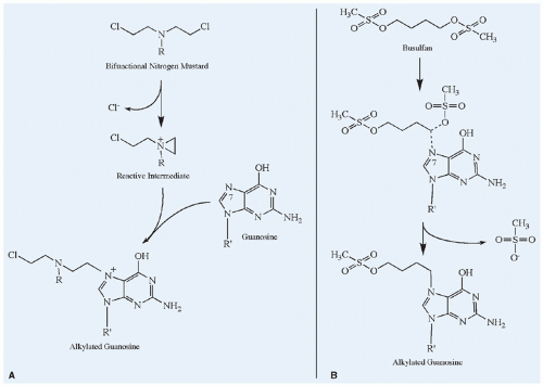
Figure 10.5 Mechanisms of alkylation of the nucleophilic N7 position of guanosine. A: The bifunctional nitrogen mustard illustrates the SN1 type of alkylation reaction in which a reactive intermediate forms spontaneously and then rapidly reacts with the nucleophilic group. The rate-limiting step for SN1 alkylation is the formation of the reactive intermediate, and thus the reaction exhibits first-order kinetics (i.e., independent of the target nucleophile concentration). If the second chloroethyl group also reacts with another nucleotide base, a cross-link is formed. B: Busulfan exemplifies an SN2 reaction, characterized by a bimolecular nucleophilic displacement. In this case, the methylsulfonate group on either end of busulfan is displaced by the nucleophilic group on guanosine. The rate of SN2 alkylation reactions depends on the concentration of the alkylating agent and the target nucleophile, and it therefore follows second-order kinetics.


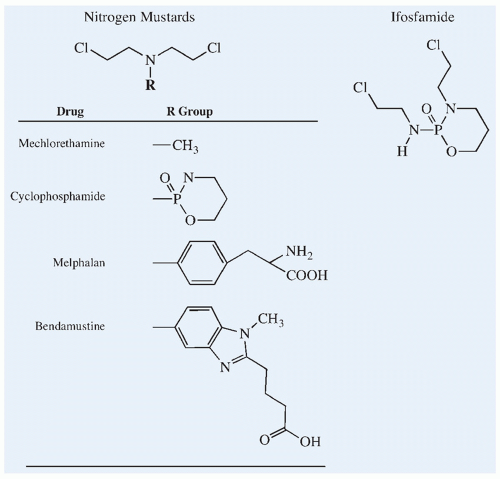
Figure 10.6 Chemical structures of the nitrogen mustard alkylating agents and the cyclophosphamide isomer, ifosfamide.


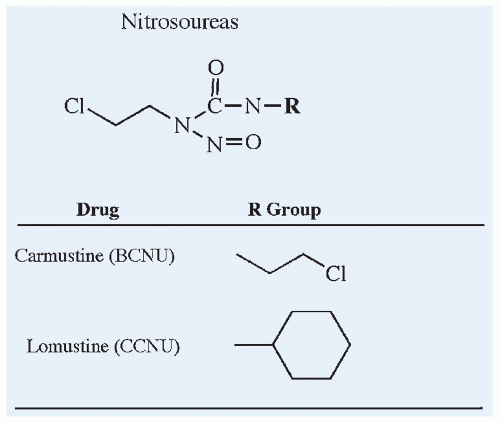
Figure 10.7 Chemical structures of the nitrosoureas, carmustine and lomustine.


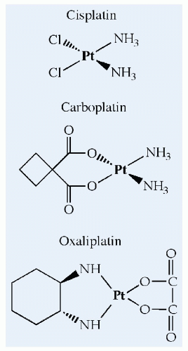
Figure 10.8 The chemical structures of cisplatin, carboplatin, and oxaliplatin, which platinate DNA in a manner analogous to alkylation by the nitrogen mustards. Reactive intermediates are formed after spontaneous elimination of chloride (cisplatin), dicarboxylate cyclobutane (carboplatin), or oxalate (oxaliplatin).


Mechlorethamine

Although the role of mechlorethamine in the treatment of cancer has declined, it is still a model for the chemical reactions of bifunctional alkylators (Fig. 10.5). The spontaneously formed alkylating intermediate is highly chemically reactive, and it rapidly undergoes hydrolysis, leading to inactivation, or it alkylates a wide variety of molecules, with a propensity to react with the N7 position on guanosine.138,145 Because of this inherent instability, even in aqueous solutions, mechlorethamine must be administered intravenously immediately after preparation to avoid significant loss of activity. Those administering the drug must take precautions, because direct contact with this reactive compound can irritate skin or mucous membranes.


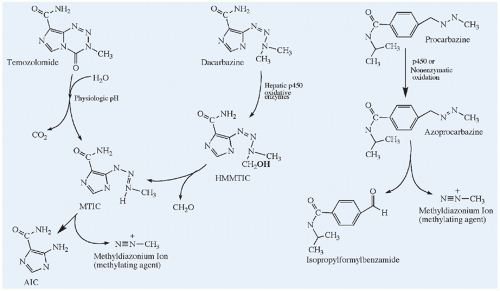
Figure 10.9 Chemical structures and activation pathways of the methylating agents, dacarbazine, temozolomide, and procarbazine, which are prodrugs. Dacarbazine requires enzymatically catalyzed activation, and temozolomide undergoes spontaneous chemical conversion in solution at physiological pH to the active metabolite, MTIC. MTIC, methyltriazenyl-imidazole carboxamide; HMMTIC, hydroxymethyl-MTIC; AIC, amino-imidazole carboxamide. The metabolic pathway for procarbazine is highly complex and incompletely shown. In addition to the methyldiazonium ion, free radicals can also be generated from azoprocarbazine.

Mechlorethamine has been used primarily in combination with vincristine, prednisone, and procarbazine (MOPP) for the treatment of Hodgkin disease, but the MOPP regimen has been supplanted as standard therapy for this disease.152 The pharmacokinetics of mechlorethamine in humans has not been well delineated. In animals, the drug disappears from plasma in seconds.145,153 In addition to its rapid spontaneous hydrolysis, mechlorethamine is rapidly metabolized (N-demethylated) in the liver.154 As a result of this rapid degradation, renal excretion is not likely to play a role in drug clearance.

In addition to its major clinical toxicities of myelosuppression, nausea, and vomiting, mechlorethamine has an anticholinergic effect, leading to diaphoresis, lacrimation, and diarrhea. It is a potent vesicant, producing a sclerosing thrombophlebitis above the site of administration and severe local tissue damage if extravasated. If extravasation occurs, sodium thiosulfate should be injected into the area as rapidly as possible to neutralize the drug.155 Neurotoxicity in the form of an acute or delayed encephalopathy has been reported with the use of high doses of mechlorethamine.


Bendamustine

Synthesized in 1963 in East Germany, bendamustine (Fig. 10.6) is a bifunctional molecule with both alkylating and antimetabolic properties. Although used for many years in Europe, bendamustine
was not studied in the United States until the 21st century, and was approved in 2008 by the U.S. FDA for the treatment of indolent B-cell lymphoma.156,157

By design, because of its bifunctionality, bendamustine was found to have clinical activity against a variety of cancers, including refractory leukemia and lymphoma.158 Because of this unique bifunctionality, bendamustine is thought to have a distinct activity and resistance profile that differentiates bendamustine from other alkylating agents.159 Bendamustine is extensively metabolized to active metabolites and excreted by both the liver and the kidneys; clearance is rapid and the steady-state volume of distribution is limited, and as such the reported half-life is less than 1 hour.160

Bendamustine is indicated for the treatment of adults with chronic lymphoblastic leukemia and non-Hodgkin lymphoma. Dose-limiting toxicity includes myelosuppression, infections, infusions reactions and anaphylaxis, tumor lysis syndrome, and skin reactions.


Oxazaphosphorines


Cyclophosphamide/Ifosfamide

The oxazaphosphorines, cyclophosphamide and ifosfamide, are inactive prodrugs that require biotransformation by hepatic microsomal oxidative enzymes before expressing alkylating activity.161 Cyclophosphamide is a true nitrogen mustard derivative with a bifunctional bischloroethylamine side chain. Ifosfamide is also bifunctional but has one chloroethyl group shifted to a ring nitrogen (Fig. 10.6). Cyclophosphamide is one of the most widely used anticancer drugs, with a broad range of clinical activity that includes the acute leukemias and a variety of solid tumors (Table 10.3). It is also used in preparative regimens before bone marrow or peripheral stem cell transplantation and as an immunosuppressant in nonmalignant disorders.162 Ifosfamide has activity as a single agent or in combination with etoposide in sarcomas (e.g., Ewing sarcoma, rhabdomyosarcoma, osteosarcoma), lymphoma, germ cell tumors, Wilms tumor, and neuroblastoma.163,164


Figure 10.10 Metabolic pathways for the oxazaphosphorines, cyclophosphamide and ifosfamide. Both compounds must undergo hydroxylation at the 4 position before expressing alkylating activity; this reaction is catalyzed by hepatic microsomal enzymes. The 4-hydroxy metabolites are in spontaneous equilibrium with the open-ring aldehydes (aldophosphamide or aldoifosfamide), which can release Acrolein and form the active alkylating mustards (phosphoramide mustard or isophosphoramide mustard). Further oxidation at the 4 position of the primary metabolites leads to the formation of inactive metabolites (ketocyclophosphamide and carboxyphosphamide or ketoifosfamide and carboxyifosfamide), which are excreted in the urine. The open-ring aldehyde metabolites can be chemically reduced to an alcohol (alcophosphamide or alcoifosfamide). Inactivation by dechlorethylation leads to formation of the potentially toxic by-product chloracetaldehyde. This is a minor pathway for cyclophosphamide but more active with ifosfamide.

Cyclophosphamide is usually administered as a single-dose bolus or in fractionated doses over 2 to 3 days. Ifosfamide is administered on a fractionated schedule over 5 days, because in the initial trials, the single-dose schedule produced intolerable nephrotoxicity, cystitis, and neurotoxicity. Ifosfamide has also been administered as a continuous 5-day infusion. The maximally tolerated total dose of ifosfamide is approximately threefold to fourfold higher than an equitoxic dose of cyclophosphamide.165

Biotransformation. The metabolic pathways of cyclophosphamide and ifosfamide are shown in Fig. 10.10. The steps in the biotransformation of these two drugs are qualitatively identical. Hydroxylation of the 4-carbon position on the ring by hepatic microsomal mixed-function oxidases yields the primary 4-hydroxy metabolites, which are in spontaneous equilibrium with the open-ring aldehydes. Hydroxylation of cyclophosphamide is catalyzed primarily by CYP2B6 with minor contributions from CYP3A4 and CYP2C9, and ifosfamide hydroxylation is catalyzed primarily by CYP3A4 with a minor contribution from CYP2A6.166 Although not chemically reactive, the 4-hydroxy metabolites are cytotoxic in vitro and are thought to be the transport forms of the active alkylating species, phosphoramide mustard and isophosphoramide mustard,
which are formed by spontaneous elimination of acrolein from the open-ring aldehydes. Quantitatively, the rate of activation of cyclophosphamide is greater than that of ifosfamide, and this difference in the rate of activation accounts for the difference in clinical pharmacokinetics and MTD of the two isomers.167,168,169

Further oxidation of the hydroxyl group at the 4-carbon position on primary metabolites by aldehyde dehydrogenase leads to inactivation. 4-ketocyclophosphamide and carboxyphosphamide are the principal urinary metabolites of cyclophosphamide. Aldehyde dehydrogenase is found in a wide variety of tissues and in cancer cells.169 The chloroethyl side chain can also be enzymatically cleaved by CYP3A4. Less than 10% of the administered dose of cyclophosphamide is metabolized via this pathway, but up to 50% of the ifosfamide is dechlorethylated, resulting in a greater rate of production of the potentially toxic by-product chloracetaldehyde compared with cyclophosphamide.167,168

Pharmacokinetics. The pharmacokinetic behavior of unchanged cyclophosphamide and ifosfamide has been well described. When administrated orally in low doses, 75% to 95% of the cyclophosphamide is absorbed.170,171,172 The minimal first-pass metabolism after oral administration indicates that the hepatic extraction ratio for cyclophosphamide is low. Plasma concentrations of the active metabolites, 4-hydroxycyclophosphamide and phosphoramide mustard, after oral administration are equivalent to those achieved with intravenous administration.172 The oral bioavailability of ifosfamide is greater than 95%.173,174,175 Peak concentrations of 4-hydroxy-ifosfamide and chloracetaldehyde were twofold higher than those achieved with the same dose administered intravenously.

Cyclophosphamide and ifosfamide are eliminated primarily by hepatic biotransformation to active and inactive metabolites, which are excreted mainly in the urine. Less than 20% of the dose is excreted as unchanged drug in the urine, and biliary excretion of unchanged drug is minimal.170,171,176,177,178 The total body clearance in adults is 30 to 35 mL/min/m2 and 60 to 80 mL/min/m2 for cyclophosphamide and ifosfamide, respectively.170,179,180 Total clearance of cyclophosphamide in children (40 to 50 mL/min/m2) appears to be higher than in adults.181,182,183 The plasma half-life in children (3 to 4 hours) is also reported to be shorter than that in adults (6 to 8 hours).170,171,180,181,182,183,184,185 Ifosfamide clearance in children ranges from 50 to 130 mL/min/m2, similar to that reported in adults, and the half-life of ifosfamide in children is 1 to 5 hours.186,187,188 The considerable interpatient variability in the disposition and metabolism of the oxazaphosphorines181,186,187,189,190 may impact patient outcome. In one study of 36 children with B-cell non-Hodgkin lymphoma, the likelihood of disease recurrence in children with low plasma clearance of the parent prodrug, and thus with decreased capacity to generate active metabolites of cyclophosphamide, was significantly higher than in children with relatively higher clearance.191

Cyclophosphamide and ifosfamide can rapidly induce their own metabolism. With infusional or fractionated dosing, there is a decrease in the plasma half-life and an increase in clearance of the parent prodrugs and an increase in metabolite concentrations.171,173,185,188,192,193 Cyclophosphamide exposure induces the expression of CYP2C9 and CYP3A4 enzyme levels in human hepatocytes.194 The increase in the rate of metabolism occurs within 12 to 24 hours of the first dose, and a new steady state is achieved by 48 to 72 hours.195 Over a 5-day course of ifosfamide, the parent drug half-life decreases, and the clearance increases by 30% to 50%.193,196 Although several studies have found that the apparent clearance of ifosfamide and its metabolites is greater when the drug is administered as a continuous infusion,197,198,199 an observation that would favor administration of drug on a fractionated schedule, a crossover study in adult patients could not find a significant difference in drug disposition between the two schedules of administration.200

The fraction of the cyclophosphamide dose that is converted to active metabolites appears to be constant (60% to 70% of the dose), and there is no evidence of saturation of the activating enzymes over a broad dosage range of 100 to 3,000 mg/m2.184,189 However, at doses of 4,000 mg/m2 used in autologous bone marrow preparative regimens, saturation of drug-activating enzymes becomes apparent.201,202 Saturation (nonlinearity) of ifosfamide metabolism has also been described at doses exceeding 2500 mg/m2. The half-life was prolonged to 15 hours, a higher percentage of the drug is excreted in the urine unchanged, and the AUC of ifosfamide metabolites does not increase in proportion to the dose.168,177,203

The activated metabolites of cyclophosphamide and ifosfamide appear in plasma rapidly, reach a peak by 2 hours after the dose, and have a half-life of approximately 4 hours.145,172,204 At equivalent doses, the plasma concentrations of alkylating metabolites of ifosfamide are approximately one-third that generated from cyclophosphamide, presumably because of a difference in the rate of enzymatic activation.167,168 Plasma concentrations of the active metabolites are considerably lower than those of the parent prodrug, because of the chemical instability and reactivity of the active 4-hydroxy metabolites. The plasma concentration of the active 4-hydroxy metabolites is approximately 1% to 3% of that of the parent drug.168,204,205,206

Patients with severe renal function impairment (i.e., creatinine clearance less than 20 mL/min) have moderately higher parent drug concentrations207 and significantly higher plasma alkylating activity.180,184,208 However, in a single anuric patient, Wagner and associates found no change in the disposition of cyclophosphamide and its activated metabolite,209 and ifosfamide disposition did not appear to be altered in an anuric child.210 The degree of cyclophosphamide-related hematological toxicity does not correlate with the severity of renal insufficiency. There is no strong evidence to support dosage modifications of cyclophosphamide in patients with renal dysfunction; however, ifosfamide dosage adjustment may be indicated because of the increased risk of neurotoxicity in patients with renal dysfunction.72,210 Cyclophosphamide and ifosfamide can be efficiently removed from the blood by dialysis.210,211 The hemodialysis extraction efficiency for 4-hydroxyifosfamide is lower than that for the parent drug.210 Hepatic dysfunction may alter the rate of drug activation and the rate of elimination. With hepatic parenchymal damage, the half-life of cyclophosphamide is prolonged, and peak concentrations of alkylating activity in plasma are lower.184

Toxicity. Myelosuppression is the major dose-limiting toxicity of the oxazaphosphorines, but unlike the lipid-soluble alkylating agents, such as the nitrosoureas, they rarely cause cumulative marrow damage. Nausea, vomiting, and alopecia occur in most patients.161

Hemorrhagic cystitis is a toxicity that is unique to the oxazaphosphorines. It may range from mild dysuria and frequency to severe hemorrhage from bladder epithelial damage. The reported incidence of this complication ranges from 5% to 10% for cyclophosphamide and 20% to 40% for ifosfamide.179 This toxic effect is dose-related and appears to be caused by the activated metabolites and by the biologically active by-products, such as acrolein (Fig. 10.10). The incidence and severity of chemical cystitis can be lessened by aggressive hydration and frequent emptying of the bladder, by bladder irrigation, or by the concurrent administration of mesna (2-mercaptoethane sulfonate). After administration, mesna is rapidly oxidized in plasma to a chemically stable and pharmacologically inert disulfide that is then rapidly excreted by the kidneys and converted back to its chemically reduced active form during tubular transport. It is therefore only active in urine and does not interfere with the antitumor effects of cyclophosphamide or ifosfamide.102, Although the dose and schedule of mesna vary, it is commonly administered at a dose equal to 60% of the total ifosfamide dose, divided into three doses and administered at 0, 4, and 8 hours after ifosfamide.102 Mesna can be administered orally or intravenously. Mesna also reduces the incidence of oxazaphosphorine-induced bladder cancers in rats, a complication that has been reported in humans.179,212

The oxazaphosphorines are also nephrotoxic. Cyclophosphamide can have a direct renal tubular effect that can result
in water retention.213,214 Ifosfamide produces proximal tubular damage resembling Fanconi syndrome, with glucosuria, amino aciduria, and phosphaturia. Animal studies suggest that it is the ifosfamide metabolite chloracetaldehyde, acting on mitochondrial NADH:ubiquinone oxidoreductase in the renal tubule, which is the primary mediator of nephrotoxicity.215 Rickets has been observed in younger children.216,217,218,219,220 Decreased glomerular filtration rate (GFR) and distal tubular damage manifested by concentrating defects and renal tubular acidosis also has been reported.221,222 Comprehensive follow-up evaluation of glomerular and tubular function in children previously treated with ifosfamide revealed dysfunction in 78%, including 28% with moderate or severe nephrotoxicity.223 Cumulative doses of 45 to 80 g/m2 or greater appear to be the primary risk factor,223,224,225 with young children appearing to be at higher risk for proximal renal tubular damage.226,227

Other toxic effects of ifosfamide include reversible neurotoxicity characterized by somnolence, disorientation, and lethargy in about 10% to 40% of patients and, more rarely, hallucinations, coma, and seizures.228,229,230 The incidence of neurotoxicity was 50% with oral administration, presumably the result of first-pass metabolism of ifosfamide to neurotoxic metabolites.168 The neurotoxicity has been attributed to the metabolite chloracetaldehyde (Fig. 10.10), which results from dechlorethylation of ifosfamide.231 The dechlorethylation pathway accounts for 50% of ifosfamide metabolism but less than 10% for cyclophosphamide. The incidence of neurotoxicity also appears to be greater in children who previously received high cumulative doses of cisplatin. Cisplatin-induced renal damage might have diminished the rate of elimination of neurotoxic metabolites of ifosfamide in these patients.232 Neurotoxicity may be reversible or preventable with methylene blue,233,234 but its actual efficacy remains uncertain.230 Transient hepatic dysfunction has also been reported with ifosfamide.179 Cardiac toxicity has been observed in patients treated with high doses (≥100 to 200 mg/kg) of cyclophosphamide. Ifosfamide has also been implicated as a cause of cardiomyopathy and arrhythmias at doses of 10 to 18 g/m2 in a transplant setting.235

Although pulmonary toxicity is not commonly associated with the oxazaphosphorines, cases of early- and late-onset interstitial pneumonitis from cyclophosphamide and ifosfamide have been reported.236,237,238 Clinical features of drug-induced lung injury typically include fever, cough, dyspnea on exertion, diffuse interstitial infiltrates on chest radiographs, and bilateral pleural thickening usually presenting within weeks to months of drug exposure. Factors that appear to augment oxazaphosphorine lung damage include administration of cyclophosphamide in combination with other cytotoxic drugs and the concurrent use of cyclophosphamide and irradiation. Inspired oxygen has also been shown to enhance lung injury in animals.239 Oxazaphosphorine-induced lung injury appears to be unresponsive to corticosteroid therapy and the prognosis is poor.

Resistance. Mechanisms of resistance to cyclophosphamide involve intracellular inactivation of the activated metabolites and enhanced repair of DNA adducts.138,161 Elevated concentrations of glutathione, resulting from increased activity of the enzyme glutathione-S-transferase, can detoxify the biologically active metabolites of the oxazaphosphorines.240,241,242,243 Sensitivity to cyclophosphamide is also inversely correlated with intracellular concentrations of the enzyme aldehyde dehydrogenase, which oxidizes activated cyclophosphamide metabolites to inactive forms. Intracellular levels of this enzyme can be estimated in tissue or tumor specimens by histochemical staining. Enhanced DNA repair by nucleotide-excision repair enzymes or O6-alkylguanine-DNA alkyltransferase may also contribute to resistance.

Drug Interactions. Compounds known to alter the activity of p450 microsomal enzymes can affect the rate of activation and elimination of the oxazaphosphorines. Phenobarbital pretreatment enhances the rate of metabolism of cyclophosphamide and its activated metabolites in animals and in humans;108 similar induction may also occur with phenytoin.244 Concurrent allopurinol appears to enhance the myelotoxicity of cyclophosphamide.245 Busulfan, which is administered with cyclophosphamide in transplant preparatory regimens, can block the conversion of cyclophosphamide to its active metabolite, when cyclophosphamide is administered less than 24 hours after a dose of busulfan.246 The neurokinin-1 receptor antagonist aprepitant, a moderate inhibitor of CYP3A4,247 can inhibit metabolism of cyclophosphamide and thiotepa, but the overall impact is small relative to the overall variability observed.248 Concurrent fluconazole can block the activation of cyclophosphamide,181 whereas concurrent itraconazole can increase cyclophosphamide clearance and generation of active metabolites.249 Dexamethasone and chlorpromazine also appear to induce the metabolism of cyclophosphamide.181


Melphalan

Melphalan (L-phenylalanine mustard, Fig. 10.6) is a rationally designed anticancer drug that has the bischloroethylamine moiety attached to the amino acid phenylalanine, with the intention that it would be taken up preferentially by melanin-producing cancers. Although this agent has a broad range of clinical activity in adult cancers (e.g., multiple myeloma, melanoma, breast and ovarian cancers, lymphoma), its use has been limited in the treatment of childhood cancers. At standard doses (35 mg/m2), melphalan is active against rhabdomyosarcoma.250 The administration of bone marrow ablative doses (140 to 220 mg/m2) of melphalan followed by rescue with autologous bone marrow transplant has resulted in high response rates in children with neuroblastoma, Ewing sarcoma, and acute leukemia,141,251,252,253 and has also been used in reduced-intensity conditioning regimens in children with acute leukemia.254 Melphalan has also been administered intra-arterially by isolated perfusion for cancers localized to an extremity or the liver,255,256 as well as intraocularly for retinoblastoma, either alone or in combination with other agents such as topotecan.257,258

Like other chemically reactive compounds, melphalan is rapidly cleared from the body. It is inactivated after spontaneous hydrolysis or alkylation reactions with plasma or tissue proteins. Melphalan does not appear to undergo any appreciable enzymatic degradation.259 The absorption of melphalan after oral administration has been reported to be incomplete and highly variable.259,260 The fraction of a dose absorbed usually ranges from 32% to 100%, but patients with no detectable drug in plasma and urine after an oral dose have been reported.260 Melphalan bioavailability is higher and less variable when the drug is administered in the fasting state.261 The incidence of myelosuppression is lower with oral than with intravenous melphalan, and poor therapeutic response may be attributable in part to poor absorption in some patients receiving oral melphalan.260 The disposition of melphalan after intravenous administration in children and adults is similar.262 With standard parenteral doses, the terminal half-life ranges from 60 to 120 minutes, with a total clearance exceeding 200 mL/min/m2.250,263 Pharmacokinetic parameters in patients receiving high-dose therapy (up to 220 mg/m2) are similar to those found at standard doses.262,264,265,266,267,268 Wide interindividual variation in melphalan AUC and clearance has been observed in most studies and has led to the development of pharmacokinetically guided dosing strategies for melphalan.145

Renal excretion is a minor route of melphalan elimination, accounting for 20% to 30% of total drug clearance.72,250,269 However, patients with renal dysfunction have a higher incidence of hematological toxicity.270 In a group of patients with a wide range of renal function, drug clearance after high-dose melphalan was correlated with creatinine clearance, but the decrease in melphalan clearance in patients with renal dysfunction was insignificant compared with the high degree of interindividual variation in drug disposition. In children previously treated with carboplatin, melphalan clearance was approximately two-thirds of that observed in other children.271,272

At standard doses (5 to 35 mg/m2), myelosuppression is the primary toxicity, and cumulative marrow damage has been observed with repeated doses. Pulmonary fibrosis and secondary
leukemia are late effects associated with the chronic administration of melphalan. At high doses with autologous bone marrow or stem cell reinfusion, gastrointestinal toxicity (e.g., mucositis, esophagitis, diarrhea) becomes dose limiting.141,251,268


Nitrosoureas


Carmustine (BCNU)/Lomustine (CCNU)

The nitrosoureas are a group of lipid-soluble alkylating agents (Fig. 10.7) that are highly active in experimental tumor models, including intracranially implanted tumors. The 2-chloroethyl derivatives, carmustine (BCNU) and lomustine (CCNU), are the nitrosoureas most widely used in pediatric oncology.273 Rapid spontaneous chemical decomposition of these compounds in solution generates an alkylating intermediate (chloroethyldiazohydroxide) and an isocyanine moiety that can carbamoylate amine groups on proteins. Alkylation, including cross-linking of DNA by the monofunctional lomustine and the bifunctional carmustine, is generally accepted as the primary mechanism of action of the nitrosoureas.274,275,276 However, the isocyanates can inhibit DNA repair of alkylator damage and may contribute to the antitumor activity and the toxicity of the nitrosoureas.273 The nitrosoureas alkylate the N3 position on cytidine and the N7 and O6 positions on guanosine,138 but the primary factor determining tumor cell resistance to the nitrosoureas is the capacity to enzymatically repair O6-alkyl-guanosine.277,278 The combination of carmustine and O6-benzylguanine, an inhibitor of the DNA repair protein O6-alkylguanine-DNA-alkyltransferase, has been evaluated in phase I and II studies in adults279,280,281,282,283 and in a phase I study in children.284 Overall, O6-benzylguanine increased the myelosuppressive effects of carmustine, resulting in no apparent net improvement in its therapeutic index.

The nitrosoureas have been used primarily to treat patients with brain tumors or lymphomas, and high-dose carmustine has been incorporated into transplant-preparative regimens. Delayed and cumulative myelosuppression and other serious long-term cumulative renal and pulmonary toxic effects, which are particularly concerning in children, limit the clinical utility of these agents in combination regimens.285,286 Carmustine has been incorporated into biodegradable polymer wafers that can be implanted into the tumor cavity after surgical resection for brain tumors. Drug is released slowly from the polymer wafer over 2 weeks, providing prolonged sustained exposure to high concentrations of carmustine locally with a lower risk of systemic toxicity.287,288

Biotransformation and Pharmacokinetics. In addition to their rapid spontaneous decomposition, nitrosoureas undergo significant hepatic metabolism. The cyclohexyl ring of lomustine is hydroxylated at the 4-position to yield two isomeric derivatives that are more soluble and have greater alkylating activity than the parent drug.273 Carmustine is inactivated by denitrosation through the action of microsomal enzymes and glutathione conjugation. As a result of this rapid spontaneous and enzymatic degradation, the clearance of nitrosoureas from plasma is extremely rapid. In early studies of carmustine and lomustine, parent drug could not be detected in plasma after intravenous or oral administration.289,290 With high-dose carmustine administered by intravenous infusion, the half-life was 22 minutes, and clearance exceeded 2,000 mL/min/m2. Similar results have been reported with standard doses of the drug (half-life, 22 minutes; clearance, 1,700 mL/min/m2).291 The half-life of the active 4-hydroxylated metabolites of lomustine is 3 hours.292 When administered orally, the nitrosoureas are well absorbed, and lomustine is extensively converted to hydroxylated metabolites presystemically during its first pass through the liver.293 These results confirm that the metabolites of lomustine are primarily responsible for the drug’s antitumor activity. Although carmustine is also well absorbed, severe vomiting after oral administration frequently precludes adequate absorption.

The lipid-soluble nitrosoureas are widely distributed and readily penetrate into the CNS. After equilibration, drug concentrations in the cerebrospinal fluid (CSF) approximate those in plasma, which in part accounts for the activity of this group of drugs in treating brain tumors.290,294 Implantation of carmustine-containing polymer wafers into the tumor bed for brain tumors bypasses the blood-brain barrier and provides local drug concentrations that are higher than those achieved with systemic administration. However, the depth of penetration into the brain parenchyma from the wafer is very limited (5 mm at 30 hours) owing to the rapid diffusion of drug into the capillaries.295

Toxicity. Gastrointestinal toxicity (i.e., nausea and vomiting) and cumulative delayed myelosuppression are the most consistent side effects of the nitrosoureas. The nadir of blood counts occurs 4 to 5 weeks after administration, and the platelet count tends to be the most affected. With repeated dosing, chronic marrow hypoplasia develops.273 With cumulative doses of more than 1500 mg/m2, progressive renal atrophy has been reported.296,297 Although in children this complication has been primarily associated with semustine (methyl-CCNU), it has also been reported after high cumulative doses of lomustine. Mitchell and Schein recommend that if nitrosourea therapy continues for more than 15 months or if cumulative doses of greater than 1,000 mg/m2 are reached, patients should be evaluated for nephrotoxicity and therapy discontinued if renal size or GFR is significantly decreased.273 Similar cumulative doses (≥1,500 mg/m2) of carmustine are associated with progressive and frequently fatal pulmonary toxicity characterized by cough, dyspnea, tachypnea, and a restrictive-type ventilatory defect.239,285,298,299 Carmustine-induced pulmonary toxicity can vary substantially in manifestations, outcome, and histopathologic appearance,285 with the risk of developing significant pulmonary symptoms remaining elevated for many years following completion of therapy.300 Long-term follow-up of 17 children with brain tumors treated with carmustine revealed that 6 (35%) had died of pulmonary fibrosis and that all of the surviving patients studied had radiographic abnormalities or restrictive defects on spirometry.286 Four of the six patients who died presented with pulmonary symptoms 8 to 13 years after treatment. Females appear to be more susceptible to the complication than males,285,301 and a history of atopy may increase the risk of pulmonary complications.302 Pulmonary fibrosis appears less frequent with lomustine, but cases have been reported.239 CNS toxicity has been reported rarely.273 High-dose carmustine (300 to 750 mg/m2) can produce hypotension, tachycardia, flushing, and confusion.291

Drug Interactions. In animals, phenobarbital enhances the microsomal metabolism of the nitrosoureas and significantly reduces the antitumor activity of carmustine and, to a lesser extent, that of lomustine.303 This potential interaction has not been studied in humans. Carmustine, an inhibitor of glutathione reductase, potentiates the hepatotoxicity of high doses of acetaminophen in animals. Liver damage results from the depletion of intrahepatocyte glutathione by a minor but reactive quinone metabolite of acetaminophen.304


Dimethanesulfonates


Busulfan/Treosulfan

The bifunctional alkylating agent busulfan is an alkyl alkane sulfonate (Fig. 10.5). The busulfan alkylation reaction occurs by nucleophilic displacement of the methylsulfonate group on either end of the molecule (Fig. 10.5). Busulfan has a greater propensity to alkylate thiol groups on amino acids and proteins than the nitrogen mustards, but it can also alkylate the N7 position on guanosine.

Busulfan is not water-soluble and is commercially available as an oral formulation (2 and 25 mg tablets) and as an intravenous formulation (Busulfex). Busulfan has been used in conventional doses (1.8 mg/m2/day) as palliative therapy for chronic myelogenous leukemia, and high-dose busulfan (16 mg/kg or 600 mg/m2, in 16 divided doses every 6 hours) is an important component of
many bone marrow transplant preparative regimens, usually in combination with cyclophosphamide.140

The pharmacokinetics of oral busulfan is highly variable and age-dependent.305 Oral busulfan is rapidly absorbed, peaking 1 to 2 hours after the dose, with an average bioavailability of 70% (range, 40 to >90%).306,307,308 Pharmacokinetic studies of the intravenous formulation in children suggest that interpatient variability is decreased with this route of administration.309,310,311 Children heterozygous or homozygous for the glutathione S-transferase variant GSTA1*B appear to have decreased busulfan clearance,312 but this finding requires confirmation in larger studies.

Busulfan is a small lipophilic compound that penetrates well across the blood-brain barrier. CSF concentrations at steady state are equivalent to those in plasma.313,314 The primary route of elimination of busulfan appears to be glutathione conjugation, which is catalyzed by an isoform of glutathione-S-transferase (GSTA1-1).315,316 Busulfan has a short half-life of 2.5 hours and a clearance in children of 80 mL/min/m2.306 These pharmacokinetic parameters appear to be linear over the wide dosage range used. Compared with adults, busulfan apparent clearance is more rapid in children, especially children who are ≤5 years of age.306,317,318 The higher apparent clearance in young children is the result of more rapid glutathione conjugation rather than lower bioavailability.318

The variability in the disposition of busulfan after oral dosing can result in up to a 20-fold range in systemic drug exposure among patients treated with a fixed dose.305,319 Factors contributing to this variability include the age-dependent clearance, variable bioavailability, hepatic dysfunction, drug interactions including phenytoin,320 and circadian rhythmicity.319 The busulfan AUC in young children treated with 1 mg/kg is less than half the AUC in adults receiving the same dose (Fig. 10.11).305,314,321 On the every-6-hour oral dosing schedule, busulfan trough plasma concentrations exhibited a marked circadian rhythm, with the highest troughs occurring at 6:00 AM.322


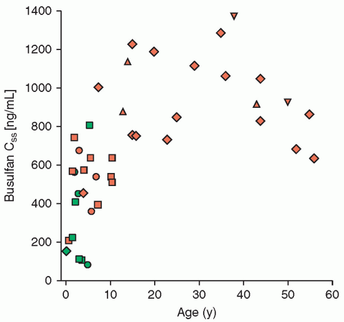
Figure 10.11 Plasma busulfan steady-state concentrations (Css) as a function of age. Css is derived by dividing the AUC by the dosing interval (6 hours). Patients were treated with 16 to 30 mg/kg of busulfan in combination with cyclophosphamide prior to bone marrow transplant. Triangles represent patients who rejected their graft or had a mixed chimera. Patients who experienced grade 0 treatment-related toxicity are designated in green, grade 1 toxicity in dark blue, grade 2 toxicity in orange, grade 3 toxicity in red, and grade 4 toxicity in black. Young children had substantially lower Css, less toxicity, and were at greater risk for graft rejection. (From data presented in Tables 1, 2, and 3 in Slattery JT, Sanders JE, Buckner CD, et al. Graft rejection and toxicity following bone marrow transplantation in relation to busulfan pharmacokinetics. Bone Marrow Transpl 1995;16:31-42.)

In the transplant setting, busulfan plasma concentrations appear to be predictive of hepatic toxicity and graft rejection, and in at least one model, may also predict for efficacy.305,323 In adults, the risk of developing severe hepatic veno-occlusive disease (VOD) is higher when the busulfan AUC exceeds 1,500 µM • min (Css of 1000 ng/mL).317,324,325 In children, targeting a Css of 600 to 900 ng/ml has been associated with improved engraftment,326,327 but the upper threshold for increased risk of toxicity has not been well defined. The busulfan AUC or Css associated with VOD or graft rejection appears dependent on the prior therapy administered,328 the preparative regimen, and the underlying disease.305 Therapeutic drug monitoring is now commonly performed following the initial dose of busulfan, as this appears to successfully maintain Css or AUC in a safe and effective range.320,327,329 Estimate of an initial starting busulfan dose, ranging from 0.8 to 1.2 mg/kg, can be based on a combination of age and weight, and model-based dosing nomograms have been proposed in order to predict exposure across a range of ages and weights.310,330,331,332

Myelosuppression is the primary toxicity from busulfan. Gastrointestinal toxicity, which is observed only at high doses, includes nausea, vomiting, and mucositis. Busulfan can rarely produce pulmonary toxicity (busulfan lung) that is characterized by diffuse interstitial fibrosis and bronchopulmonary dysplasia. Busulfan lung presents with cough, fever, rales, and dyspnea and usually progresses to respiratory failure.239 Hepatic VOD is observed in up to 40% of patients who are treated with high-dose busulfan without pharmacokinetically guided dosing, and the VOD is severe in 10% of patients.319,333,334,335 Seizures have also been reported with high-dose therapy, but they are preventable with prophylactic anticonvulsants.336 Girls who receive high-dose busulfan have a high incidence of severe and persistent ovarian failure.337

Treosulfan is an alkylating agent related to busulfan, available commercially in the EU, where it is labeled for the treatment of ovarian cancer in adults. Treosulfan is converted in vivo to an active epoxide compound, which is thought to provide the basis for its activity. Treosulfan has also been used in conditioning regimens for children with nonmalignant diseases.338,339 The variability in treosulfan exposure following dosing in children may be less than that seen with busulfan, but only limited data are available.340


NONCLASSICAL ALKYLATING AGENTS


Platinum Compounds


Cisplatin/Carboplatin/Oxaliplatin

Cisplatin, carboplatin, and oxaliplatin are heavy metal coordination complexes (Fig. 10.8) that exert their cytotoxic effects by platination of DNA, a mechanism of action that is analogous to alkylation. Reactive equated intermediates are formed in solution in a manner similar to the nitrogen mustards (Fig. 10.5). Chloride is the leaving group that is replaced by a water molecule in cisplatin. Dicarboxycyclobutane is the leaving group in carboplatin, and oxalate is the leaving group in oxaliplatin. These reactive intermediates covalently bind to DNA (N7-positoin of adenine and guanine) and form intrastrand and interstrand DNA cross-links.341 The rate of reaction of these platinum analogs with water to form reactive intermediates is an important determinant of the stability of the compounds in solution and influences the drugs’ pharmacokinetics.342,343 Cisplatin is more reactive than carboplatin and is less stable in aqueous solution. The stability of oxaliplatin is intermediate. Chloride-containing solutions such as 0.9% NaCl are required to stabilize cisplatin prior to administration.

Cisplatin is an effective agent for the treatment of testicular tumors and has demonstrated activity against osteosarcoma, neuroblastoma, Wilms tumor, germ cell tumors, and brain tumors.344,345,346,347 The drug is administered intravenously on a variety of schedules, including a single dose, infused over 4 to 6 hours; divided doses, usually daily for 5 days; and by continuous infusion for up to
5 days. The divided dose and continuous-infusion schedules may lessen the gastrointestinal and renal toxicities.344,348 Cisplatin has been administered regionally in a number of trials, including intraperitoneally for ovarian cancer, intravesicularly for bladder cancer, intrapleurally for the malignant pleural effusions, and intra-arterially for brain tumors and for sarcomas of the extremity, including osteosarcoma.349,350

The spectrum of antitumor activity of carboplatin is similar to that of cisplatin in adults, though it may be less efficacious in several solid tumors including testicular cancer.341,351 Carboplatin is active against brain tumors, neuroblastoma, sarcomas, and germ cell tumors.352 The pharmacokinetic and toxicity profiles of cisplatin and carboplatin are quite different (Tables 10.3 and 10.4).342,353 In children, carboplatin is administered as a bolus dose of 400 to 600 mg/m2 or in divided doses of 400 mg/m2 on 2 consecutive days or 160 mg/m2 daily for 5 days, every 4 weeks. Adaptive dosing formulas that individualize carboplatin dose based on the GFR have also been developed for children and are described below.

Oxaliplatin and 5-fluorouracil are an active combination for the treatment of adults with colorectal carcinoma.354 As a single agent, oxaliplatin is usually administered at a dose of 130 mg/m2 every 3 weeks, a dose that is also the recommended phase II dose in children when oxaliplatin is delivered on an every 3-week schedule.355 Objective responses in children have been seen in rare patients with CNS tumors following treatment with oxaliplatin as a single agent356 or in combination with etoposide.357

Pharmacokinetics. The chemical stability (reactivity) of the platinum analogs is a critical determinant of their pharmacokinetics. The reactive intermediates of cisplatin and carboplatin are rapidly and covalently bound to plasma protein and tissue.358 After binding with plasma or tissue proteins, the reactive platinum intermediates are inactivated. Only the free (unbound) platinum species (including the parent drug) are cytotoxic.359,360 This interaction of platinum compounds with protein is a time-dependent reaction. For cisplatin, more than 90% of total platinum in plasma is protein bound and inactivated within 2 to 4 hours.361 This represents the major route of drug elimination. Oxaliplatin, like cisplatin, is highly protein bound. More than 80% of platinum species were bound to plasma proteins 1 hour after administration of oxaliplatin to pediatric patients enrolled on a phase I trial of this agent.362 The major route of excretion of oxaliplatin is renal, and there is no evidence of cytochrome p450-mediated metabolism.343 Carboplatin is more chemically stable than cisplatin and oxaliplatin. Only 20% to 40% of total platinum is protein bound at 2 hours following administration of carboplatin, and this slowly increases to 50% over 24 hours.363,364 Tissue-bound platinum may be retained in the body for a prolonged time and is still measurable in plasma for 10 to 20 years after treatment.365

The pharmacokinetic behavior of bound and unbound, active forms of platinum differ appreciably. For cisplatin, after an initial rapid decay, total platinum (≥95% protein bound) persists in plasma and can be detected in urine for many days. The terminal half-life of total platinum ranges from 1 to 5 days.359,361,366 In contrast, the unbound, active platinum species have a much more rapid decline, with a half-life of less than 1 hour, which is primarily a reflection of the chemical reactivity of cisplatin and the avid binding of the reactive intermediates to tissue and plasma protein.359,367 In children receiving cisplatin, the half-lives of total and ultrafilterable (unbound) platinum are 44 hours and 40 minutes to 1.5 hours, respectively.347,368

Approximately 50% of the platinum administered as cisplatin is excreted in the urine over 4 to 5 days, primarily in an inactive form.361,369,370 Initially, total platinum clearance equals or exceeds creatinine clearance, reflecting excretion of unbound platinum species, but as protein binding becomes extensive, renal clearance of total platinum drops to only a small fraction of creatinine clearance.359 The renal clearance of the unbound, ultrafilterable species of platinum can actually exceed creatinine clearance, suggesting tubular secretion.368,371,372 It has been noted that tubular secretion rather than glomerular filtration is increased in obese adults,373 and the absolute clearance of cisplatin is significantly increased in obese adults compared with lean controls.374 In children, the clearance of cisplatin was not related to the GFR.368 Approximately 25% of unbound platinum species is excreted in the urine, and the degree of renal excretion is schedule-dependent (greater with short infusions).375 In patients with impaired renal function, the peak concentration of active, unbound platinum was elevated, but the terminal half-life was not prolonged, presumably because of the rapid reaction of these active species with plasma and tissue protein leading to inactivation.369,376 However, dosage reductions in patients with renal dysfunction may be indicated because of the drug’s nephrotoxic effects, which could further impair renal function.311

The disposition of carboplatin is characterized by a lower rate and degree of protein binding than for cisplatin. As a result, the terminal half-life of unbound carboplatin is longer (2 to 3 hours), and renal excretion is the primary route of elimination.342,364,370,377 By 24 hours, as much as 70% of the total platinum from carboplatin is excreted in the urine, most as parent drug. Carboplatin is dialyzable in patients with severe renal insufficiency.378,379

Pharmacokinetic parameters for carboplatin in children are similar to those in adults. The total clearance in children with a normal creatinine clearance is approximately 70 mL/min/m2, and the half-life is 2 to 3 hours.380,381,382 In children under 5 years of age, carboplatin clearance is 120 mL/min/m2, but in children <1 year of age, the clearance is 75 mL/min/m2.383 These age-related differences in carboplatin clearance appear to be related to differences in the GFR. The variability in carboplatin clearance supports the use of the adaptive dosing formulas based on GFR described subsequently.

The total clearance of carboplatin is highly correlated with creatinine clearance (Fig. 10.12),67,342,382,384 and patients with renal dysfunction and higher carboplatin AUCs have a greater probability of experiencing dose-limiting hematological toxicity. These associations allowed the development of adaptive dosing formulas for individualizing carboplatin dose based on creatinine clearance in adults and children (Table 10.5).67,380,382,384,385,386,387 The use of these formulas to calculate an individualized dose decreases the variability in systemic drug exposure (AUC) and reduces the incidence of severe thrombocytopenia.388 Caution
must be exercised when using these formulas, however, as the results are expressed either as an absolute dose (mg) or as a dose normalized to body surface area (mg/m2).389 When administered as a single dose in combination with ifosfamide and etoposide, a targeted carboplatin AUC of up to 10 mg • min/mL was tolerable.388 In ovarian and testicular cancers in adults, a carboplatin AUC of 5 to 7 mg • min/mL was associated with a higher response rate and a lower risk of disease recurrence.384 Additional study of the pharmacokinetics of carboplatin in infants is needed, as an analysis of carboplatin exposures achieved in patients less than 1 year of age suggests that clearance of this drug may be different in very young patients.390


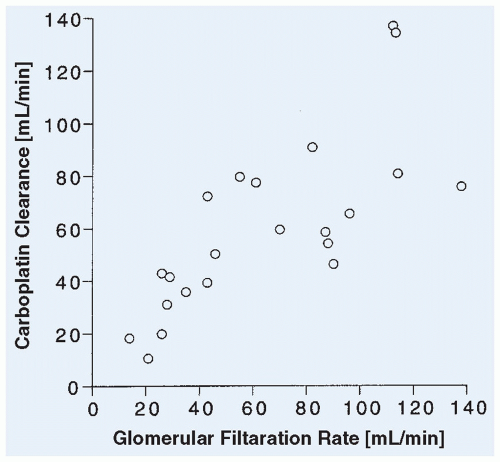
Figure 10.12 Relation between carboplatin clearance and GFR as measured by 51Cr-EDTA clearance in 22 children. (Adapted from Newell DR, Pearson ADJ, Balmanno K, et al. Carboplatin pharmacokinetics in children: the development of a pediatric dosing formula. J Clin Oncol 1993;11:2314.)


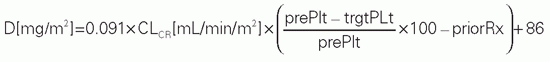
TABLE 10.5 Adaptive Dosing Formulas for Targeting Carboplatin Dose to Achieve a Desired Nadir Platelet Count or AUC, with the Target AUC Ranging from 7 to 10 mg • min/mL


Population


Formulaa


Adultsb


image


Adultsc


D[mg]=trgtAUC[mg•min/mL]×(GFR[ml/min]+25)


Childrend


D[mg/m2]=trgtAUC[mg•min/mL]×(0.93×GFR[ml/min/m2]+15)


Childrene


D[mg]=trgtAUC[mg•min/mL](GFR[ml/min]+(0.36×BW[kg]))


a D, dose; CLCR, creatinine clearance; prePlt, pretreatment platelet count; trgtPlt, target nadir platelet count; priorRx, 0 for previously untreated and 17 for previously treated; trgtAUC, target systemic drug exposure (AUC); GFR, glomerular filtration rate estimated by radioisotopic method; BW, body weight. Units for each parameter are listed in the brackets.

b Egorin MJ, Van Echo DA, Tipping SJ, et al. Pharmacokinetics and dose reductions of cis-diammine(1,1-cyclobutanedicarboxylato)platinum in patients with impaired renal function. Cancer Res 1984;44:5432.

c Calvert AH, Newell DR, Gumbrell LA, et al. Carboplatin dosage: prospective evaluation of a single formula based on renal function. J Clin Oncol 1989;7:1748.

d Marina NM, Rodman J, Shema SJ, et al. Phase I study of escalated targeted doses of carboplatin combined with ifosfamide and etoposide in children with relapsed solid tumors. J Clin Oncol 1993; 11:554.

e Newel DR, Pearson ADJ, Balmanno K, et al. Carboplatin pharmacokinetics in children: the development of a pediatric dosing formula. J Clin Oncol 1993; 11:2314.


The pharmacokinetic behavior of oxaliplatin in children appears to be similar to that in adults.355,356,362 Oxaliplatin is rapidly hydrolyzed in a nonenzymatic fashion to a large number of reactive intermediates. The volume of distribution in adults has been shown to exceed 500 L, compared with approximately 20 L for cisplatin and carboplatin. This suggests that the diaminocyclohexane ligand may enhance tissue distribution of oxaliplatin.343 Adaptive oxaliplatin-dosing formulas based on GFR have not been developed, although pediatric pharmacokinetic data identify covariates such as weight and renal function that could potentially be incorporated into a dosing nomogram.356 Dose reduction does not appear to be necessary in patients with moderate renal or hepatic dysfunction.391,392

Toxicity. The toxicity profiles of the platinum analogs are strikingly different. Cisplatin is associated with only mild myelosuppression, but produces significant and potentially irreversible nephrotoxicity, ototoxicity, and neurotoxicity. The dose-limiting toxicity of carboplatin is hematological toxicity, primarily thrombocytopenia, and the nonhematological toxicities observed with cisplatin are only seen at doses of carboplatin exceeding 800 mg/m2.342,353,393 Dose-limiting toxicities of oxaliplatin include neurotoxicity, thrombocytopenia, and neutropenia.

Nephrotoxicity, manifested as azotemia and electrolyte disturbances (especially hypomagnesemia requiring oral supplementation), was the dose-limiting toxicity in the initial clinical trials with cisplatin.393,394 The exact mechanism of cisplatin nephrotoxicity is not defined, but patients experience a reduction in renal blood flow and GFR and a loss of tubular function. Pathologic changes are seen primarily in the renal proximal and distal tubule epithelium and collecting ducts.393,395,396 Renal damage from cisplatin is cumulative. As a result of its nephrotoxic effects, cisplatin can alter its own elimination rate and that of other drugs, such as methotrexate, that rely on renal excretion.397 In one series, the renal clearance of ultrafilterable platinum fell from almost 500 mL/min with the first course to 150 mL/min by the fourth course in patients receiving repeated doses, probably because of decreased renal tubular secretion of the drug.398

Intravenous fluid hydration with normal saline prior to and after the infusion of cisplatin reduces the severity of nephrotoxicity associated with this agent. Diuresis with mannitol and furosemide has been used in an effort to decrease cisplatin-induced nephrotoxicity, but randomized studies have not shown a clear and reproducible benefit associated with the use of diuretics.399 The use of hypertonic sodium chloride solutions to promote chloruresis and the coadministration of amifostine remain controversial.348,400,401 Because cisplatin-associated renal dysfunction may be augmented in patients exposed to concomitant aminoglycosides, concurrent administration of nephrotoxic medications such as nonsteroidal anti-inflammatory drugs, iodinated contrast agents, and aminoglycosides should be avoided in patients receiving cisplatin.399 While these measures have reduced the incidence and severity of cisplatin-induced nephrotoxicity, moderate and permanent reductions in the GFR of patients receiving cisplatin have been documented.402,403,404 However, in a long-term follow-up study of 40 children who received a median of 500 mg/m2 of cisplatin, 22 of the 24 patients with abnormally low end-therapy GFRs partially recovered, with a median increase in GFR of 13 mL/min/m2.405 The 211 patients participating in the German Late Effects Surveillance System (LESS) had received a somewhat lower cumulative dose of cisplatin (360 mg/m2), but were followed longitudinally (median follow-up time 2 years). None of these patients developed an elevation in creatinine exceeding 1.5 times the upper limit of normal, and the hypomagnesemia detected in 12% of these patients following cisplatin therapy was mild.406 The long-term nephrotoxic effects of cisplatin in infants are similar to those reported in older children.407

As methods to prevent nephrotoxicity have allowed the administration of higher single and cumulative doses of the drug, ototoxicity and peripheral neuropathy have become more prominent.393 Cisplatin causes a reversible sensory peripheral neuropathy (i.e., numbness, tingling, and paresthesias) at cumulative
doses of 300 to 600 mg/m2.229,393 Lhermitte sign (an electric shock sensation when the neck is flexed) is common at high cumulative doses of cisplatin.408 Symptoms may progress after discontinuation of cisplatin and persist for months to years. Seizures and encephalopathy have also been reported in children receiving intensive cisplatin therapy.409 The irreversible hearing loss is in the high-frequency range and appears to be related to a cumulative dose of cisplatin of greater than 400 mg/m2.410,411,412 Children under 5 years of age also appear more likely to develop cisplatin-related hearing loss compared with older children.413 Genetic differences may explain some of the variability in platinum-associated neurotoxicity in adults, though the significance of germline variants in genes such as TPMT and catechol-O-methyltransferase (COMT) is a subject of debate in the literature.414,415,416 Amifostine decreases the incidence and severity of platinum-related neurotoxicity and ototoxicity in adults.417 In a study of children with average-risk medulloblastoma, amifostine appeared to have provided some otoprotection,418 but protective effects have yet to be observed in other pediatric studies.419,420,421 More recently, sodium thiosulfate has been studied as an otoprotectant.422 Although it appears to lessen the ototoxicity of cisplatin, the lack of selectivity appears to confer a tumor-protective effect that may curtail further clinical development. Additional toxic effects associated with cisplatin include prominent nausea and vomiting, mild myelosuppression, Raynaud phenomenon, and hypersensitivity reactions.344

Carboplatin’s myelosuppressive effects are delayed, affecting the frequency by which the drug can be administered. Platelet nadirs are typically seen up to 3 weeks after the dose, and milder granulocyte nadirs are observed 3 to 4 weeks after carboplatin administration. Some patients require 5 to 6 weeks for complete count recovery.423 Not only are the nephrotoxicity, ototoxicity, and peripheral neuropathy from carboplatin milder than that associated with cisplatin, but the nausea and vomiting, which can be dose-limiting with cisplatin, are also less severe.342,423,424 High cumulative doses of carboplatin are associated with a small drop in GFR and serum magnesium, but these changes are usually not clinically significant.425 Hypersensitivity reactions to carboplatin are relatively common, and the risk increases after multiple cycles of therapy.426,427

Myelosuppression due to oxaliplatin is usually mild, and the dose-limiting toxicity in adults is a cumulative peripheral neuropathy. Oxaliplatin is also associated with an unusual acute neurologic toxicity, pharyngolaryngeal dysesthesia, in which patients report difficulty in breathing or swallowing in the absence of laryngeal obstruction, probably related to transient sensory disturbances.428 The sensory neuropathy associated with oxaliplatin is exacerbated by cold in children as in adults.355 Although more than one-third of patients enrolled on a phase II study of oxaliplatin in children with CNS tumors developed a sensory neuropathy, it was severe in less than 5% of the patients.356

Resistance. Studies in preclinical tumor models have implicated several possible mechanisms of resistance to platinum compounds.429,430 Decreased drug accumulation may be related to altered drug uptake or the presence of a membrane efflux pump. Increased intracellular levels of thiol-containing compounds, such as glutathione and metallothionein, can react with and inactivate the active equated forms of cisplatin and carboplatin. The enhanced repair of platinum-DNA adducts by the nucleotide-excision repair pathway removes the cytotoxic lesion produced by the platinum analogs. Platinum-induced DNA damage activates apoptosis, and expression of cellular proteins that suppress the apoptotic response to this damage or loss of mismatch repair activity may alter sensitivity to the platinum analogs.137,431


Hydrazines


Procarbazine

Procarbazine is a methylhydrazine analog that was originally synthesized as a monoamine oxidase inhibitor, but was discovered to have antitumor activity in animals. Procarbazine is currently used for the treatment of Hodgkin disease432 and is also active against brain tumors.433 Procarbazine is a prodrug that requires metabolic activation in vivo to express its antitumor activity. This activation yields methylating and free-radical intermediates, which appear to produce the drug’s antitumor effect.

The spontaneous chemical decomposition and biotransformation of procarbazine is complex. Metabolic activation probably occurs in the liver and is catalyzed by the cytochrome P-450 enzyme complex (Fig. 10.9).434 In liver perfusion studies, procarbazine is extensively converted to its active azo-metabolite.435

The disposition of procarbazine and its active intermediates has not been well characterized in humans. The drug is rapidly and completely absorbed from the gastrointestinal tract,436 and it undergoes complete first-pass conversion to cytotoxic metabolites, which probably accounts for the activity of the drug when administered orally. After intravenous administration, procarbazine is rapidly metabolized and has a half-life of less than 10 minutes. The metabolites of procarbazine are excreted primarily in the urine. Procarbazine or unidentified metabolites enter the CSF readily. Drugs such as phenobarbital and phenytoin that are capable of inducing hepatic microsomal enzymes can increase the rate of procarbazine activation. Procarbazine can inhibit the biotransformation of the barbiturates, phenothiazines, and other sedatives, resulting in potentiation of their sedative effects. The inhibition of monoamine oxidase by procarbazine can put patients at risk for hypertensive reactions from foods high in tramline (e.g., bananas, wine, cheese). Procarbazine also appears to alter its own metabolism over a 14-day course of therapy. The plasma concentrations of procarbazine metabolites differ markedly between days 1 and 14 of treatment.437

The primary toxicities of procarbazine include nausea, vomiting, and myelosuppression. Some patients develop evidence of neurotoxicity consisting of paresthesias, somnolence, depression, or agitation. Neurotoxicity is prominent with high-dose intravenous administration.438 Patients are also at risk for the long-term toxicities, including azoospermia, ovarian failure, and teratogenic and carcinogenic effects.


Tetrizines


Dacarbazine

Although dacarbazine (Fig. 10.9) was originally developed as an inhibitor of purine biosynthesis, it does not exert its antitumor effects as an antimetabolite. Dacarbazine is a prodrug that undergoes hepatic microsomal metabolic activation (N-demethylation), which is catalyzed primarily by CYP1A2, to the active metabolite, methyltriazenyl imidazole carboximide (MTIC).439 MTIC then spontaneously decomposes into a reactive methylating species (methyldiazonium ion) and the primary circulating metabolite aminoimidazole carboxamide (AIC). The methyldiazonium ion can methylate nucleophilic sites, including the O6 and N7 positions on guanosine, but it cannot form cross-links.

Dacarbazine is generally administered intravenously (150 to 250 mg/m2) on a divided once-daily dosage schedule for 5 days. Absorption after oral administration is slow, incomplete, and variable.440 After intravenous administration, the drug is rapidly cleared from the plasma, with a terminal half-life of 40 minutes and a total clearance of 450 mL/min/m2. One-half of the dose is excreted unchanged in the urine, and renal clearance exceeds the GFR, suggesting the drug is also eliminated by renal tubular secretion.441 The remainder of the dose presumably undergoes biotransformation. The half-life and renal clearance of the metabolite AIC are similar to that of the parent drug.441 Methylated DNA adducts in white blood cells of patients treated with dacarbazine (250 to 800 mg/m2) increase rapidly during the first hour after treatment, but then decline with a more prolonged half-life (72 hours) than the parent drug.

When dacarbazine was administered as a 1,000 mg/m2 infusion over 24 hours, the steady-state plasma concentration was 8.6 µg/mL.442 Other pharmacokinetic parameters derived from the study of this
schedule included a total clearance of 110 mL/min/m2, a volume of distribution at steady state of 23 L/m2, and a terminal half-life after infusion of 3 hours.

Gastrointestinal toxicity, consisting of moderate-to-severe nausea and vomiting, is the primary toxicity and is frequently dose-limiting. Tolerance usually develops over the 5-day course of administration. At standard doses, myelosuppression is mild. Other side effects include a flulike syndrome with malaise, fever, and myalgias; mild hepatic dysfunction; and local pain at the site of intravenous injection. Rare cases of liver failure and death from VOD and hepatic vein thrombosis (Budd-Chiari syndrome) have been associated with the use of this drug.443


Temozolomide

The methylating agent temozolomide is structurally and mechanistically related to dacarbazine. Like dacarbazine, temozolomide is a prodrug, but temozolomide does not require enzymatic activation in the liver. In solution at physiological pH, temozolomide spontaneously decomposes to MTIC, the same active metabolite that is derived by enzymatic N-demethylation of dacarbazine (Fig. 10.9).444,445

Temozolomide is insoluble in aqueous solution and was initially only available in capsules for oral administration. However, a study comparing a 90-minute intravenous infusion with an equivalent oral dose of temozolomide demonstrated exposure equivalence. In addition, testing has shown that solutions made from the intravenous formulation are sufficiently stable to permit oral administration. This agent is now available for intravenous administration and as an oral solution for patients who are unable to tolerate capsules.446,447 On the basis of preclinical studies444,448 that demonstrated divided dosing schedules had greater antitumor effect than a single bolus dose, and on the initial phase I clinical trial449 in which responses were only observed on the divided dose schedule, temozolomide is administered as a single daily dose for five consecutive days. The recommended dose for children is 200 mg/m2/day (1,000 mg/m2/course) when administered as a single agent, though doses as high as 260 mg/m2/day given daily for 5 days have been well tolerated in children with leukemia.450,451,452 A continuous daily dosing schedule is also being investigated, and a dose of 75 mg/m2/day appears to be tolerable for 6 to 7 weeks in adults.453 Temozolomide is used in children primarily for the treatment of brain tumors, but has also been studied as part of combination regimens for a number of childhood solid tumors.454,455,456,457,458,459

Absorption of temozolomide from the gastrointestinal tract is rapid and complete.449,460 The peak concentration of temozolomide is achieved in plasma within 1.5 hour of the dose.451 When administered with food, the bioavailability is slightly lower but remains >90%.461 Temozolomide is also rapidly eliminated. Its halflife (1.8 hours) is similar to the drug’s half-life in a pH 7.4 phosphate buffer solution in vitro,445 suggesting that decomposition to the active metabolite, MTIC, is the primary route of elimination for temozolomide. A pharmacokinetic study of radiolabeled temozolomide confirmed that AIC, which is the end product of temozolomide decomposition to MTIC, is a primary urinary metabolite.462 In children, 5% to 15% of the dose of temozolomide was recovered in urine as unchanged drug.451 The apparent clearance of temozolomide in children is approximately 100 mL/min/m2, and the terminal half-life is similar to that observed in adults.452 The active metabolite, MTIC, is much less stable and has an estimated halflife of 2.5 minutes and clearance exceeding 5,000 ml/min/m2.462,463 There is some evidence that temozolomide clearance is lower in younger children.464 Temozolomide is widely distributed in tissues and penetrates well across the blood-brain barrier,460 and could therefore be considered the transport form for MTIC.

Myelosuppression is the dose-limiting toxicity of temozolomide. Nadir neutrophil and platelet counts typically occur 21 days after the start of therapy, and recovery of blood counts may take 7 to 10 days.445,450,451 This delayed myelosuppression necessitates administering temozolomide on a 28-day schedule. The myelosuppression from temozolomide does not appear to be cumulative.449 Nonhematological toxicities are mild and include nausea and vomiting, which can be controlled by pretreatment with standard antiemetics, headache, fatigue, constipation, and serum transaminase elevations.460

The DNA repair protein O6-alkylguanine-DNA alkyltransferase (MGMT) removes the methyl adduct from the O6-position of guanine. Although this adduct accounts for only 5% of DNA adducts formed by temozolomide,444 it is thought to be the primary cytotoxic lesion. Tumor cell lines with high levels of this repair protein are resistant to the cytotoxic effect of temozolomide.444,445,465 Administration of temozolomide itself depletes MGMT.466 In addition, depletion of MGMT by coadministration of the modulating agent O6-benzylguanine markedly enhances the cytotoxic effects of temozolomide.467 Loss of DNA mismatch repair capacity enhances to resistance to temozolomide. Coadministration of O6-benzylguanine increases the myelosuppression associated with temozolomide. The combination of O6BG (120 mg/m2/day × 5 days) and temozolomide (75 mg/m2/day × 5 days) is well tolerated, and objective responses in patients with CNS tumors have been observed.468


ANTIMETABOLITES

The antimetabolites are structural analogs of vital cofactors or intermediates in the biosynthetic pathways of DNA and RNA. By acting as fraudulent substrates for the enzymes in these pathways, antimetabolites inhibit synthesis of the nucleic acids and their building blocks or are incorporated into DNA or RNA, resulting in a defective product. Antimetabolites that are used in the treatment of pediatric cancers include the folate analog methotrexate (Fig. 10.13), the pyrimidine analogs cytarabine, gemcitabine, and fluorouracil (Fig. 10.14), and the purine analogs mercaptopurine, thioguanine, fludarabine, cladribine, clofarabine, and nelarabine (Fig. 10.15).

In general, the clinical pharmacology of these agents is similar to that of the endogenous compounds that they structurally resemble. The absorptive, metabolic, and excretory pathways are frequently shared by the endogenous compound and the antimetabolite.

The rate of elimination of the antimetabolites is usually rapid. Most of the antimetabolites are prodrugs that require metabolic activation within the target cell to express their cytotoxic effects. The purine and pyrimidine analogs, for example, require intracellular conversion to phosphorylated nucleotides, which are the active forms of these drugs. Because most antimetabolites interfere directly with DNA synthesis, they are cell-cycle and S-phase specific; the maximum cytotoxic effect occurs in cells that are synthesizing DNA. This partially explains the schedule-dependence of this class of anticancer drugs. More prolonged drug exposure
that results from administering these agents by continuous infusion or by chronic daily dosing increases the chance of exposing a higher proportion of the tumor cell population to the drugs during active DNA replication.


Figure 10.13 Chemical structures of the antifolate methotrexate compared with the structure of folic acid.


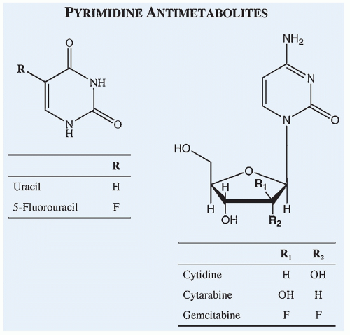
Figure 10.14 Chemical structures of commonly used pyrimidine antimetabolites compared with the structures of corresponding endogenous compounds of which they are analogs.


Antifolates


Methotrexate

Methotrexate is the most widely used antimetabolite in childhood cancers. It is effective in the treatment of ALL, non-Hodgkin lymphoma, the histiocytoses, and osteosarcoma. Methotrexate is administered on an intermittent schedule by a variety of routes, including oral, intramuscular, subcutaneous, intrathecal, and intravenous. Chronic oral or intramuscular therapy is administered weekly at a dose of 20 mg/m2. With intravenous therapy, an extraordinarily wide range of doses has been employed, ranging from a 10-mg bolus to 33,000 mg/m2 as a 24-hour infusion. Doses above 300 mg/m2, which are usually administered by continuous infusion, must be followed by a course of the rescue agent leucovorin (5-formyl-tetrahydrofolate) to prevent the development of severe toxicities.


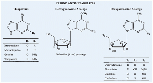
Figure 10.15 Chemical structures of commonly used purine antimetabolites compared with the structures of corresponding endogenous compounds of which they are analogs.

The loading and infusion doses required to achieve a desired steady-state plasma concentration ([MTX]plasma) can be estimated from the following formulas:469

Loading dose (mg/m2) = 15 • [MTX]plasma (µM)

Infusion dose (mg/m2/h) = 3 • [MTX]plasma (µM)

For example, to achieve a steady-state plasma concentration of 10 µM, the loading dose would be 150 mg/m2, followed by an infusion of 30 mg/m2 per hour. Infusion durations of up to 42 hours are tolerable when followed by leucovorin rescue. In clinical practice, infusion durations range from 4 to 36 hours depending on the type of cancer being treated. Patients who are treated with a high-dose methotrexate infusion must be adequately hydrated and alkalinized to prevent precipitation of methotrexate in acidic urine, and routine monitoring of urinary output, serum creatinine, and plasma methotrexate concentrations is mandatory to determine the duration of leucovorin rescue. For most infusion regimens, 12 to 15 mg/m2 of leucovorin should be continued every 6 hours until plasma methotrexate concentration decreases to 0.05 to 0.1 µM.

Mechanism of Action. Methotrexate is a structural analog of folic acid, a required cofactor for the synthesis of purines and thymidine. As a result of the substitution of an amino group for the hydroxyl group at the 4-position on the pteridine ring of folic acid (Fig. 10.13), methotrexate is a tight-binding inhibitor of DHFR, the enzyme responsible for converting folates to their active, chemically reduced (tetrahydrofolate) form.470 10-formyltetrahy-drofolate acts as the single carbon donor in the de novo purine synthetic pathway, and 5,10-methylenetetrahydrofolate donates its
single-carbon group and is oxidized to dihydrofolate in the conversion of deoxyuridylate (dUMP) to thymidylate (dTMP) by thymidylate synthase. In the presence of methotrexate, intracellular tetrahydrofolate pools are depleted, leading to depletion of purines and thymidylate and inhibition of DNA synthesis. Accumulation of partially oxidized dihydrofolic acid, resulting from the inhibition of DHFR, appears to contribute to the inhibition of de novo purine synthesis.471,472 A critical determinant of methotrexate cytotoxicity is the rate of thymidylate synthesis, because the synthesis of thymidylate from uridylate is the only reaction that oxidizes the tetrahydrofolate cofactor to the inactive dihydrofolate form. Another determinant is achieving an intracellular methotrexate concentration that is in excess of DHFR-binding sites, because intracellular levels of this target enzyme are 20-fold to 30-fold higher than that required to maintain tetrahydrofolate pools.470,473,474

Methotrexate shares membrane-transport processes and intracellular metabolic pathways with the naturally occurring folates. It competes with the tetrahydrofolates for an energy-dependent transport system for cell entry. On entry, methotrexate is rapidly and tightly bound to DHFR, and uptake into the target cell is essentially unidirectional until the enzyme binding sites are saturated, allowing for even greater intracellular accumulation of drug.470

With the accumulation of free intracellular drug in excess of DHFR-binding sites, methotrexate, like the naturally occurring folates, is metabolized intracellularly to polyglutamated derivatives, which cannot readily efflux from the cell. Methotrexate polyglutamate formation enhances the cytotoxicity of the drug by allowing greater accumulation of free intracellular drug and retention of the drug within the cell, even after extracellular drug is cleared. Methotrexate polyglutamates are also more potent inhibitors of DHFR and are capable of directly inhibiting other enzymes in the synthetic pathways for thymidine (thymidylate synthase) and purines.473,475,476 Methotrexate polyglutamate formation is optimal in vitro when cells are exposed to high concentrations for prolonged periods, and children with ALL randomized to receive high-dose methotrexate as initial induction therapy had higher methotrexate polyglutamate levels in their lymphoblasts than patients randomized to low-dose methotrexate.477,478 The lymphoblasts from children with ALL and good prognostic features, such as B-lineage immunophenotype, hyperdiploidy, young age, low presenting white cell count, and female sex, tend to accumulate methotrexate polyglutamates more efficiently than blasts from higher risk patients, suggesting that ALL in lower risk patients may be more sensitive to methotrexate’s antileukemic effect.478,479,480,481

Pharmacokinetics. At oral doses of 7.5 to 20 mg/m2, the rate and extent of absorption of methotrexate is highly variable.58,482,483,484,485 Peak plasma concentrations can occur from 0.5 to 5 hours after oral administration, and the percentage of the dose that is absorbed ranges from 5% to 97%.482 The AUC of oral methotrexate ranged from 0.63 to 12 µM • hour at a dose of 18 to 22 mg/m2, and over a broader dosage range, the AUC correlated poorly with the dose.58 In patients who are studied after multiple doses, there was also considerable intrapatient variation in the AUC.58 Absorption of methotrexate is saturable, and as the dose is increased, the fraction of the dose that is absorbed diminishes.486,487,488,489 Simply increasing the dose in patients who have low plasma concentrations after standard oral doses may not overcome poor bioavailability. The bioavailability of oral methotrexate can also be significantly reduced when administered with food.490 Despite this variability with oral dosing, there was no relation between the relapse rate and methotrexate pharmacokinetic parameters, such as peak concentration, AUC, and erythrocyte methotrexate concentrations.29,58,491 When administered intramuscularly or subcutaneously, methotrexate is completely absorbed.488,489,492,493

The disposition of methotrexate in children differs from that in adults.494,495,496,497 In one study, children had lower plasma concentrations of methotrexate and excreted the drug in the urine more rapidly after a 6-hour infusion than did adults.498 The volume of distribution was also greater in children. Within the pediatric age group, the clearance of methotrexate is also age-dependent.499

Children under 10 years of age (n = 94) had a clearance of 160 mL/min/m2, compared with 110 mL/min/m2 in those over 10 (n = 21). Infants (<1 year old) have a slightly lower clearance rate than children, with somewhat more pronounced differences observed in very young (<3 months) infants.500,501

The plasma disappearance of methotrexate is multiphasic, with a terminal half-life of 8 to 12 hours.469 Retention of the drug in large extravascular fluid collections, such as ascites or pleural fluid, is associated with prolongation of the half-life as a result of slow release of retained drug into the circulation. This prolonged exposure to the drug can increase the risk for toxicity. Patients who have large extravascular fluid collections and are receiving methotrexate should have their methotrexate concentrations monitored closely.

Methotrexate is eliminated primarily by renal excretion, undergoing glomerular filtration and renal tubular reabsorption and secretion.502,503 Approximately 70% to 90% of a dose is excreted unchanged in the urine, most within the first 6 hours. Mutations in the drug transporter ABC gene ABCC2 have been associated with impaired methotrexate elimination.504,505 The renal clearance of methotrexate can exceed the rate of creatinine clearance. In patients with significant renal dysfunction, methotrexate clearance is delayed, resulting in prolonged drug exposure and a greater risk of severe toxicities. High-dose methotrexate should not be given to patients with a creatinine clearance of less than 50% to 75% of normal. Low-dose therapy should be withheld in patients with a serum creatinine level greater than 2 mg/dL. Any patient who is suspected of having renal dysfunction and who receives methotrexate should have the plasma concentrations closely monitored and receive leucovorin if drug clearance is delayed.506

Methotrexate is also metabolized in the liver to 7-hydroxy-methotrexate.507 Although this is a minor route of elimination, plasma concentrations of 7-hydroxy-methotrexate can be equivalent to or exceed those of methotrexate after high-dose infusions, because of the slower clearance of the metabolite.508,509,510 7-Hydroxy-methotrexate may compromise the cytotoxicity of methotrexate by competing for membrane transport and polyglutamation. However, once polyglutamated, 7-hydroxy-methotrexate appears to be able to bind to and inhibit DHFR.470 Methotrexate clearance is not significantly altered with hepatic dysfunction, but modification of the methotrexate dose in patients with abnormal liver function tests may be indicated to avoid additional hepatic damage.

Total renal and metabolic methotrexate clearance is approximately 100 mL/min/m2, but it may vary widely among patients.510,511 In patients with normal creatinine clearance, there is not a good correlation between methotrexate clearance and creatinine clearance.511 Renal tubular dysfunction, which is not measured by creatinine clearance, may account for this disparity. A small test dose of methotrexate can accurately predict the kinetics and steady-state concentration of a high-dose infusion.512 Optimal management dictates that each course of high-dose methotrexate be closely monitored by following renal function and plasma methotrexate concentration to determine the dose and duration of leucovorin rescue.

Penetration of systemically administered methotrexate into CSF is only 3% in patients without meningeal tumor spread,513,514 but is 20% in patients with leptomeningeal carcinomatosis.514 At infusion rates exceeding 3,500 mg/m2 over 24 hours, the CSF methotrexate concentration is typically >1 µM, 514 and highdose methotrexate infusion regimens are effective for treating and preventing leptomeningeal leukemia.515

Toxicity. The primary toxic effects of methotrexate are myelosuppression and orointestinal mucositis, which occur 5 to 14 days after the dose. The development of toxic reactions is related to the concentration of drug and the duration of exposure.469 In patients receiving a 6-hour infusion of methotrexate, a 48-hour methotrexate concentration above 1 µM was associated with the development of
significant toxicity. These toxicities can be prevented by administration of leucovorin. With the use of therapeutic drug monitoring and continuation of leucovorin rescue until plasma methotrexate concentration has fallen below 0.05 to 0.1 µM, the toxicity of high-dose methotrexate can be avoided in most patients.469 Despite these measures, however, nephrotoxicity still occurs in almost 2% of patients receiving HDMTX infusions.516

Nephrotoxicity observed with high-dose methotrexate can delay methotrexate clearance and markedly intensify the drug’s other toxic effects.517,518 An early rise in serum creatinine (1.5 times baseline) within the initial 24 hours can help identify a population of patients at increased risk for delayed MTX elimination.519 The renal damage may be related to precipitation of methotrexate or 7-hydroxy-methotrexate in acidic urine or to direct toxic effects on the renal tubule.518 Aggressive hydration and alkalinization520 as well as increasing the sodium content of the hydration fluids521 can prevent drug precipitation and result in enhanced excretion of MTX.

The development of renal dysfunction during high-dose methotrexate is a medical emergency. Patients must be closely monitored and the leucovorin dose increased in proportion to the plasma methotrexate concentration. Hemodialysis and charcoal hemoperfusion have not proved useful for drug removal in patients with renal dysfunction,522,523,524 unless they are used repeatedly.525 Glucarpidase (carboxypeptidase-G2), a recombinant bacterial enzyme that catabolizes methotrexate to the inactive metabolite, 4-amino-4-deoxy-N10-methylpteroic acid (DAMPA),526 rescues patients who develop methotrexate nephrotoxicity by providing an alternative route of elimination.100,527 Glucarpidase is well tolerated and results in a 95.6% to 99.6% reduction in plasma methotrexate concentrations within minutes. Unlike dialysis, there is minimal rebound of plasma drug concentrations after glucarpidase.528

Hepatic toxicity consisting of transient elevations of serum transaminase and, less commonly, hyperbilirubinemia, has been associated with standard and high doses of methotrexate, but is more common and more severe with high-dose therapy. Hepatic fibrosis has been observed primarily in patients receiving chronic low-dose methotrexate.495,529 Other side effects include a dermatitis characterized by erythema and desquamation, allergic reactions, and acute pneumonitis.529,530 Methotrexate osteopathy is a cumulative toxicity that causes bone pain, osteoporosis, and an increased risk for fractures. Neurotoxicity from high-dose methotrexate includes an acute, stroke-like encephalopathy, seizures, and chronic leukoencephalopathy, particularly in association with cranial irradiation.229,531,532,533

Resistance. Mechanisms of resistance to methotrexate identified experimentally include decreased membrane transport, increased levels of the target enzyme DHFR, altered affinity of DHFR for methotrexate, decreased polyglutamation of methotrexate, and decreased thymidylate synthase activity.473 Increases in target enzyme levels have been associated with amplification of gene encoding for DHFR, a phenomenon that has also been documented in lymphoblasts from patients whose disease was clinically resistant to methotrexate.534,535 Flow cytometric analysis of lymphoblasts from 29 children with newly diagnosed and relapsed ALL demonstrated heterogeneous expression of elevated DHFR in 11 of 29 specimens and impaired methotrexate transport in 3 of 29 specimens.536 Newly diagnosed patients whose marrow specimens contained DHFR overproducing subpopulations of lymphoblasts had shorter remission durations than comparable patients whose lymphoblasts only expressed lower DHFR levels. Impaired methotrexate uptake and decreased expression of the reduced folate carrier (the membrane transport protein involved in cellular uptake of methotrexate) appears to occur frequently in osteosarcoma.537,538,539 Although inactivating reduced folate carrier mutations do not appear to be a major mechanism of MTX resistance in ALL,540 a case-control study of gene expression in children with ALL has found an association between higher transcript levels of the human reduced folate carrier gene (heft) and relapse.541

Drug Interactions. Several drugs have been associated with increased toxicity when coadministered with methotrexate.61,108,109 The most significant interactions involve agents that interfere with methotrexate excretion, primarily by competing for renal tubular secretion. These drugs include probenecid, salicylates, sulfisoxazole, penicillins, ciprofloxacin; the nonsteroidal anti-inflammatories indomethacin, ketoprofen, and ibuprofen; and the proton pump inhibitors omeprazole, rabeprazole and pantoprazole.61,108,542,543,544,545,546,547,548 Nephrotoxic drugs, such as the aminoglycosides, vancomycin, and cisplatin, may also alter the clearance of methotrexate.108,549 Pharmacodynamic interactions resulting in synergistic cytotoxic effects have been reported with methotrexate and fluorouracil or cytarabine.550 The synergistic effects of methotrexate and asparaginase are sequence dependent: asparaginase administration should always follow methotrexate administration. Administering asparaginase prior to or concomitant with methotrexate can directly antagonize methotrexate’s effectiveness.473,551,552


PURINE ANTIMETABOLITES


Thiopurines


Mercaptopurine/Thioguanine

Mercaptopurine and thioguanine are thiol-substituted derivatives of the naturally occurring purine bases hypoxanthine and guanine (Fig. 10.15). Mercaptopurine has been used in the treatment of ALL for 5 decades, primarily for the maintenance of remission. It is also used in the treatment of chronic myelogenous leukemia, histiocytosis, and inflammatory bowel disease. In standard maintenance regimens, mercaptopurine is administered orally at a dose of 60 to 75 mg/m2/day with upward or downward dose adjustments based on the degree of myelosuppression. Ensuring that patients are receiving their MTD of mercaptopurine appears to be an important factor in the outcome for children with ALL.29 In a retrospective analysis, when the actual dose of mercaptopurine received increased by 22% as a result of more aggressive prescribing guidelines, the relapse-free survival improved by 18%. Although high-dose intravenous infusions of mercaptopurine (1,000 mg/m2 over 6 to 24 hours) have been evaluated as an approach to circumvent the pharmacokinetic limitations of oral dosing,553,554,555 this route of administration does not offer an advantage over oral dosing in children with ALL.556 Thioguanine is primarily used in the treatment of acute nonlymphocytic leukemia and is administered orally in doses of 75 to 100 mg/m2 daily for 5 to 7 days or in doses of 40 to 60 mg/m2 daily for more prolonged courses.

The thiopurines are prodrugs that must be converted intracellularly to thioguanine nucleotides to exert a cytotoxic effect. The metabolic pathways for activation of mercaptopurine and thioguanine are outlined in Fig. 10.16. The active intracellular metabolites are phosphorylated thiopurine nucleotides, which inhibit de novo purine synthesis and purine interconversion and are incorporated into DNA.557,558 Incorporation of thioguanosine into DNA appears to be the critical determinant of thiopurine cytotoxicity,559 but there is evidence that for mercaptopurine methylated metabolites also appear to contribute to its overall antiproliferative effects.560 Thioguanine is tenfold more potent and less schedule-dependent than mercaptopurine against lymphoblastic leukemia cell lines and lymphoblasts from patients with ALL in vitro,561 and can achieve cytotoxic drug concentrations within the CSF with oral dosing.562 Despite preliminary clinical data suggesting an advantage to thioguanine over mercaptopurine for the treatment of children with ALL,563 randomized clinical trials failed to demonstrate an overall event-free survival advantage.564,565

Only gold members can continue reading. Log In or Register to continue

Stay updated, free articles. Join our Telegram channel

Aug 25, 2016 | Posted by in ONCOLOGY | Comments Off on General Principles of Chemotherapy

Full access? Get Clinical Tree

Get Clinical Tree app for offline access