##
Adrenal venous sampling (AVS) is technically demanding. The success rate of sampling both adrenal veins is ≈95% at centers of excellence. However, at medical centers with low AVS case volume or where multiple interventional radiologists perform AVS, the success rate can be as low at 30%. When AVS is not successful, it is almost always due to lack of successful sampling of the right adrenal vein. The right adrenal vein is small and enters the inferior vena cava (IVC) at an acute angle. When the right adrenal vein is not sampled, but the left adrenal production of aldosterone is suppressed, it can be inferred that the right adrenal gland is the source of aldosterone hypersecretion.
Case Report
The patient was a 58-year-old man with a 20-year history of hypertension that accelerated over the past 1 year. Two months previously he was admitted to the hospital with a blood pressure of 240/113 mmHg and a serum potassium level of 2.6 mEq/L. He was treated with a two-drug program: β-adrenergic blocker (nebivolol 5 mg twice daily) and a calcium channel blocker (nifedipine 60 mg extended release twice daily). Blood pressure control was not optimal, with systolic blood pressures typically in the 140–150 mmHg range. At the time of referral to Mayo Clinic he was taking 80 mEq of potassium chloride daily. He was recently diagnosed with type 2 diabetes mellitus. He had no signs or symptoms of Cushing syndrome.
INVESTIGATIONS
The baseline laboratory test results are shown in Table 14.1 . The patient had positive case detection testing for primary aldosteronism (PA) with plasma aldosterone concentration (PAC) >10 ng/dL and plasma renin activity (PRA) <0.6 ng/mL per hour. In addition, PA was confirmed because when a patient has spontaneous hypokalemia and the PAC >20 ng/dL, there are no other differential diagnostic possibilities beyond PA. , Thus formal confirmatory testing (e.g., oral sodium loading or a saline infusion test) was not needed (see Table 14.1 ).
| Biochemical Test | Result | Reference Range |
| SodiumPotassiumCreatinineeGFRAldosteronePlasma renin activity | 1403.01.2>6023.8<0.6 | 135–1453.6–5.20.8–1.3>60 mL/min per BSA≤21 ng/dL≤0.6–3 ng/mL per hour |
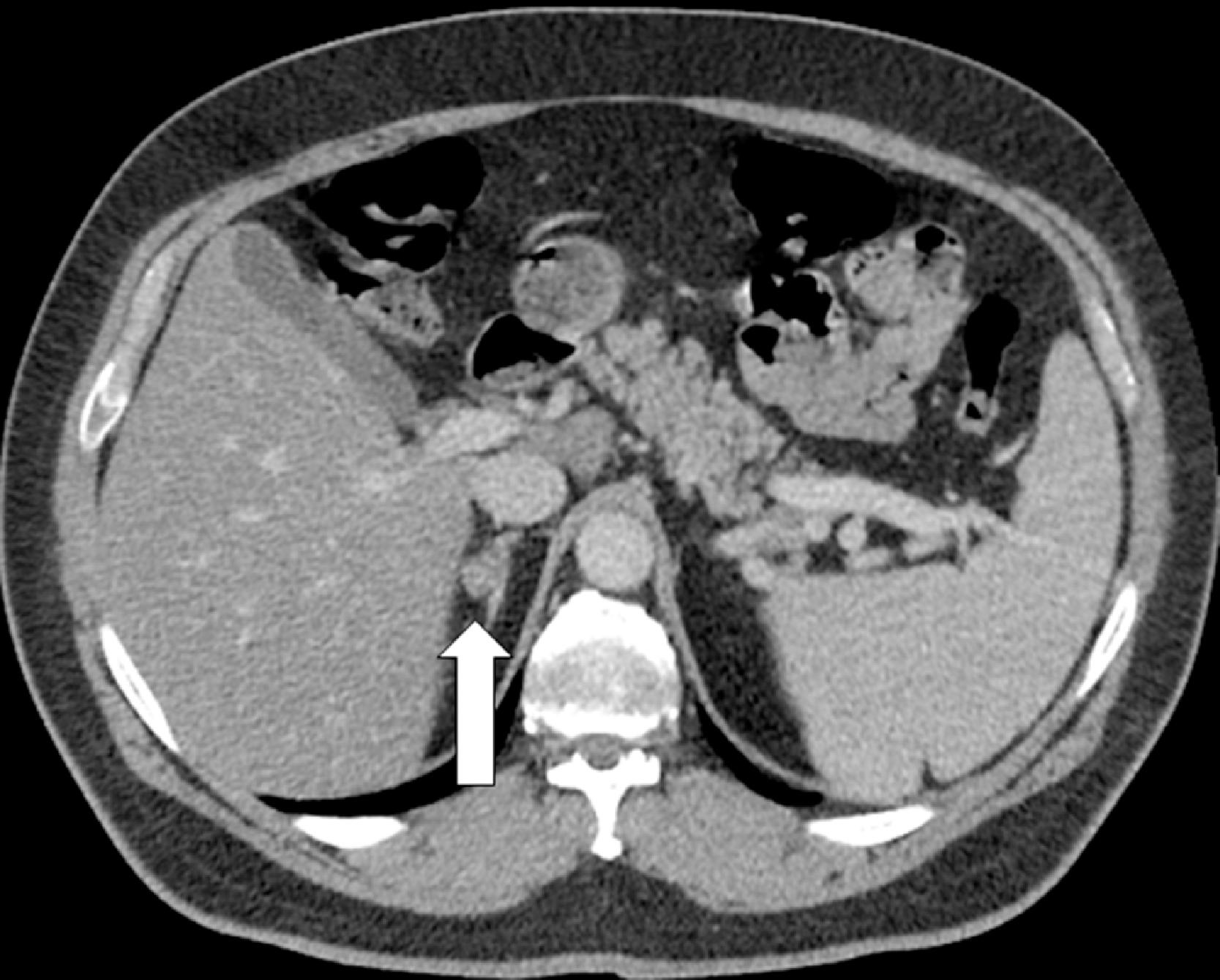
A contrast-enhanced abdominal computed tomography (CT) scan showed a 1.4-cm nodule in the lateral limb of the right adrenal gland ( Fig. 14.1 ); some mild thickening and micronodular changes were seen in the left adrenal gland.

Stay updated, free articles. Join our Telegram channel

Full access? Get Clinical Tree


