Eye
ANATOMY
The ocular structures consist of eyelids, cilia, lacrimal glands, drainage apparatus, and conjunctiva.
The globe is composed of three tunicae: the outer coat (the cornea and sclera), the middle coat (the uvea), and the inner layer (the retina).
The vascular supply is derived from the central retinal artery and the ciliary system.
The lens is located posterior to the iris and is suspended from the ciliary body.
The ocular groove, nerves, vessels, orbital fat, and ocular muscles are encompassed within the bony orbit.
OCULAR MALIGNANCIES
Basal and Squamous Cell Carcinomas of Eyelid
Meibomian Gland Carcinoma
Meibomian gland carcinomas are sebaceous gland carcinomas that may be multicentric, resulting in local recurrences.
Radiation therapy is an equivalent alternative to surgery, with acceptable cosmesis for both primary and recurrent disease.
High radiation doses of 60 to 65 Gy in 6 to 7 weeks are highly recommended.
UVEAL TUMORS
Metastatic Tumors of Posterior Uvea
Metastatic carcinoma to the eye is the most common malignant disease involving the eye.
Metastatic uveal lesions account for 15% of all cases. The most common primary sites are the breast or lung in women and the lung or gastrointestinal tract in men.
Uveal metastases are most commonly unifocal, although multifocal disease within the same eye is not uncommon.
Metastatic uveal tumors can cause visual symptoms that should be treated. The aim of therapy is to return visual function to patients.
Observation for small lesions may be appropriate if the patient is undergoing systemic therapy; however, lack of response to systemic therapy mandates local treatment to the involved eye with radiation therapy (8).
For patients with active systemic disease, palliative irradiation of 30 to 35 Gy over 3 weeks to the entire ocular structure is recommended.
In the absence of active systemic disease, and in patients with projected long-term survival, a more aggressive approach, delivering 45 to 50 Gy in 4.5 to 5.5 weeks, is required.
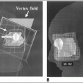
Radiation therapy can be delivered with lateral portals, with the shielding of the lens and cornea.
Treatment can be delivered with 15- to 18-MeV electrons or with low-energy photons (4 to 6 MV).
The lateral field should be tilted posteriorly 5 to 10 degrees to avoid irradiation of the contralateral lens and cornea, and parallel to the base of the skull (when possible) to avoid the brain.
Plaque radiation therapy is an option for treating solitary uveal metastasis, particularly when external-beam irradiation fails to control the disease.
Malignant Melanoma of Uvea
Malignant melanoma represents 75% of primary malignant tumors involving the eye (2,500 new cases annually).
Melanoma of the anterior uvea is usually detected earlier than posterior tumors.
Anterior uveal melanoma may be removed by iridectomy or iridocyclectomy. Posterior uveal melanoma traditionally has been treated by enucleation of the affected eyeball, although there is controversy about the optimal management of this disease. Some investigators question the role of tumor enucleation because it may promote seeding of tumor cells, affecting a patient’s prognosis.
Brachytherapy techniques using various sources, including60Co,125I,192Ir,109Ru, and198Au seeds, have been used (2, 14).
Indications for plaque radiation therapy are: (a) selected small melanomas that are growing, (b) potential for preservation of vision with medium-sized choroidal and ciliary body melanomas, or (c) actively growing tumor that occurs in the patient’s only useful eye.
For tumors that exceed 15 mm in diameter and 10 mm in thickness, radiation therapy can cause significant morbidity, and enucleation is preferred.
The visual outcome of eye treatment with radiation therapy depends on tumor size and location relative to the fovea or the optic disc. Large tumors and tumors in proximity to the fovea or optic disc place patients at high risk of radiation retinopathy and papillopathy after treatment.
Studies have shown increased risk of visual loss when doses higher than 50 Gy are delivered to the fovea or the optic disc (5).
Combined plaque irradiation and laser photocoagulation, transpupillary thermotherapy (20), or chemotherapy has been used to increase local control, particularly in tumors close to the optic disc.
There is general agreement that large ocular melanomas and tumors with extrascleral extension at diagnosis are not readily amenable to radiation therapy. In this group of patients, enucleation or exenteration is the preferred option.
In view of the continued poor survival of patients treated with enucleation, several studies have evaluated the role of preoperative irradiation and its impact on survival. However, most studies have not shown long-term survival benefit with preoperative irradiation, compared with enucleation alone (8).
The Collaborative Ocular Melanoma Study (COMS) is a prospective randomized study that, among other aims, was designed to compare survival in patients treated with either brachytherapy or enucleation. Thousand three hundred and seventeen patients with mediumsized choroidal melanoma (height 2.5 to 10.0 mm and size ≤ 16.0 mm) accrued from 1987 to 1998 and were randomly assigned to enucleation (n = 660) or to125I plaque brachytherapy (n = 657). Mortality rates following125I brachytherapy did not differ from those following enucleation through 12 years of follow-up (25). The 12-year overall survival with enucleation
was 59% versus 57% with brachytherapy (p = NS).The risk of death from metastatic disease was 17% versus 21%, respectively.
Two randomized trials using particle therapy for uveal melanoma have been published. A randomized trial of protons using 50 cGE versus 70 cGE in the treatment of choroidal melanoma concluded that the lower dose resulted in similar local and distant control but had an improved toxicity profile with respect to visual field loss (10). A randomized trial of helium ion therapy versus125I plaque brachytherapy for uveal melanoma reported improved local control with helium ion treatment but no difference in overall survival (4).
The prognostic factors associated with poor visual acuity or vision loss include increasing tumor thickness, proximity of the plaque to foveola of less than 5 mm, and patient age over 60 years (21).
RETINAL TUMORS
Retinoblastoma
Retinoblastoma is the most common intraocular malignancy in children. There are approximately 250 new cases diagnosed in the United States each year, retinoblastoma is bilateral in one third of patients.
Retinoblastoma is hereditary in approximately 40% of diagnosed cases and is transmitted as an autosomal-recessive trait (13).The genetic abnormality involves deletion or mutation of tumor suppressor gene (RB gene) on the long arm of chromosome 13 (13q).
In general, the hereditary form is diagnosed earlier than the nonhereditary form of the disease.
Most patients with the hereditary form have bilateral disease.
Most children with this tumor are diagnosed before 3 to 4 years of age.
Leukocoria (white papillary reflex), strabismus (squint), and a mass in the fundus are the common presenting signs and symptoms, which are commonly noticed at 6 to 24 months of age.
Diagnostic workup of retinoblastoma requires a history, a physical examination (including a complete ophthalmologic examination with retinal drawings and photographs), an ultrasound for documentation of tumor location and size, and a cranial computed tomography scan.
Routine cerebrospinal fluid study and bone marrow examination are not recommended, except when there are signs and symptoms suggestive of extraocular extension.
Factors that carry a poor prognosis include orbital invasion, involvement of the optic nerve, central nervous system dissemination, and heritable bilateral tumors. Tumor parameters such as size, growth pattern (endophytic or exophytic), and differentiation do not significantly influence the systemic prognosis (24).
Historically, the most widely used grouping system for retinoblastoma was the Reese-Ellsworth classification system (Table 12-1). More recently, the International Classification System (15) has been employed for risk stratification and is used in current Children’s Oncology Group (COG) protocols (Table 12-2). The AJCC devotes several chapters of its seventh edition (1) to ocular cancers. Space does not permit reproduction of these extensive TNM tables, and the reader is referred to the new AJCC manual for details. See Table 12-3 for a synopsis.
The goal of therapy is both cure and preservation of vision.
Stay updated, free articles. Join our Telegram channel

Full access? Get Clinical Tree


