ESTABLISHMENT OF THE CAUSE OF NONTOXIC GOITER
Part of “CHAPTER 38 – NONTOXIC GOITER“
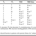
Serum antithyroid antibody titers should be measured in all patients with nontoxic goiter. Elevated antibody titers (e.g., anti-thyroglobulin and anti–thyroid peroxidase [anti-TPO, “microsomal”] antibodies) are prima facie evidence of an autoimmune basis for goiter formation in the euthyroid patient (e.g., those with Hashimoto thyroiditis). Elevated titers occur in up to 90% of such patients.48
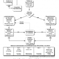
In antibody-negative patients with nontoxic goiter, thyroidal radioiodide uptake and scan (iodine-123) may be helpful diagnostically. An elevated uptake is consistent with increased TSH action (e.g., antibody-negative Hashimoto thyroiditis or iodide deficiency) or with the action of non-TSH thyroid stimulators. Extremely low or undetectable radioiodide uptake by the thyroid is an important finding in the goitrous patient; the differential diagnosis includes excessive iodide intake, lymphocytic thyroiditis (“painless thyroiditis,” usually with transient hyperthyroidism), and granulomatous thyroiditis. Struma ovarii and factitious hyperthyroidism are also associated with extremely low or undetectable radioiodide uptake, but no goiter is present. The normalization of a previously high iodine intake in patients with iodine-induced hypothyroidism may result in an acute elevation of thyroidal radioiodide uptake, although serum T4 values remain low. A “patchy” uptake pattern on scan is frequently encountered in Hashimoto thyroiditis. Localized areas of reduced or absent uptake are difficult to interpret. They are associated with a 0% to 30% risk of carcinoma in various reports but most commonly represent adenoma or cyst. Areas of increased uptake are associated with an extraordinarily low risk of cancer (see Chap. 34). The estimation of iodide intake by measurement of 24-hour urinary iodide excretion is seldom necessary in the United States.
Related posts:

Stay updated, free articles. Join our Telegram channel

Full access? Get Clinical Tree


