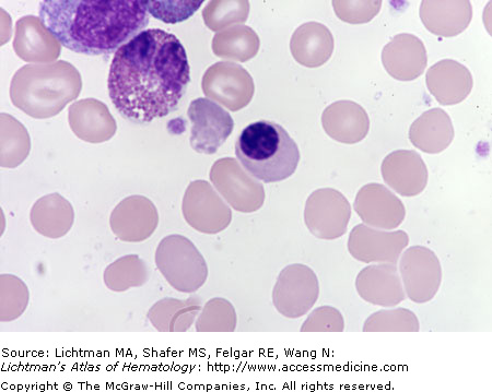
V.C.001 Erythroblasts
V.C.001
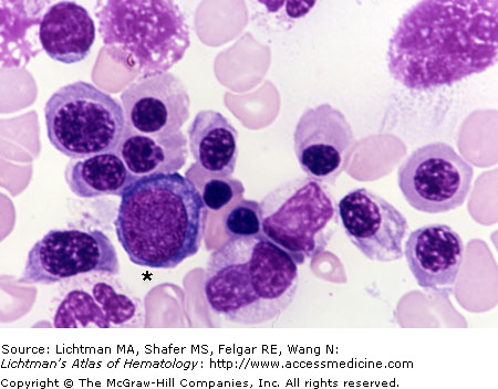
Erythroblasts. Marrow film. A basophilic erythroblast (asterisk) is surrounded by polychromatophilic and orthochromatic erythroblasts. A portion of a segmented neutrophil, two neutrophil precursors, a lymphocyte (upper left), and a plasma cell are also in the field. Three degenerating cell nuclei are also evident.
V.C.002 Erythroblast, Basophilic
V.C.002
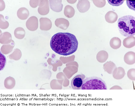
Basophilic erythroblast. Marrow film. This cell is smaller than the proerythroblast, about 15-20 μm in diameter. The nuclear to cytoplasmic ratio is high but usually a bit less than the proerythroblast. The paranuclear Golgi zone is a bit smaller than the proerythroblast and the cytoplasm is intensely basophilic as a result of a large population of polyribosomes. Peroxidase staining indicates that hemoglobin is being synthesized (not shown) but its concentration does not yet lessen the intense basophilia.
V.C.003 Erythroblast, Enucleation
V.C.004 Erythroblast, Enucleation (Transmission Electron Micrograph)
V.C.004
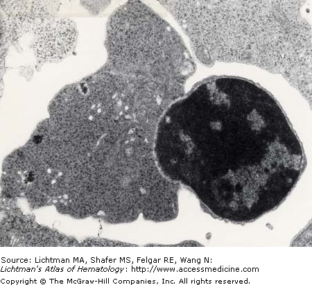
Enucleation of an erythroblast: transmission electron micrograph. An erythroblast with the nucleus in the final phase of extrusion. Note the characteristic rim of cytoplasm encircling the extruded nucleus. The erythroblast (moments away from being an erythrocyte) still contains mitochondria and siderosomes. It also shows a serrated margin. Marrow reticulocytes have surface folds that get remodeled after egress from the marrow.
V.C.005 Erythroblast, Orthochromatic
V.C.005
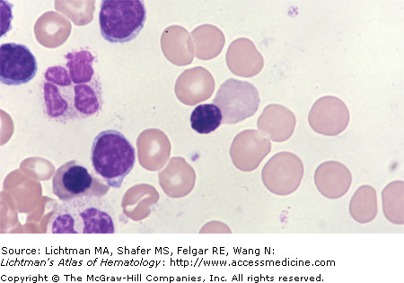
Orthochromatic erythroblast. Marrow film. The cell size has been decreased from the polychromatophilic erythroblast to about 10-15 μm. The cytoplasm approaches the red cell in color (“orthochromatic”) as a result of a rich concentration of hemoglobin but in many cases a faint bluish-purple color persist, reflecting a small concentration of ribosomes and a few mitochondria. The nucleus is condensed and featureless. The nucleus is usually eccentrically positioned and the nuclear-cytoplasmic ratio is much lower than in earlier precursors. A typical mature eosinophil with a bilobed nucleus is in the upper left.


