Erythema Multiforme, Stevens-Johnson Syndrome, and Toxic Epidermal Necrolysis
Anju T. Peters
Neill T. Peters
Erythema multiforme (EM), Stevens-Johnson syndrome (SJS), and toxic epidermal necrolysis (TEN) represent a spectrum of immunologically mediated diseases most often due to hypersensitivity to drugs or infections. There is no uniformly accepted definition or classification of these diseases, and understanding of their exact immunologic basis is lacking.
Historical Background
Erythema multiforme (EM) is a term originally attributed to Ferdinand von Hebra. In 1866, he wrote about “erythema exudativum multiforme,” a single cutaneous eruption with multiple evolving stages of lesions (1). Von Hebra described EM as a mild cutaneous syndrome featuring symmetric acral lesions, which resolved without sequelae, and had a tendency to recur. In 1922, Stevens and Johnson described a generalized eruption in two children characterized by fever, erosive stomatitis, and severe ocular involvement (2). This eruption became known as Stevens-Johnson syndrome. Thomas, in 1950, proposed that the milder von Hebra form of EM be called EM minor, and the more severe mucocutaneous eruption of SJS be called EM major (3). According to Thomas, fever and severe ocular involvement were the main points of distinction between the two types. The term toxic epidermal necrolysis was first introduced in 1956 by Lyell to describe patients with extensive epidermal necrosis that resembled scalded skin (4).
In 1993, an international consensus conference attempted to classify severe EM, SJS, and TEN on the basis of skin lesions and the extent of epidermal detachment (5). Using an illustrated atlas to standardize the diagnosis of acute severe bullous disorders attributed to drugs and infectious agents, the researchers defined bullous EM, SJS, SJS-TEN overlap, and TEN (Table 16.1). Unfortunately, the ultimate classification of severe forms of EM remains a matter of debate and confusion for both primary care physicians and specialists. Given the rarity of SJS and TEN, it has been difficult to create a universally accepted standard of care for the management of these patients. Nonetheless, several concepts regarding EM, SJS, and TEN and their therapy have been proposed. It is well accepted that SJS and TEN represent varying severity of the same disease spectrum, with TEN being the most severe form (6,7).
Table 16.1 Classification of Erythema Multiforme, Stevens-Johnson Syndrome and Toxic Epidermal Necrolysis | |||||||||||||||||||||
|---|---|---|---|---|---|---|---|---|---|---|---|---|---|---|---|---|---|---|---|---|---|
| |||||||||||||||||||||
Erythema Multiforme
Erythema multiforme, or the classic von Hebra type of EM, is a symmetric eruption with a predilection for the extremities. The characteristic primary lesion is a “target” comprised of three zones (8) (Figs. 16.1 and 16.2). Centrally there is a disk surrounded by an elevated, pale ring. A zone of erythema then borders the pale ring (5). Mucosal involvement occurs in a majority of cases of EM; however, it is usually limited to the oral or ocular mucosa and typically is not severe (9,10). EM is often associated with herpes simplex virus (HSV) infections and follows an outbreak of HSV by 1 to 3 weeks (11,12). The eruption is self-limited, lasts 1 to 4 weeks, and requires only symptomatic management. HSV-induced EM may be recurrent, and in such cases, recurrences can be prevented with suppressive antiherpetic therapy (13,14). More severe variants of EM have been also described and are often caused by infections including HSV and Mycoplasma pneumoniae (15,16). Drugs may cause a small proportion of EM cases (17,18). Discontinuation of the implicated medication and supportive therapy results in complete resolution of the skin eruption. Short courses of oral corticosteroids have been used in treatment protocols without any significant side effects (16). In some cases of EM, no obvious cause may be elicited (18).
 Figure 16.1 Target lesion characteristic of erythema multiforme. (Courtesy of Dana Sachs, MD.) |
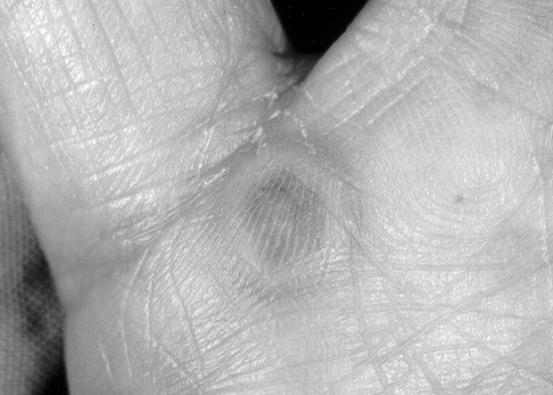 Figure 16.2 Target lesion. (Courtesy of Dana Sachs, MD.) |
Stevens-Johnson Syndrome and Toxic Epidermal Necrolysis
SJS and TEN are severe, mucocutaneous eruptions involving two or more mucosal surfaces, with or without visceral involvement (Figs. 16.3 and 16.4). Both SJS and TEN are rare, with an incidence of 1.89 cases per million people per year (19). The majority of the cases are attributed to drug exposures (18–26) (Table 16.2). Infections, especially with Mycoplasma pneumoniae, are also known to produce SJS (27,28). Vaccines and viral infections such as varicella zoster virus have also been reported to cause SJS (29–31). TEN has significant morbidity, and a mortality rate of about 30% (23,25,32). Most deaths are attributed to sepsis (22,25,32).
 Figure 16.3 Stevens-Johnson syndrome secondary to trimethoprim-sulfamethoxazole. |
 Figure 16.4 Stevens-Johnson syndrome. Same patient as shown in Fig. 16.3. |
Related posts:
 Allergens and Other Factors Important in Atopic Disease
Diagnosis of Immediate Hypersensitivity
Drug Allergy Part A Introduction, Epidemiology, Classification of Adverse Reactions, Immunochemical Basis, Risk Factors, Evaluation of Patients with Suspected Drug Allergy, Patient Management Considerations
Asthma Clinical Trials
Contact Dermatitis
Delivery Devices for Inhaled Medications
Allergens and Other Factors Important in Atopic Disease
Diagnosis of Immediate Hypersensitivity
Drug Allergy Part A Introduction, Epidemiology, Classification of Adverse Reactions, Immunochemical Basis, Risk Factors, Evaluation of Patients with Suspected Drug Allergy, Patient Management Considerations
Asthma Clinical Trials
Contact Dermatitis
Delivery Devices for Inhaled Medications
Stay updated, free articles. Join our Telegram channel

Full access? Get Clinical Tree


