Epithelial Polyps and Inflammatory Bowel Disease-Related Dysplasia


Epithelial Polyps and Inflammatory Bowel Disease-Related Dysplasia


Nicole C. Panarelli


Colorectal epithelial polyps comprise a heterogeneous group of dysplastic and nondysplastic neoplasms, many of which confer risk of metachronous or subsequent colorectal adenocarcinoma. Dysplastic polyps (i.e., adenomas) are widely recognized precursors to adenocarcinoma, but until recently, nondysplastic serrated polyps were believed to lack malignant potential. Better characterization of serrated polyps and emerging molecular evidence have improved our understanding of the biologic potential of serrated polyps and led to changes in their clinical management. This chapter discusses the clinical, pathologic, immunohistochemical, and molecular features of epithelial colorectal polyps and reviews current evidence regarding their biologic importance.


HYPERPLASTIC POLYPS


Clinical and Endoscopic Features of Hyperplastic Polyps

Nondysplastic serrated polyps are classified in two broad groups: hyperplastic polyps without proliferative abnormalities and sessile serrated polyps (sessile serrated adenomas) with proliferative abnormalities. Hyperplastic polyps are the most common type of polyps in the colorectum, are small (usually <0.5 cm), and show a predilection for the distal colorectum. They develop earlier than adenomas and are found in 23% of patients 20 to 54 years old, but their prevalence also increases with age.1 Alcohol consumption, smoking, obesity, and low dietary folate are all associated with an increased risk of hyperplastic polyps.2 Hyperplastic polyps have been historically considered to be nonneoplastic, and prospective data indicate that their presence does not confer an increased risk for colorectal cancer, despite the frequent detection of specific molecular changes in these lesions.3 Hyperplastic polyps are pale, sessile polyps with a smooth contour that may flatten upon air insufflation or show a layer of mucus on the surface.4 They display a crypt pattern of orderly circular dots by chromoendoscopy, which distinguishes them from adenomatous polyps.5


Pathologic Features of Hyperplastic Polyps

Hyperplastic polyps are composed of crypts that show prominent infolding of the epithelium, producing a “sawtooth” or serrated appearance in histologic sections (Figure 4.1A). Crypts tend to be narrower at their bases compared to the surface and contain a mixed population of goblet cells, nongoblet mucinous columnar cells, and endocrine cells in varying proportions. Mild cytologic atypia may be seen, but is limited to the basal proliferative crypt region, and dysplasia is absent. Multinucleated cells are also occasionally observed and likely represent a degenerative phenomenon (Figure 4.1B). They do not reflect viral infection or abnormal proliferation.6 Some left-sided hyperplastic polyps also contain a thickened basement membrane.


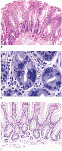
FIGURE 4.1: Microvesicular hyperplastic polyps are composed of serrated crypts that are narrow at their bases with wide openings at the surface. The crypts contain goblet and nongoblet mucinous cells with abundant cytoplasm and small basally oriented nuclei (A). Cytologic atypia, including multinucleated cells, is occasionally encountered (B). Goblet cell-rich hyperplastic polyps display serration mostly on the surface and are largely composed of goblet cells (C).


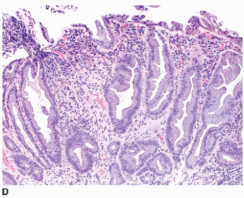
FIGURE 4.1: (Continued) Mucin-poor hyperplastic polyps display a serrated architecture similar to microvesicular hyperplastic polyps, but contain somewhat mucin-depleted epithelial cells and decreased goblet cells (D).

Three morphologic subtypes of hyperplastic polyp are described: microvesicular, goblet cell rich, and mucin poor. Microvesicular hyperplastic polyps are most common and display prominent crypt and surface serration. They contain a dual population of goblet cells and more numerous nongoblet mucinous epithelial cells with “microvesicular” cytoplasm (Figure 4.1A). Goblet cell-rich hyperplastic polyps expand the mucosa and display minimal crypt serration with numerous goblet cells (Figure 4.1C). Mucin-poor hyperplastic polyps are architecturally similar to microvesicular hyperplastic polyps and contain numerous nongoblet mucin-depleted epithelial cells with hyperchromatic nuclei and rare goblet cells (Figure 4.1D).7 These lesions share immunohistochemical and molecular features with microvesicular hyperplastic polyps and likely represent the latter with superimposed regenerative features. Notably, subclassification of hyperplastic polyps has not been shown to be clinically important and is not recommended at this time.


Immunohistochemical and Molecular Features of Hyperplastic Polyps

Immunohistochemical Ki-67 staining of hyperplastic polyps reveals a symmetrically expanded proliferative zone that extends approximately 1/3 to 1/2 up the crypt length.8, 9 and 10 Cytokeratin 20 staining is present in deep and surface epithelium compared to superficial staining of the normal colon, and uniform nuclear immunostaining for the intestine-specific caudal homeobox protein, CDX2, is limited to the basal 1/3 to 2/3 of crypt epithelial cells.10,11 Hyperplastic polyps stain for colon-specific apomucin (MUC2) and gastric mucin (MUC5AC) and are reportedly negative for pyloric mucin (MUC6). However, a substantial number of hyperplastic polyps of the abdominal colon show MUC6 positivity, thereby limiting the utility of this marker in the distinction between hyperplastic polyps and sessile serrated polyps.7,11

Many hyperplastic polyps have mutually exclusive KRAS and BRAF mutations, challenging the notion that they are nonneoplastic and lack malignant potential. KRAS mutations occur in up to 47% of hyperplastic polyps and are more frequent among distally located and goblet cell-rich examples.12, 13 and 14 BRAF mutations are detected in 40% to 76% of hyperplastic polyps, particularly those of the proximal colon with microvesicular morphology.12,14 Approximately 40% to 60% of hyperplastic polyps reportedly show the CpG island methylator phenotype (CIMP), which is more common among microvesicular (70%) than goblet cell-rich (40%) hyperplastic polyps.14 The CIMP is inversely correlated with KRAS mutations, as discussed in Chapter 11.15 Loss of immunohistochemical staining for O6-methylguanine-methyltransferase (MGMT) and MLH1 is sometimes present in the nonproliferative superficial epithelium of hyperplastic polyps, but does not reflect underlying mutations, promoter methylation, or silencing of these genes from transcription.14


SESSILE SERRATED POLYPS


Clinical and Endoscopic Features of Sessile Serrated Polyps

Sessile serrated polyps are nondysplastic polyps that show a predilection for the abdominal colon and, unlike most hyperplastic polyps, are more common among women. These polyps have been termed “serrated polyps with abnormal proliferation,” “giant hyperplastic polyps,” and “sessile serrated adenomas,” but are now classified as “sessile serrated adenomas/polyps” by the World Health Organization in order to avoid confusion with hyperplastic polyps and serrated adenomas.16 Sessile serrated polyps represent 2% to 9% of all endoscopically resected polyps and 7% to 21% of all nondysplastic serrated polyps.7,17 Sessile serrated polyps are plaques that merge imperceptibly with the adjacent nonlesional mucosa, and, thus, their size maybe difficult to appreciate endoscopically. They are often covered with a thick layer of mucus and contain round dilated crypts that are visible by chromoendoscopy. A growing body of evidence indicates that these polyps represent precursors to some sporadic colonic adenocarcinomas characterized by a high degree of microsatellite instability (MSI-H). They are believed to confer a biologic risk approaching that of conventional adenomas of comparable size. Thus, screening guidelines parallel those for conventional adenomas with overt dysplasia.


Pathologic Features of Sessile Serrated Polyps

Sessile serrated polyps are broad-based polyps that contain numerous elongated crypts with prominent infolding of the crypt epithelium (Figure 4.2A). The crypts of a sessile serrated polyp show persistent dilatation at their bases, often with branching or budding above the muscularis mucosae (Figure 4.2B). The crypts contain variable numbers of goblet cells and nongoblet mucinous columnar cells, similar to hyperplastic polyps, but they tend to show a greater degree of cytologic atypia that extends in an asymmetric fashion from the crypt bases (Figure 4.2C).7,18 Distinction of sessile serrated polyp from hyperplastic polyp is subject to a high degree of interobserver variability, particularly when polyps are smaller than 1 cm in diameter and occur in the left colon. Diagnostic challenges stem from the fact that there are no “hard” criteria for a diagnosis of sessile serrated polyp. Rather, the diagnosis is based on a constellation of seven findings, four or more of which should be present in order to classify a polyp as a sessile serrated polyp (Table 4.1).7,18 Unfortunately, quantifying these features is somewhat subjective, and many are frequently present to a variable extent among small, serrated polyps of the descending colon and rectum, resulting in substantial overlap between the diagnostic criteria for hyperplastic polyp and sessile serrated polyp.8 Indeed, hyperplastic polyps of the abdominal colon also show immunohistochemical and molecular features that substantially overlap with those of sessile serrated polyps, as discussed subsequently.19

Immunohistochemical and Molecular Features of Sessile Serrated Polyps

Sessile serrated polyps show Ki-67 nuclear staining of approximately 33% of the nuclei, which is slightly more than hyperplastic polyps, but less than that of conventional adenomas. Ki-67-labeled nuclei are not confined to the basal epithelial cells, but are randomly distributed within the crypts, reflecting abnormal proliferation.10 Cytokeratin 20 and CDX2 immunostains demonstrate randomly distributed positive cells within the crypts.10 Sessile serrated polyps show immunostaining for MUC2, MUC5AC, and MUC6.11,17,20 Early studies suggested that MUC6 positivity may be a helpful marker to distinguish between sessile serrated polyps and hyperplastic polyps, but better designed studies have since refuted this notion.20 MUC6 immunostains are often positive in hyperplastic polyps of the abdominal colon, suggesting that it is a marker of location (i.e., proximal colon) rather than phenotype among serrated polyps.21

Seventy-eight to 100% of sessile serrated polyps harbor BRAF mutations, whereas KRAS mutations are uncommon, being observed in less than 10% of cases.8,13,14,22 These polyps frequently
(60% to 70%) show the CIMP.14,22 Loss of MGMT and MLH1 immunoexpression is limited to the superficial epithelial cells, but reflects the nonproliferative nature of the polyp surface, rather than underlying microsatellite instability (MSI). Early investigations reported MGMT and MLH1 promoter methylation in 40% and 72% of sessile serrated polyps, respectively.7,14,17,23 Unfortunately, these studies utilized qualitative, rather than quantitative, assays, and more recent data indicate that sessile serrated polyps are microsatellite stable with preserved MLH1 function.24


FIGURE 4.2: Sessile serrated polyps are broad-based plaques composed of elongated crypts that show persistent dilatation at their bases (A). They also display architectural abnormalities, including laterally branched, or budding, crypts oriented parallel to the muscularis mucosae (B). Atypical, hyperchromatic nuclei with pseudostratification may extend beyond the crypt bases into the midcrypt region (C).


Table 4.1 Diagnostic Criteria of Sessile Serrated Polyp


Feature


Description


Crypt dilatation


Elongated crypts with exaggerated luminal serration Persistent dilatation of crypts into the deep mucosa Inverted L- or T-shaped crypts


Crypt orientation


Horizontally oriented crypts above the muscularis mucosae


Mucin production


Extracellular mucin in crypt lumina and on polyp surface


Cytoplasmic eosinophilia


Clusters of cells with eosinophilic cytoplasm within irregularly shaped, mucin-poor crypts


Decreased endocrine cells


Contiguous crypts with absent or rare endocrine cells


Abnormal proliferation


Proliferative zone extends to the middle or upper third of crypts


Nuclear atypia


Enlarged, oval or round, basally located nuclei


Hyperchromasia


Prominent nucleoli


Data from Torlakovic E, Snover DC. Serrated adenomatous polyposis in humans. Gastroenterology 1996;110(3):748—755.


SERRATED (HYPERPLASTIC) POLYPOSIS

Serrated (hyperplastic) polyposis is defined by the presence of (1) at least five hyperplastic polyps located proximal to the sigmoid colon, two of which span at least 1 cm, (2) twenty or more hyperplastic polyps of any size distributed throughout the colorectum, or (3) any number of hyperplastic polyps proximal to the sigmoid colon in a first-degree relative of a patient with serrated (hyperplastic) polyposis.25 The molecular features of these phenotypes are variable. Patients with large polyps of the abdominal colon tend to have fewer serrated polyps that harbor BRAF mutations and show the CIMP. They also have a variety of other types of dysplastic colorectal polyps with, or without, serrated morphology. In contrast, other patients have numerous small polyps with KRAS mutations and, possibly lower overall cancer risk. Approximately 5% of serrated polyposis cases have a familial association, although the precise genes involved and modes of inheritance are not clear. Retrospective data suggest an overall colorectal cancer prevalence of 35% to 50%, although prospective data estimate cancer risk to be 5% to 10% at 7 years.26


CONVENTIONAL ADENOMAS


Clinical and Endoscopic Features of Conventional Adenomas

Conventional adenomas occur throughout the colorectum and are usually asymptomatic, although large polyps may ulcerate causing rectal bleeding and iron deficiency anemia. They account for 40% to 60% of all endoscopically removed colorectal polyps, and their incidence increases with age, such that they are detected in approximately 12% of adults over 50 years of age.27 The number and size of adenomatous polyps is associated with a risk of future cancer
development, as discussed in Chapter 3. Patients with a few, small adenomas are at low cancer risk, whereas those with more numerous adenomas, large polyps (>1 cm), or high-grade dysplasia are at a higher risk for cancer development and undergo more rigorous surveillance.


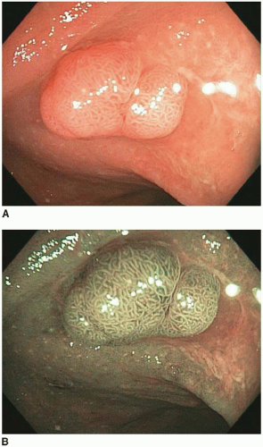
FIGURE 4.3: A sessile tubular adenoma of the right colon is slightly more erythematous than the background mucosa (A). Narrow band imaging accentuates the irregular pit pattern characteristic of dysplastic (adenomatous) polyps (B).

May 22, 2016 | Posted by in ONCOLOGY | Comments Off on Epithelial Polyps and Inflammatory Bowel Disease-Related Dysplasia

Full access? Get Clinical Tree

Get Clinical Tree app for offline access