II.D.001 Eosinophils, Normal
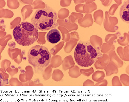
II.D.001
Eosinophils, normal. Buffy coat (white cell concentrate). The field contains two eosinophils, two neutrophils, and a lymphocyte. The eosinophils are typical cells; bilobed nucleus, present in virtually all normal eosinophils, and a cytoplasm filled with eosinophilic granules. The granules do not obscure the nucleus as is characteristic in basophils. Some small areas without granules (degranulation) are present. Note that the neutrophil granules are too small to be resolved by the light microscope, whereas the large eosinophilic granules can be resolved. This is especially evident if eosinophilic granules are released from disrupted cells and are free in the surrounding area.
II.D.002 Eosinophil, Normal
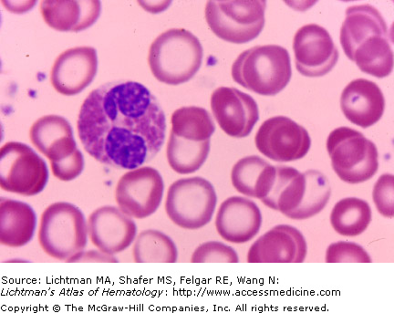
II.D.002
Eosinophil, normal. The field contains an eosinophil with characteristic bilobed nucleus, present in virtually all normal eosinophils, and a cytoplasm filled with eosinophilic granules. The granules do not obscure the nucleus as is characteristic in basophils. Some very small areas without granules (degranulation) are present—a common artifact of blood film preparation. Note severely hypochromic red cell as a result of iron deficiency.
II.D.003 Eosinophil, Normal
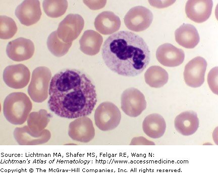
II.D.003
Eosinophil, normal. The field contains an eosinophil with characteristic bilobed nucleus, present in virtually all normal eosinophils, and a cytoplasm filled with eosinophilic granules. The granules do not obscure the nucleus as is characteristic in basophils. Some very small areas without granules (degranulation) are present—a common artifact of blood film preparation.
Related posts:

Stay updated, free articles. Join our Telegram channel

Full access? Get Clinical Tree


