Embryonal and Pineal Region Tumors


Embryonal and Pineal Region Tumors


Murali M. Chintagumpala

Arnold Paulino

Ashok Panigrahy

Cynthia Hawkins

Andrew Jae

Williams D. Parsons


EMBRYONAL TUMORS

Embryonal tumors constitute approximately 25% of all primary central nervous system (CNS) tumors occurring in patients 18 years or younger,1,2,3 and nearly 40% of all malignant CNS neoplasms. Although embryonal tumors occur throughout the pediatric age spectrum, they often arise early in life, with one-fifth or more diagnosed in the first 3 years, making diagnosis and management even more challenging.1,2,3,4 In the World Health Organization (WHO) classification of CNS tumors, embryonal tumors constitute a collection of biologically heterogeneous lesions that are malignant and share the tendency to disseminate in the nervous system via the cerebrospinal fluid (CSF) pathways early in the course of illness.1 The most common embryonal tumor is medulloblastoma (Table 26B.1), with others including CNS primitive neuroectodermal tumors (PNETs) and atypical teratoid/rhabdoid tumors.1,5,6

Tumors of the pineal region are considered a distinct subset of neoplasms in the WHO classification, although they are composed of a number of biologically and clinically different tumor types, including pineoblastoma, which histologically most closely resembles embryonal tumors, such as medulloblastoma1,7 (Table 26B.2). Although primary CNS germ cell tumors may occur anywhere within the neuroaxis, they most commonly arise in the pineal region; and for this reason, the subgroup of germ cell tumors will also be discussed in this chapter.1,8 General considerations and the approach to a child with a brain tumor is covered separately in Chapter 26A.


Medulloblastoma


Introduction

Medulloblastoma, the most common malignant brain tumor of childhood, occurs in the posterior fossa and, like other embryonal tumors, may metastasize either early or late in the disease course.1,3,4,9 Multiple subtypes of medulloblastoma have previously been recognized based on histopathologic features, as noted in Table 26B.1. The classical medulloblastoma makes up the majority of cases, and the diagnosis of other subtypes, such as the anaplastic or desmoplastic form, is often subjective, as tumors may share overlapping histologic features or only focally display a specific histologic feature.10,11,12 Recent advances in our understanding of medulloblastoma biology have divided medulloblastoma into four distinct molecular subtypes, which are each associated with characteristic genetic and clinical features that offer the potential for tailored therapies in the future.13,14,15,16 This is of great significance because although institutional and consortium studies have demonstrated an improved survival rate for medulloblastoma,3,4,16,17 specific subsets of tumors remain resistant to therapy and disease- and treatment-related morbidities remains a major issue, especially in young children.18,19


TABLE 26B.1 WHO Classification of Medulloblastoma and Other Embryonal Tumors1


Medulloblastoma


Desmoplastic/nodular medulloblastoma


Medulloblastoma with extensive modularity


Anaplastic medulloblastoma


Large-cell medulloblastoma


CNS primitive neuroectodermal tumor


CNS PNET, NOS


CNS neuroblastoma


CNS ganglioneuroblastoma


Medulloepithelioma


Ependymoblastoma


Atypical teratoid/rhabdoid tumor


CNS, central nervous system.


TABLE 26B.2 WHO Classification of Pineal and Germ Cell Tumors1


Tumors of the Pineal Region


Pineocytoma


Pineal parenchymal tumor of intermediate differentiation


Pineoblastoma


Papillary tumor of the pineal region


Demographics

Medulloblastomas constitute approximately 20% of all primary CNS tumors in children between the ages of 0 and 14 years.1,2 They are less common in patients between 15 and 19 years of age, constituting 6% of brain tumors in this age group. Medulloblastomas do also arise in adulthood; however, they comprise less than 2% of all adult brain tumors.20,21,22 Medulloblastomas have a bimodal age distribution, peaking at 3 to 4 years, and then again between 8 and 10 years of age. Fifteen percent or more of tumors are diagnosed in infancy. As will be further discussed later in the chapter, specific molecular subtypes of medulloblastoma have been associated with characteristic demographic features, such as the finding that sonic hedgehog (SHH) pathway tumors are typically found in either infants and young children (<3 years of age) or older adolescents and adults.

Medulloblastomas are more likely to occur in Caucasians, as compared with other ethnic groups, at a ratio of 1.75:1. In most epidemiologic studies in the United States, nearly 80% of all medulloblastomas have been diagnosed in non-Hispanic White children.1,23,24,25 This ethnic pattern is even more marked in cooperative group trials, raising issues concerning disparity of access or utilization of health care.23,24,25 However, the relative ratio of medulloblastoma to other childhood brain tumors is similar in other ethnic groups.26,27,28,29


Etiology

The etiology of medulloblastoma is unknown for the majority of patients. Several familial syndromes have been associated with an
increased risk of developing medulloblastoma, including Turcot syndrome, Gorlin syndrome, and Li-Fraumeni syndrome.30,31,32,33,34,35,36,37 The associations with Gorlin syndrome and Turcot syndrome have stimulated research into the molecular pathways active in medulloblastoma.

Gorlin syndrome, also known as the nevoid basal cell carcinoma syndrome, is diagnosed by characteristic dermatologic and skeletal features, including multiple basal cell carcinomas and odontogenic keratocysts of the jaw.30,31 However, some stigmata may be present only later in life. This association accounts for less than 2% of all patients with medulloblastoma but has led to a better understanding of the molecular underpinnings of a subset of these tumors. Gorlin syndrome is caused by an inherited germ line mutation of the PATCHED1 (PTCH1) gene on chromosome 9, which encodes the SHH receptor PATCHED1 that normally suppresses SHH signaling.30,31,37 In addition to these germ line mutations in Gorlin syndrome patients, somatic (tumor-specific) mutations of PTCH1 have found to be characteristic of the SHH subtype of medulloblastoma.38 An important consideration in the association of medulloblastoma with Gorlin syndrome is that these children are predisposed to development of basal cell carcinoma years after treatment, especially in the fields of radiation utilized to treat medulloblastoma.30,31 This makes the use of craniospinal radiotherapy problematic in children with the syndrome. Diagnosis of Gorlin syndrome early in life is quite difficult, although the presence of skeletal abnormalities, including bifid or fused ribs, macrocephaly, or early calcification of the falx cerebri, should raise concerns. Overall, 3% to 5% of patients with Gorlin syndrome will develop medulloblastoma.

Type II Turcot syndrome is an autosomal dominant disorder caused by mutations of the adenomatous polyposis coli gene (type 2) of the Wnt signaling pathway. The syndrome is associated with extensive colorectal adenomas and extra-colonic manifestations such as osteomas, desmoid tumors, jaw cysts, and supernumerary teeth, as well as an increased risk of developing medulloblastoma.33 Somatic alterations of CTNNB1 and other genes in the Wnt molecular pathway have been identified in approximately 10% of medulloblastoma patients and define the Wnt subset of these tumors, which carries a relatively good prognosis.38,39

Patients with Li-Fraumeni syndrome, caused by germ line mutations in the TP53 gene, may develop multiple CNS cancer types, including medulloblastoma, although gliomas are more common.35,36,37


Clinical Presentation

Medulloblastoma, by definition, arises in the posterior fossa, predominantly in the region of the roof of the fourth ventricle, and causes signs and symptoms by obstruction of the CSF pathways or by direct damage to the cerebellum or other brainstem structures.3,4 The most common presentation of medulloblastoma is vomiting and headache, occurring in nearly 80% of patients by the time of diagnosis, usually associated with obstruction of cerebrospinal CSF flow at the outlet of the third or fourth ventricle and secondary hydrocephalus. Symptom duration is usually 1 to 3 months before diagnosis, and the majority of children are diagnosed within 45 and 60 days after the onset of symptoms. There is significant overlap between the symptoms caused by medulloblastoma and other posterior fossa tumors, such as ependymomas, cerebellar astrocytomas, and atypical teratoid/rhabdoid tumors (Table 26B.3).


TABLE 26B.3 Clinical Presentation of Posterior Fossa Tumors


Tumor Type


Peak Age (y)


Duration of Symptoms (mo)


Early Signs/Symptoms


Medulloblastoma


Two peaks: 3-5; 7-10


1-3


Headaches, vomiting; truncal unsteadiness and gait disturbance


Cerebellar astrocytoma


6-10


2-5


Dysmetria; gait disturbance; later headaches


Brain stem glioma


5-15


1-6


Diplopia, facial weakness, swallowing difficulties, unsteadiness; cranial neuropathies, crossed hemiparesis


Ependymoma


5-9, though 40% less than 3


2-4


Ataxia, diplopia, headaches, nausea; cerebellopontine (sixth, seventh, eighth) neuropathies


AT/RT


Less than 2


1-3


Vomiting, cranial neuropathies, ataxia


AT/RT, atypical teratoid/rhabdoid tumor.


The headache pattern in children with medulloblastoma is often nonspecific early in the course of illness. Later in the course of disease, especially when hydrocephalus is present, the headaches are more likely to be those classically associated with increased intracranial pressure, occurring upon wakening, with accompanying morning nausea and vomiting. Unsteadiness is noted in 50% to 80% of children by the time of diagnosis. Because of the tumor’s primary midline location, unsteadiness is more frequently truncal than lateralized. Sixth nerve palsies, manifest by turning in of one or both eyes, occur in 90% of children with medulloblastoma. Other ophthalmologic findings such as nystagmus are less well characterized in reports but are also frequent. Papilledema is present in approximately three-fourths of patients at the time of diagnosis. Nonspecific findings such as head tilt, stiff neck, and weight loss may also occur.4

Medulloblastomas may present acutely, especially when there is hemorrhage within the tumor.40 This characteristically results in acute alternations of consciousness, including coma, probably secondary to both acute hydrocephalus and direct brainstem compression.

Diagnosis in infants and very young children may be more difficult and delayed. Symptoms and signs in infants include macrocephaly, unexplained intermittent lethargy, head tilt, and poorly characterized ophthalmologic findings.4,41 The classical “setting sun” sign, manifest by downward deviation of the eyes due to tectal pressure and loss of upgaze may occur, but is documented in less than 10% of infants at diagnosis.

Presentation in older children and adults does not differ dramatically from that seen in younger patients. In adult series, the time to diagnosis has been slightly longer than in series reporting children alone. The majority of older patients do have headaches, nausea, and ataxia. Cerebellopontine involvement due to laterally placed lesions with the resultant sixth, seventh, and possibly eighth nerve paresis is somewhat more common in older patients, including adults.42,43,44


Neuroimaging

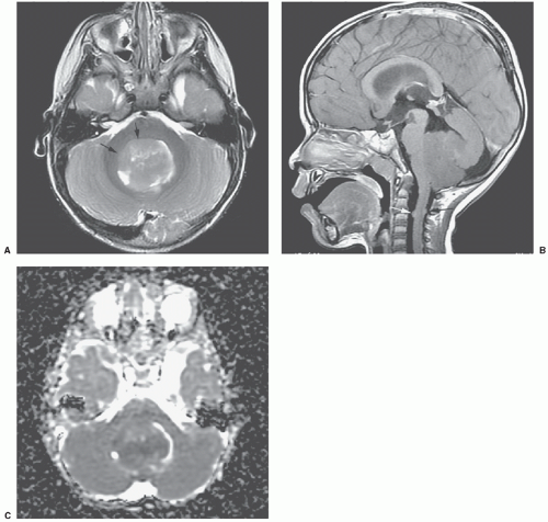
Medulloblastomas are characteristically relatively well-defined mass lesions that arise in the inferior medullary velum/roof of the fourth ventricle, and grow anteriorly into the fourth ventricle; they can invade the middle cerebellar peduncle or the dorsal brain stem (Fig. 26B.1). In older children and adolescents, medulloblastomas have the tendency to present either in the
lateral cerebellar hemisphere or near the cerebellopontine angle cistern (Fig. 26B.2). Ninety percent of tumors demonstrate some hyperattenuation compared with normal cerebellar attenuation on computed tomography (CT), a reliable imaging feature that distinguishes medulloblastomas from pilocytic astrocytomas.45 Calcifications are present in 10% to 20% of cases.


Figure 26B.1 Typical medulloblastoma in a 3-year-old child. Axial T2 (A) and postcontrast sagittal T1 (B) images show a midline tumor filling the fourth ventricle, with suspect invasion of the right middle cerebellar peduncle and the dorsal right hemipons (arrow, A). T2 signal is hypointense (A); the mass does not enhance, though small, weakly-enhancing metastatic nodules are evident in the upper cervical canal (arrow, B). The tumor shows reduced apparent diffusion coefficient (ADC) with low signal on the ADC map (C).

On magnetic resonance imaging (MRI), medulloblastomas are usually homogeneous with iso- to hypointense signal on T1-weighted images, and generally hypointense signal (between gray matter and white matter) on T2-weighted images; signal is often isointense to gray matter on FLAIR images and hyperintense on diffusion-weighted images.46 Although most medulloblastomas show moderate to intense contrast enhancement, approximately 5% to 10% of tumors do not enhance, and another 10% to 15% show only minimal (<25%) contrast enhancement (Fig. 26B.1). In infancy, “medulloblastomas with extensive nodularity” may occur, which display multiple, discrete, contrast-enhancing masses in almost a grape-like cluster.

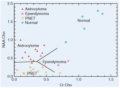
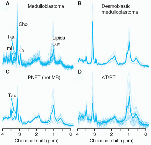
Both diffusion imaging and MR spectroscopy can be helpful in the differentiation of medulloblastomas from other posterior fossa tumors, namely, juvenile pilocytic astrocytomas and ependymomas. The apparent diffusion coefficient values of medulloblastoma are significantly lower than those of ependymoma and pilocytic astrocytomas, a reflection of the high cellularity and large nuclear areas of medulloblastoma.47,48,49 Using a discriminant analysis, single-voxel magnetic resonance spectroscopy of pediatric cerebellar tumors has been proven capable of separating medulloblastomas from pilocytic astrocytomas and ependymomas and from normal cerebellar tissue based on a plot of creatine-choline (Cr:Cho) ratios against N-acetyl aspartate (NAA):Cho ratios50 (Fig. 26B.3). Medulloblastomas have very high levels of choline and very low, lower, or absent NAA peak (decreased NAA-to-creatine ratio). High choline levels indicate a high degree of membrane metabolism, which usually takes place in rapidly proliferating malignant tumors. Lactate and lipid peaks can also be identified as a result of metabolic acidosis and tissue breakdown. Taurine (Tau) has been consistently observed by several groups in medulloblastoma51,52,53,54 and is an important differentiator of medulloblastoma from other tumors of the posterior fossa (Fig. 26B.4). A possible caveat is that it has been observed that taurine levels are low in some medulloblastomas with desmoplastic nodular histology.


Figure 26B.2 Teenage boy with a medulloblastoma in the left cerebellopontine angle. Axial T2 (A) and postcontrast axial T1 (B) images reveal a predominately T2 isointense mass in the CP angle cistern, with a mild amount of surrounding cerebellar edema, and fairly homogeneous, intense enhancement following gadolinium (B). Enhancement in the right foramen of Luschka represents normal choroid, not metastatic disease (arrow, B).

Atypical teratoid/rhabdoid tumors (AT/RTs) can also mimic medulloblastomas but have a greater propensity to present with hemorrhagic components. In the posterior fossa, AT/RTs are often located in the cerebello-pontine angle, in contrast to medulloblastomas, which favor the midline (vermis, fourth ventricle) in the first decade. Leptomeningeal invasion, surrounding edema, and enhancement from breakdown of the blood-brain barrier are also common. Ependymomas may be distinguished by their different anatomic location and pattern of spread through the foramina of Luschka and Magendie, and their characteristic appearance of speckled calcification on CT. MRs can give additional support to the diagnosis, especially in those cases in which the size and extent of the mass render difficult the identification of its point of origin. Solitary cerebellar hemangioblastoma usually will demonstrate serpiginous flow voids, reflecting the vascularity of the lesion, in addition to intense enhancement after intravenous contrast.55


Figure 26B.3 Nacetyl aspartate-choline (NAA:Cho) versus creatine-choline (Cr:Cho) scattergram for astrocytoma, ependymoma, medulloblastoma, and normal cerebellar tissue. The straight lines are boundaries between the three tumor types found by discriminant analysis. PNET, primitive neuroectodermal tumor.


Pathologic Classification of Medulloblastoma

PNETs arising in the cerebellum are termed medulloblastoma and are classified into five histologic groups in the current WHO classification of CNS tumors1: classic, desmoplastic nodular, medulloblastoma with extensive nodularity (MBEN), anaplastic, and large cell.1,56 Of note, there is significant morphologic overlap
between PNETs arising in different regions of the CNS as well as in extra-CNS locations. The nomenclature used by the WHO classification relies on both the location of the tumor and the histologic appearance to render a diagnosis.


Figure 26B.4 Unfiltered individual spectra (thin lines) and the averaged spectrum (thin lines), respectively, of embryonal tumors. All spectra were acquired with single-voxel PRESS, TE = 35 ms. Spectra are scaled to measured concentration to allow direct comparison. Taurine is readily detected in classic medulloblastoma (A) and in CNS primitive neuroectodermal tumors (PNET) (C). There is no evidence of taurine in the mean spectra of the desmoplastic subtype of medulloblastoma (B) or in atypical teratoid/rhabdoid (AT/RT) (D). Choline levels in AT/RT are more moderate; however, there is considerable variation in individual spectra.


Figure 26B.5 Typical histologic features of medulloblastoma (primitive neuroectodermal tumor). Tumor formed by apparently undifferentiated, basophilic, round-to-oval nuclei with minimal perceptible cytoplasm (hematoxylin and eosin, ×3,400).

Classic medulloblastomas are highly cellular, soft, friable tumors composed of cells with deeply basophilic nuclei of variable size and shape, little discernible cytoplasm, and, often, abundant mitoses (Fig. 26B.5) although they may exhibit surprising histologic heterogeneity. Homer Wright rosettes and pseudorosettes are variably present. Various degrees of glial or neuronal differentiation are noted, suggesting that the primitive cell of origin possesses the capacity for bipotential differentiation. A histologic variant with an abundant stromal component, desmoplastic nodular medulloblastoma (Fig. 26B.6), occurs dominantly in the lateral cerebellar areas of adolescents and adults and in the setting of Gorlin syndrome in infants and young children.35,36 These may have a nodular appearance. A variant of desmoplatic nodular medulloblastoma, called medulloblastoma with extensive nodularity (MBEN), occurs predominantly in very young children.

Aggressive variants of medulloblastoma, termed large-cell and anaplastic (frequently grouped together as large-cell anaplastic), have also been described. As the names imply, the histologic features that distinguish this subset of medulloblastoma are large round nuclei with prominent nucleoli, frequent mitoses, abundant apoptosis, and, in the anaplastic subset, nuclear pleomorphism. These tumors typically express synaptophysin and chromogranin. Monosomy 22 has not been seen in the cases described, but they tend to be associated with MYC or MCYN amplification.57 The large-cell anaplastic variant represented 4% of the nearly 500 cases of medulloblastoma reviewed by Brown et al.58,59


Figure 26B.6 Desmoplastic medulloblastoma showing linear arrangement of cells along delicate background fibers (hematoxylin and eosin, ×3,400).


Biologic Classification and Prognosis

Correlations have been noted between histologic subtypes and clinical outcomes, prominently including an improved prognosis for nodular desmoplastic medulloblastoma and a poor prognosis for large-cell anaplastic tumors. More recently, genomic methods have enabled the identification of biologically distinct subsets of medulloblastoma and provided a biologic basis for those clinical observations. These studies have led to a current consensus that medulloblastoma consists of four molecular subtypes with characteristic genetic, demographic, and clinical features: Wnt, SHH, Group 3 and Group 4 (Table 26B.4).60,61,62 Groups 3 and 4 medulloblastomas are frequently categorized together as “non-Wnt, non-SHH” tumors. Retrospective analyses have revealed this classification to have important prognostic significance in patients with medulloblastoma. Patients with tumors demonstrating Wnt pathway activation have an overall survival rate greater than 90%, while SHH subtype and Group 4 tumors have an intermediate survival of approximately 75% and Group 3 tumors have the poorest outcome with overall survival ranging from 40% to 60%.62 Classification of medulloblastomas into Wnt, SHH and non-Wnt, non-SHH subtypes (which can be performed using immunohistochemical methods on Formalin-Fixed Paraffin-Embedded [FFPE] specimens) has facilitated the subtyping of medulloblastomas for the conduct of future prospective clinical trials.63


Wnt Subtype

Medulloblastomas with activation of the Wnt signaling pathway constitute approximately 10% of medulloblastomas overall and have the best prognosis (>90% survival) (Fig. 26B.4). This subtype is most common in older children and adolescents (median age of 10 years) and is generally associated with tumors with classic histology. Greater than 90% of Wnt medulloblastomas have mutations in the β-catenin gene (CTNNB1) that result in a mutant protein that is resistant to degradation and accumulates in the nucleus of tumor cells. These tumors consistently show deletion of one copy of chromosome 6, allowing monosomy 6 and nuclear β-catenin accumulation to serve as specific and sensitive markers for Wnt pathway medulloblastoma. Mutations of the DDX3X gene are also frequent, occurring in approximately 50% of Wnt tumors.64,65,66,67,68 Of note, mutations in TP53, although associated with a poor prognosis in SHH pathway tumors, do not have a significant effect on the prognosis for this group of patients.69


SHH Subtype

Approximately 30% of medulloblastomas are defined by upregulation of the SHH signaling cascade through a variety of genetic mechanisms: most frequently, mutation of the PTCH1 tumor suppressor gene (a negative regulator of SHH signaling) and less often, alterations of other pathway members such as SMO, SUFU, and GLI2 (Fig. 26B.4). Bimodal peaks of SHH medulloblastoma incidence are seen: the first in children less than 3 years of age, and the second in older adolescents and young adults. Mutations in the SUFU gene are more common in tumors occurring in children less than 3 years of age, and SMO mutations are more frequent in adult tumors, which have potential implications for the use of molecularly-targeted therapies targeting SHH tumors in those populations. Although the prognosis for the group of patients with SHH tumors is intermediate (˜75%), TP53 gene mutation in an SHH pathway tumor carries a particularly poor prognosis and
may frequently be a germ line rather than somatic (e.g., in a child with Li-Fraumeni syndrome).70 Although SHH tumors do occur in the midline location, a cerebellar hemispheric location almost always indicates a tumor of SHH subtype.


Group 3 Subtype

This subtype of medulloblastoma accounts for approximately 25% of tumors and has both the highest rate of metastases and the worst survival rate (˜50%). Unlike Wnt and SHH medulloblastomas, no defining pathway alterations have been identified, although recurrent amplifications of the MYC and OTX2 genes and mutations of SMARCA4 are seen. Group 3 medulloblastomas occur more commonly in males (male:female ratio of 2:1) and primarily affect infants and young children rather than teenagers.


Group 4 Subtype

This subtype of medulloblastoma accounts for approximately 35% of tumors and has an intermediate survival rate (˜75%) although metastatic disease is relatively frequent (˜35%). As for Group 3 tumors, the biologic basis of Group 4 tumors is poorly understood, with amplification of MYCN found in a minority of tumors and no recurrent mutations occurring in more than 10% to 15% of tumors. Isochromosome 17q is seen more commonly in Group 4 than in Group 3 tumors and is rarely seen in Wnt or SHH subtypes. Group 4 tumors are seen in children of all ages but relatively rarely in infants. Like Group 3 medulloblastoma, Group 4 tumors occur more frequently in males (male:female ratio of 3:1).

Additional genetic and biologic studies are required to further clarify the basis of medulloblastoma, and in particular Group 3 and Group 4 tumors. Future prospective studies will incorporate this subtype information for risk stratification, treatment assignment, and selection of agents to target specific subtypes of medulloblastoma with the ultimate goal of improving outcomes and decreasing treatment-related morbidities.


Staging and Risk Stratification

Although the future holds great promise with the possible incorporation of the biologic findings described earlier, clinical studies so far have relied on clinical staging and risk stratification.4 Staging remains predominantly based on determination of the extent of tumor at the time of diagnosis and the degree of surgical resection. Complete staging requires pre- or postoperative MRI of the entire brain and spine, postoperative MRI of the tumor site, and CSF analysis.14

As with other CNS tumors of presumed primitive neuroepithelial origin, widespread seeding of the subarachnoid space may occur. The reported frequency of CNS spread outside the area of the primary tumor at diagnosis is 11% to 43%, and such spread eventually occurs in as many as 93% of patients who come to necropsy.71,72


TABLE 26B.4 Demographic, Clinical, and Genetic Characteristics of Medulloblastoma Subtypes.


Wnt


SHH


Group 3


Group 4


MBs (%)


10%


30%


25%


35%


Peak ages affected


Older children and adolescents (median 10 y)


Older children and adults (<3 or >16 y)


Infants and young children (<10 y)


Children of all ages (median 9 y)


M:F ratio


1:1


1.5:1


2:1


3:1


Survival


>90%


75%


50%


75%


Metastatic at diagnosis (%)


5%-10%


15%-20%


40%-45%


35%-40%


Key alterations


CTNNB1, DDX3X, SMARCA4, TP53


PTCH1, SMO, SUFU, GLI1, TP53


MYC, OTX2, SMARCA4


KDM6A, MYCN


Extraneural spread has been observed in 20% to 35% of patients in smaller institutional studies, but more recent larger series suggest that the rate of such events is less than 4%. Bone and bone marrow are the most common extraneural sites, accounting for more than 80% of extraneural metastases; lymph nodes, liver, and lung are the other reported sites.73,74,75,76

Historically, the Chang staging system, utilizing preoperative imaging studies, the surgeon’s intraoperative impression, and CSF cytology, was used to assign the stages of primary (T stage) and metastatic (M stage) disease14 (Table 26B.5). This system has been significantly modified and the assessment of the preoperative size of the tumor (T stage) has been supplanted by a postoperative evaluation of the extent of resection or, more specifically, the amount of postoperative residual disease, at times, modified by the impression of the surgeon at the time of diagnosis (Table 26B.6).

The M stage of the disease remains the single, most prognostic clinical parameter.77,78 The prognostic significance of M1 disease, indicating positive CSF cytology, without radiographic evidence of disseminated disease, has been debated and has been found to be predictive in some, but not all, studies. Part of this difference may be due to the means to assess the presence of free-floating tumor cells. Lumbar CSF cytology has been shown to be the most sensitive, but in some studies, either ventricular and/or spinal fluid have been used for assessment. Gajjar et al.79 found lumbar CSF to be more sensitive in the detection of disseminated disease than ventricular fluid and found cytologic assessment to be complementary to neuroaxis neuroimaging. In the German HIT 1991 study, 13% of patients had M1 disease and their overall survival (OS) rate of 65% did not differ from those with M0 disease.77 A similar finding was noted for a small number of patients with M1 disease in the French M7 study.78 In contradistinction, the Children’s Cancer Group (CCG)-921 study found that 18% of patients had M1 disease, utilizing almost exclusively lumbar CSF, and found a 5-year and progression-free survival of 57% for those patients with positive cytology, a rate lower than that for those with M0 disease, and higher than that for patients with M2 or M3 disease, although differences were of borderline significance.80

CSF for staging is most predictive if obtained 2 weeks or more following tumor resection.4,79 Preoperative lumbar puncture may be contraindicated if there is mass effect of the tumor and the potential for cerebellar herniation. Cisternal fluid, obtained at the time of diagnosis, has been variably related to outcome. Arachnoid biopsy, at the time of surgery, is presently being evaluated as a possible independent prognostic factor.

Neuroradiographically confirmed metastatic disease (M2 or M3) has been prognostic in all series.77,78,79,80,81 Extraneural spread at diagnosis (M4 disease) is rare outside of infancy, and assessment for extraneural disease by bone scans and bone marrow analysis is now not recommended, in most prospective studies, as part of initial staging. The sensitivity of MRI for the detection of metastatic disease is much greater than that of CT or myelography. Because of this, it is likely that patients who in the past would have been
diagnosed as nonmetastatic, or with M1 disease, are now being classified as having M2 disease.


TABLE 26B.5 Chang Staging System for Posterior-Fossa Medulloblastoma


Stage


Definition


Tumor


T1


Tumor, 3 cm in diameter and limited to the midline position in the vermis, the roof of the fourth ventricle, and, less frequently, to the cerebellar hemispheres


T2


Tumor, 3 cm in diameter, further invading one adjacent structure or partially filling the fourth ventricle


T3


Divided into T3a and T3b


T3a


Tumor invading two adjacent structures or completely filling the fourth ventricle with extension into the aqueduct of Sylvius, foramen of Magendie, or foramen of Luschka, thus producing marked internal hydrocephalus


T3b


Tumor arising from the floor of the fourth ventricle or brain stem and filling the fourth ventricle


T4


Tumor further spreading through the aqueduct of Sylvius to involve the third ventricle or midbrain, or tumor extending to the upper cervical cord


Metastases


M0


No evidence of gross subarachnoid or hematogenous metastasis


M1


Microscopic tumor cells found in cerebrospinal fluid


M2


Gross nodule seedings demonstrated in the cerebellar-cerebral subarachnoid space or in the third or lateral ventricles


M3


Gross nodule seedings in the spinal subarachnoid space


M4


Extraneuraxial metastasis


Despite the accepted use of MRI for staging, performance and interpretation of neuroimaging studies for the evaluation of disease dissemination at the time of diagnosis remains imperfect. In a recent Children’s Oncology Group (COG) study and French prospective studies, upon central review, inadequate imaging studies, due to patient movement or incomplete staging, were noted in up to 10% of patients.17 Another 5% to 10% of patients, upon central neuroimaging review, were found to have significant residual postoperative disease or evidence of dissemination not appreciated by the treating institutions. Patients with evidence of dissemination, upon central review, had a particularly poor prognosis if treated on reduced-radiotherapy protocols. Patients with inadequate imaging had intermediary outcomes between those with evidence of disease dissemination and those with no evidence of disseminated disease upon central review. Determination of the extent of disease on neuroimaging studies is particularly challenging in those with nonenhancing tumors, as spinal metastasis can be quite difficult to appreciate. Similarly, spread to the third ventricle and cisterns around the brain stem was problematic to appreciate.


Extent of Resection

A near-total (arbitrarily defined as more than a 90% resection) or a total resection is now achieved in approximately 80% of children with medulloblastoma, utilizing contemporary microsurgical techniques, entered in cooperative group studies.82 Evidence suggesting that the extent of resection correlates with outcome has been provided by several single institutions and by multi-institutional studies.83,84 For example, in the series reported by Jenkin, a 93% 5-year progression-free survival rate was noted in children undergoing gross-total resection as comparedwith only 45% in those undergoing a partial resection.85 In one CCG study, a clear relationship was found between the presence of greaterthan-1.5-cm2 residual disease on postoperative imaging and a significantly lower progression-free survival rate in patients with nonmetastatic disease.80,83 Arbitrarily, most cooperative group studies have continued to utilize some measure of the extent of residual disease after surgery as a staging criteria, as children with less than 1.5 cm2 of postoperative residual disease have been included in the average-risk category, while those with a greater amount of disease are classified as high-risk. It is widely accepted that patients who undergo only a minimal degree of resection fare poorly.


TABLE 26B.6 Stratification of Medulloblastoma


Standard Risk


High Risk


Age at diagnosis


>3 y


≤3 y


Extent of surgical resection


>1.5 cm2 residual tumor


≤1.5 cm2 residual tumor


Extent of disease


Stage M0


Stage M1-M4


Brain stem involvement at the time of diagnosis was found in older studies, in which patients were predominantly treated with radiation therapy alone, to be predictive of poor outcome.9,80 However, in most studies that have coupled chemotherapy with radiation therapy, the presence of brain stem involvement has not been found to be a significant prognostic variable.20,77,86


Histology

The significance of different histologic patterns on survival has been variable. In infants, the desmoplastic variant of medulloblastoma has been related to improved survival.11,12 More recently, the anaplastic or large-cell variant had been linked with more extensive and/or disseminated tumors and poorer prognosis, even in cases without dissemination.12,22,44 This latter finding has been demonstrated predominantly by retrospective reviews and in at least one prospective study.93 In the most recent COG prospective national studies, patients with nonmetastatic disease but with extensive or “diffuse” anaplasia have been included on high-risk protocols. Most of the children with large-cell anaplastic medulloblastoma fall into the Group 3 and 4 subtypes and tend to have a poorer prognosis. The significance of anaplasia as an outcome measure has been confounded by its subjectivity and relationship to biologic markers (such as higher MYC expression) that have been related to poorer prognosis.22

Aug 25, 2016 | Posted by in ONCOLOGY | Comments Off on Embryonal and Pineal Region Tumors

Full access? Get Clinical Tree

Get Clinical Tree app for offline access