ELECTRON MICROSCOPY OF THE THYROID
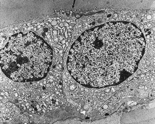
Ultrastructural studies of normal thyroid disclose that the follicular cells are arranged in a single layer. The apical surface contains microvilli that extend into a central lumen (Fig. 29-4); one to four cilia project from the central portion of each follicular cell. The cilia may alter the physical properties of the colloid, allowing for its ingestion by the apical portion of the follicular cell.20,21 and 22 Within the cytoplasm, lysosomes are prominent
and endoplasmic reticulum and small mitochondria are seen. Well-developed desmosomes and terminal bars are found between cells. The basal surface is separated from the interfollicular space by a basement membrane that is 35 to 40 nm thick. In the interstitium, fenestrated capillaries and collagen fibers are noted.
and endoplasmic reticulum and small mitochondria are seen. Well-developed desmosomes and terminal bars are found between cells. The basal surface is separated from the interfollicular space by a basement membrane that is 35 to 40 nm thick. In the interstitium, fenestrated capillaries and collagen fibers are noted.
 FIGURE 29-4. A normal thyroid follicle at the ultrastructural level. Note the cytoplasm, which is rich in organelles. Microvilli are visible at the cell–colloid interface (arrow) and in the basement membrane (arrowheads). ×10,600
Stay updated, free articles. Join our Telegram channel
Full access? Get Clinical Tree
 Get Clinical Tree app for offline access

|


