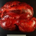
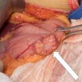
Most patients with corticotropin (ACTH)-dependent Cushing syndrome (CS) will have an ACTH-secreting pituitary microadenoma. Approximately 15% of patients with ACTH-dependent CS have an ectopic source of ACTH secretion, with the most common source being a bronchial carcinoid tumor (≈25%). The tumor may not be found in up to 9%–27% of patients with ectopic ACTH secretion. Although resecting the culprit neuroendocrine tumor should be attempted when possible, it is important not to delay therapy for severe hypercortisolism when this proves difficult. In these cases, bilateral adrenalectomy can be lifesaving. Here we present a case in which an alternative therapy for a suspected neuroendocrine tumor avoided bilateral adrenalectomy.
Case Report
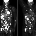
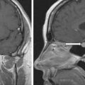
The patient was a 69-year-old woman who was referred for management of ACTH-dependent CS. Evaluation was prompted by progressive symptoms of generalized weakness, inability to stand up from a sitting position, lower extremity edema, and easy bruisability. The patient and her family have also noticed some weight loss (mainly as a result of muscle loss) and rounding of the face. At one point, hypokalemia was diagnosed and potassium supplements were initiated. Several months before evaluation at the Mayo Clinic, she was also diagnosed with type 2 diabetes mellitus and required additional medications for hypertension control. CS was suspected, and initial workup suggested ACTH-dependent hypercortisolism ( Table 59.1 ). Localizing studies that included magnetic resonance imaging (MRI) of the pituitary gland, computed tomography (CT) of the chest and abdomen, In-111 pentetreotide scintigraphy, as well as F-18 fluorodeoxyglucose (FDG) positron emission tomography (PET) scan were obtained but failed to localize the culprit lesion. The patient was referred to Mayo Clinic for further management. On physical examination, the patient was sitting in a wheelchair. Skin was found to be thin, with multiple bruises. Her fingernails revealed onychomycosis. She had moon facies without plethora. She had no striae. She had proximal myopathy on examination in both upper and lower extremities. Mild edema was noted.
| Biochemical Testing at a Local Institution | Biochemical Testing at Our Institution | ||
| Biochemical Test | Result (6 months Prior) | Result (At the Time of Evaluation) | Reference Range |
| 8 am serum cortisol, mcg/dL | 56 | 18 | 7–25 |
8-mg overnight DST, mcg/dL | 34 | <1 | |
ACTH, pg/mL | 155 | 108 | 10–60 |
DHEA-S, mcg/dL | 115 | 110 | 15–157 |
24-Hour urine free cortisol, mcg (low volume of 800 mL) | 47 | <45 |
Medications included glipizide, metformin, hydrochlorothiazide, spironolactone, lisinopril, metoprolol, and potassium chloride.
INVESTIGATIONS
ACTH-dependent hypercortisolism was confirmed (see Table 59.1 ). Inferior petrosal sinus sampling was performed to determine the subtype of ACTH-dependent hypercortisolism ( Box 59.1 ) and pointed toward an ectopic source. , Given that a CT of the patient’s chest was performed locally, the images were obtained and reexamined. On CT, a 7 × 5–mm right lower lobe nodule was demonstrated ( Fig. 59-1 ).
| ACTH, pg/mLCortisol, mcg/dL | |||||
| Right IPS | PV | Left IPS | PV | ||
| −5 minutes | 122 | 94 | 111 | 19 | |
| −1 minute | 120 | 103 | 117 | 18 | |
| +2 minutes | 195 | 100 | 106 | 19 | |
| +5 minutes | 233 | 108 | 111 | 18 | |
| +10 minutes | 183 | 113 | 125 | 19 | |
| +30 minutes | 108 | 19 | |||
| +45 minutes | 109 | 20 | |||
| +60 minutes | 109 | 19 | |||
Stay updated, free articles. Join our Telegram channel

Full access? Get Clinical Tree


