Fig. 8.1
Congenital orbital rhabdomyosarcoma (a) and leukemia (b) previously misdiagnosed as hemangiomas, received an unsuccessful course of propranolol
Diagnosis of hemangiomas is based first on the medical history. Two questions are of paramount importance:
Was the lesion present at birth? Did proportional or disproportional growth of the lesion after birth occur?
A careful history that includes age of onset, color, and location, along with physical examination findings, is generally sufficient in determining the pathological condition. The presence of the lesion at birth supports the diagnosis of vascular malformation or congenital hemangioma. Infantile hemangiomas are characterized by a postnatal rapid proliferation phase in the first 6–9 months of life, followed by a slow involution phase, and in many cases complete regression. Congenital hemangiomas fully developed before birth present as erythematous warm vascular plaques or nodules with a rim of pallor and do not undergo postnatal growth. Congenital hemangiomas are divided into two categories – rapidly involuting congenital hemangiomas (RICH) and non-involuting congenital hemangiomas (NICH). As the name implies, RICH lesions begin to involute almost immediately after birth and in many cases fully involute by 1 year of age. A NICH may partially involute or soften, but full resolution does not occur (Fig. 8.2). Despite RICH involute much faster than common hemangiomas, some patients may develop significant health problems (e.g., cardiac failure or bleeding) due to arteriovenous shunting [1–3]. After physical examination hemangiomas should be classified as superficial, deep, or mixed type, according to their location in the skin and subcutaneous tissue and focal or segmental according to their extension. Superficial infantile hemangiomas may be mistaken for capillary malformations (port-wine stains). Deep infantile hemangiomas typically present as a fluctuant, compressible bluish mass and may resemble lymphatic, venous, or mixed (venous and lymphatic) malformations [4, 5]. Congenital hemangiomas may be confused with venous malformations or mixed malformations. RICH may not be distinguished from a NICH at birth and only clinical observation would allow differentiation. In the newborn, lesions may not be evident or may present as a red, white, telangiectatic, or blue patch. A peripheral rim of pallor due to vasoconstriction may be seen at this stage. As the tumor proliferates, the lesion may take on the more classic appearance, which depends on the depth of lesion and stage of evolution [6].
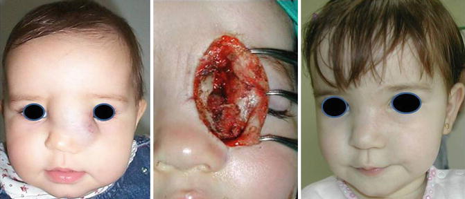
Fig. 8.2
Ultrasound and MRI findings were unable to determinate the diagnosis of glioma versus hemangioma. Excisional biopsy leads to final diagnosis of hemangioma
In cases in which clinical examination alone is inconclusive, other diagnostic modalities such as ultrasonography and magnetic resonance imaging (MRI) may be utilized. Ultrasound Doppler can assess the flow of hemangiomas, characterized by a shunt pattern with decreased arterial resistance and increased venous velocity. In deep cutaneous or visceral hemangiomas, contrast-enhanced MRI demonstrates the extent of the lesion and helps differentiation between hemangiomas and other disorders presenting with similar findings on US examination. Hemangiomas have a typical solid appearance with intermediate intensity on a T1-weighted spin-echo image, which is more intense compared with venous or lymphatic malformations. During the proliferative stage, hemangiomas show a relatively low intensity in a T2-weighted spin-echo image, while in the involution phase, they have a very low intensity. Contrast-enhanced T1-weighted MRI shows moderate intensity with prominent flow voids during the proliferative stage because of the high flow at this stage. In contrast, hemangiomas show low intensity during involution as a result of the low flow at that stage. The appropriate diagnostic tests for congenital hemangiomas are ultrasonography with Doppler (the lesions are uniformly hypoechoic, mostly confined to the subcutaneous fat and diffusely vascular, being traversed by multiple tubular vascular channels) and magnetic resonance imaging (RICH has areas of inhomogeneity and larger flow voids). Angiography is only indicated if embolization has to be performed (large and irregular feeding arteries in disorganized patterns, arterial aneurysms, direct arteriovenous shunts, and intravascular thrombi are common features in RICH and are rarely seen in infantile hemangiomas) [7].
The type of lesion can usually be determined based on the medical history and clinical examination. Imaging is mostly useful for confirming the clinical diagnosis, estimating the extent of the lesion and determining the feasibility of surgical resection.
Related posts:
 Coagulation Disorders Associated with Vascular Anomalies
Other Medical Treatments for Infantile Hemangioma and Congenital Vascular Tumors
Conclusions
Dermatological Manifestations of Vascular Malformations
Head and Neck Congenital Vascular Malformations: Sclerosis Treatment
Veno-lymphatic Vascular Malformations: Medical Therapy
Coagulation Disorders Associated with Vascular Anomalies
Other Medical Treatments for Infantile Hemangioma and Congenital Vascular Tumors
Conclusions
Dermatological Manifestations of Vascular Malformations
Head and Neck Congenital Vascular Malformations: Sclerosis Treatment
Veno-lymphatic Vascular Malformations: Medical Therapy
Stay updated, free articles. Join our Telegram channel

Full access? Get Clinical Tree


