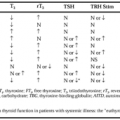
DIAGNOSIS
Part of “CHAPTER 65 – PAGET DISEASE OF BONE“
The diagnosis of Paget disease is based on a combination of the medical history and physical examination (particularly when increased heat or bone deformity is present), laboratory tests (measurement of serum alkaline phosphatase, and in some instances, a urinary marker of bone resorption), and radiographic studies (bone scans and conventional radiographs). Bone biopsy is indicated only occasionally in this disorder, primarily as a means of distinguishing between Paget disease and malignant conditions.
Perhaps the most specific and straightforward diagnostic test in the evaluation of Paget disease is a radiograph of an involved site, because the changes in the appearance of bone are characteristic (see Fig. 65-1). These include enlargement or expansion of bone, coarsening of trabecular markings, and pathognomonic patterns of lytic and sclerotic change. A bone scan is not a specific diagnostic test for Paget disease but is extremely useful, primarily for its sensitivity in identifying active sites of involvement. Radiographs of these areas then can be obtained to confirm the diagnosis and to provide information on the appearance of the process. For example, radiographs of an involved femur will reveal the extent of sclerotic versus lytic change, provide data on the status of the hip joint, identify areas of fissure fractures if present, and demonstrate bowing.
Related posts:

Stay updated, free articles. Join our Telegram channel

Full access? Get Clinical Tree


