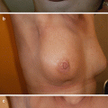
Fig. 47.1
Axial T2-weighted magnetic resonance imaging (MRI) image showing desmoid fibromatosis (DF) of the left breast of a 28-year-old female with bilateral breast implants in situ (2 years post implant surgery)
47.3 Assessment
All potential cases of DF should undergo full radiological and histopathological assessment prior to any management decision. All patients presenting with symptomatic breast lumps should undergo triple assessment, including physical examination, breast imaging and tissue sampling.
47.3.1 Radiological
Every suspected case of DF should ideally undergo ultrasonography (US) and magnetic resonance imaging (MRI) that includes the chest wall prior to intervention. Mammography is not particularly helpful in the diagnosis of DF and may miss a significant proportion of lesions (up to 62% [5]) although it is still routinely used to exclude breast cancer and other breast lesions. It should be noted that DF can also mimic carcinoma on mammography and can rarely present with dense calcifications.
US allows many characteristics of DF to be deduced including tumour size, location, echogenicity, homogeneity and vascularity [8]. The widespread availability of US means it is often the first-line investigation used in cases of DF. Despite this MRI remains the gold standard modality to assess and monitor DF [9]. Over time desmoid tumours undergo a temporal change in their histological makeup which correspond to an evolution in their appearance on MRI [10, 11]. Early in their evolution desmoid tumours are highly cellular with a low collagen component. This corresponds to a low signal intensity on T1-weighted images and high signal intensity on T2-weighted images. As they evolve the relative cellular component of DF decreases, whilst the relative collagen component increases. This corresponds with an increasingly heterogeneous picture on T2-weighted MRI sequences. Finally, in their later stages of evolution, desmoid tumours become fibrous in nature. This results in low signal intensity on both T1- and T2-weighted sequences.
If MRI is contraindicated, computed tomography with intravenous contrast should be performed to provide cross-sectional anatomical information. Some form of cross-sectional imaging is advisable due to the possibility of chest wall invasion.
47.3.2 Histopathological (Biopsy)
All potential cases of DF should undergo a planned biopsy prior to treatment, with the resulting histopathology being reviewed by a specialist sarcoma pathologist. Soft tissue tumours should be biopsied using a core needle biopsy (CNB) as this is more sensitive than other minimally invasive biopsy techniques, i.e. fine needle aspiration (FNA) [5], and provides tissue samples suitable for architectural, immunohistochemical and molecular analyses.
47.4 Management
DF’s unique biological behaviour and rarity mean that every biopsy-proven case should be referred to and managed by a soft tissue tumour (sarcoma) multidisciplinary team (MDT). This MDT will by definition include soft tissue sarcoma surgeons, musculoskeletal radiologists, histopathologists and medical and clinical oncologists with expertise covering the radiological and histological appearances and management of DF. There are a number of different scenarios as to how DF may present to a breast surgeon (see ◘ Table 47.1). In the majority of cases however, the diagnosis is made following a core biopsy of a suspicious breast lesion performed to rule out carcinoma.
Table 47.1
Typical scenarios for breast desmoid fibromatosis (DF) and management strategies
Scenario | Management |
|---|---|
Core biopsy diagnostic of DF | Request MRI (including chest wall) Refer to sarcoma service |
Core biopsy suggestive but not diagnostic of DF | Review by specialist sarcoma histopathologist – if still not diagnostic excision biopsy |
Breast lump excised – histology confirms DF (incompletely excised) | Refer to sarcoma service Do not re-excise chasing margins |
Breast lump excised – histology confirms DF (completely excised) | Refer to sarcoma service for ongoing surveillance |
Implant-related DF | Refer to sarcoma service (surgery normally required because of deformity to implant). Combined surgery breast and sarcoma surgeon |
47.4.1 Natural History/Active Observation
When observed over time, a significant proportion of desmoid tumours stop growing or stabilise, with up to 50% of cases undergoing spontaneous regression [12]. Historically DF treatment has revolved around surgical resection aimed at achieving wide histological margins. In recent years however, after recognising this complex and unique natural history, the majority of soft tissue tumour clinicians have shifted to a more conservative approach and now adopt a front-line policy of active observation wherever possible [13, 14]. This approach has been shown to result in long-term progression-free survival rates comparable to other more invasive treatments [12] and avoids the high recurrence rates and significant morbidity that often complicate DF resections [15]. Recognising these advantages, all cases of histologically proven breast DF should be managed with active observation in the first instance. During observation all patients should be closely followed up clinically and radiologically (with serial MRI scans) to pick up disease progression and those with painful tumours provided adequate analgesia.
All patients should be counselled extensively about the benign nature of DF and warned that regression may take many months and that progression may occur requiring medical (or rarely surgical) treatment.
47.4.2 Surgery
Following the recent changes in approach to DF management, surgical resections should now only be routinely utilised for those cases that have undergone an inconclusive biopsy which may represent DF, those patients unwilling to opt for a nonoperative approach despite appropriate counselling or those cases of DF associated with breast implants (which may necessitate removal of the implant) [16, 17].
The association between DF recurrence and histological margins is unclear. Although intra-lesional (R2) DF resections are associated with disease progression [18], no significant relationship has been identified between microscopically incomplete (R1) resections and recurrence. It is clear that recurrence may follow a complete (R0) resection, but will not necessarily follow a microscopically incomplete (R1) resection. Considering this, in the few cases where surgery is still indicated, surgeons should not seek wide (R0) histological margins at the expense of function or cosmesis. Glandular excision should be used sparingly, and therapeutic mammoplasties or other oncoplastic techniques may be required in order to avoid significant deformities. In such cases close cooperation between sarcoma specialists and oncoplastic breast surgeons is needed to ensure an optimal cosmetic outcome. Furthermore incomplete (R1/2) resections need not be re-resected but instead a baseline MRI performed and close clinical and radiological follow-up adopted to identify any potential disease progression or recurrence.
Breast implant-associated anaplastic large cell lymphoma (ALCL) [19, 20] should be excluded in all patients with DF and silicone implants, particularly in the presence of peri-implant seroma or severe capsular contraction. If mastectomy is required, then the decision about immediate breast reconstruction should be considered cautiously in view of significant risk of local recurrence, especially within first 3 years of follow-up.
47.4.3 Nonoperative Treatment
A detailed review of the nonoperative management of DF is outside the scope of this chapter. Many guidelines are available for clinicians considering nonoperative treatment of DF [21–23]. Treatment options include non-steroidal anti-inflammatory drugs (NSAIDs), hormonal therapy with antioestrogens, radiotherapy, chemotherapy and tyrosine kinase inhibitors [24–27]. These should be instigated in a stepwise fashion in accordance with the strength of the evidence supporting their use and their associated side effects. The decision to implement nonoperative DF treatment should only be made in conjunction with a soft tissue tumour MDT and administered under the supervision of an oncologist with an interest in sarcoma.
Stay updated, free articles. Join our Telegram channel

Full access? Get Clinical Tree


