Cytokine Therapy for Cancer


Cytokine Therapy for Cancer


Henry B. Koon

David F. McDermott


Cytokines are naturally occurring glycoproteins produced in response to infections and foreign antigens. While their biologic activities are complex, postulated antitumor effects of cytokines have included immunomodulation, antiproliferative activity, and inhibition of angiogenesis. Interleukin-2 (IL-2) and interferon-α (IFNα) are the most widely used of these molecules and have demonstrated clinical efficacy against several malignancies.

This chapter reviews the basic biology of cytokines and their role in the treatment of cancer. The primary focus is on cytokines that have received regulatory approval. In addition, this chapter discusses a rational refinement of cytokine therapy through improvements in patient selection, combination regimens, and novel strategies for overcoming tumor resistance.


Cytokine Biology and Cytokine Receptors

IFN was described over 50 years ago by Isaacs and Lindermann1 as a soluble factor that “interfered” with viral infection of cells. Subsequently characterized cytokines were referred to as interleukins because they were produced by and acted on leukocytes. Although the term, interleukin, does not accurately reflect the biologic properties of all such cytokines, some of which have been shown to be synthesized by cells other than leukocytes, it has been adopted as standard nomenclature. Because of their pleiotropic effects, multiple classification systems for cytokines have been devised. A functional classification of cytokines has been proposed that segregates cytokines based on whether they affect innate immunity, adaptive immunity, or hematopoiesis.2 However, given that a number of cytokines affect both the innate and adaptive immune systems, a more practical classification system is based on homology of their cognate receptors—the Type I cytokine receptors, the Type II cytokine receptors, and the immunoglobulin superfamily receptors.

The Type I cytokines receptors are characterized by a common signaling subunit that complexes with a cytokine specific subunit or units to activate intracellular signaling pathways. The prototypic Type I cytokine receptor is the interleukin-2 (IL-2) receptor family in which the common γ chain (γc) is shared by the IL-2, interleukin-4 (IL-4), interleukin-7 (IL-7), interleukin-9 (IL-9), interleukin-15 (IL-15), and interleukin-21 (IL-21) receptors (Fig. 31-1). Additional Type I cytokine receptor subgroups include the granulocyte/monocyte colony-stimulating factor (GM-CSF) and interleukin-6 (IL-6) receptor families. IL-6, IL-11, and IL-12 share GP130 as a common subunit. Interleukin-3 (IL-3), interleukin-5 (IL-5), and GM-CSF are members of the GM-CSF receptor subfamily and share a common β chain that complexes with cytokine-specific α chain. The effects of IFNα, IFNβ, IFNγ and interleukin-10 (IL-10) are mediated by Type II cytokine receptors, which are composed of a signaling chain and a ligand-binding chain. The immunoglobulin superfamily receptors contain extracellular immunoglobulin domains and include the receptors for interleukin-1 (IL-1), interleukin (IL-18), stem cell factor, and monocyte colony-stimulating factor.


Interferons


Biology

The IFNs are grouped based on their ability to bind specific IFN receptors. The Type I, Type II, and Type III IFN receptors are Type II cytokine receptors, which are composed of a signaling chain and
a ligand-binding chain.3, 4, 5 The human Type I IFN classes, α, β, ε, κ, and ω, are intronless genes that are located on chromosome 9p21.3 IFNα is the largest class of the Type I IFNs with 12 protein forms expressed.3 All the Type I IFNs, α, β, ε, κ, and ω, mediate their effects through the Type I IFN receptor, which is composed of IFNAR1and IFNAR2. IFNγ, the only Type II IFN, is located on chromosome 12q14 and activates the Type II IFN receptor composed of IFNGR1 and IFNGR2.3,5, 6, 7 The Type III IFNs are composed of IFNγ1 (IL-29), IFNγ2 (IL-28A), and IFNγ3 (IL28B).8,9 The IFNγ is located on chromosome 19q13 and activates the Type III receptors, IL-28R1 and IL-10R2.8,9


FIGURE 31-1 The IL-2 receptor family is the prototypic Type I cytokine receptors. All members of the family share the common gamma chain (γc). IL-2 and IL-15 share a common β signaling subunit (*) but have distinct a subunit that determines their affinity for the receptor.


Type I Interferons

The Type I IFNs consist of at least five classes in humans of which IFNα and IFNβ have been used the most clinically.3 Both IFNα and IFNβ have properties that make them attractive as immunotherapies. They up-regulate major histocompatibility complex (MHC) class I molecules and induce maturation of a subset of dendritic cells (DCs).10,11 Type I IFNs also activate cytotoxic T cells (CTLs), natural killer cells (NK) cells, and macrophages.12, 13, 14 Mice with targeted deletion of the Type I IFN receptor have a higher rate of carcinogen-induced cancer compared to controls and have enhanced tumor development in transplantable tumor models, supporting the hypothesis that the Type I IFNs are important in immunosurveillance.15,16 In addition to their immunologic effects, the Type I IFNs can have a cytostatic effect on tumors cells and may be proapoptotic.17,18 They also have antiangiogenic effects on the tumor vasculature.19,20 These observations suggest that IFNs may have direct tumor effects in addition to its immune effects. Experiments using tumors that are deficient for STAT1, a critical downstream signaling molecule of IFN, demonstrated that the presence or absence of STAT1 in the tumor had no effect on response to IFN treatment.21 In contrast, the mice that were deficient in STAT1 that were inoculated with the STAT1-competent B16 murine melanoma cell line derived no survival benefit from the administration of IFNα, whereas the WT mice had improved survival.21 These data suggest that the major effect of IFN in vivo is through the host cells. This experiment does not address whether the observed effect is through immune response or inhibition of angiogenesis. Despite extensive investigation, the primary mechanism of IFN antitumor effects in humans remains to be clarified.


FIGURE 31-2 Signal transduction pathways activated by Type I and Type II IFNs.

Type I IFN can be produced by almost all cells in the body in response to viral and bacterial nucleic acids and bacterial cell wall components such as lipopolysaccharide (LPS), but it was recognized that a small subset of peripheral mononuclear cells produce the majority Type I IFN.22 Plasmacytoid dendritic cells (pDC) were identified as the elusive “natural type-I-IFN-producing cell.”23,24 On exposure to viral and bacterial products, the Toll-like receptors (TLRs) are engaged and activate multiple signaling pathways, including the NF-κB pathway, mitogen-activated protein kinase (MAPK) pathway, and IFN regulatory factors (IRFs, discussed below), which lead to maturation of the DC and induction to the Type I IFNs (reviewed in Refs [25,26]). Flt3 ligand can be used to differentiate bone marrow cells into pDC and mobilize pDC into peripheral circulation in humans.27,28 Although the mechanism for IFN induction for infections has been well characterized, how IFN is induced in the setting of malignancy and immunosurveillance is unclear. Interestingly, the IRFs have been implicated in the DNA damage response and this may provide a mechanism by which abnormal cells initiate an innate immune response.29, 30, 31, 32


Type I Interferon Signal Transduction

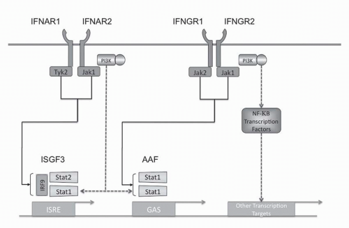
Type I IFN intracellular signaling is mediated through multiple pathways (see Fig. 31-2). The Type I IFN receptor subunits constitutively associate with members of the Janus kinase (JAK) family. IFNAR1 associates with TYK1 and IFNAR2 associates with JAK2.33,34 Ligand binding results in activation of these JAK kinases as well as phosphorylation of tyrosine residues in the receptor chains. These events lead to phosphorylation and activation of members of the signal transducer and activator of transcription (STAT) family. The Type I IFN receptor has been shown to phosphorylate all seven members of the STAT family but STAT1 and STAT2 appear to be the main effectors. The phosphorylated STATs
form homodimers or heterodimers and translocate to the nucleus. Two transcriptional complexes are formed, IFN-α-activated factor (AAF, IFN-γ-activated factor [GAF]) and IFN-stimulated gene factor-3 (ISGF3).35 AFF is a homodimer of phosphorylated STAT1, which binds to the IFNγ activated sequence (GAS), and ISGF3 is a heterodimer of STAT1 and STAT2 complexed with IRF-9 (p48, ISGF3γ) that binds to the IFN-stimulated regulatory element (IRSE).35, 36, 37 In addition to STAT dimers, CT10 oncogene homologue (avian)-like protein (CrkL) forms heterodimers with STAT5 upon receptor activation and binds with the GAS sequence.38 Although the JAK/STAT pathway is the classic mediator of IFN signaling, a number of other signaling pathways play a critical role in IFN signaling.

The Type I IFN receptor activates the phosphatidylinositol 3-kinase (PI3K)/AKT pathway as well as members of the MAPK family.39, 40, 41 PI3K activation generates the phosphatidylinositols, PIP2 and PIP3.42 These second messengers activate downstream targets such as 3-phosphoinositide-dependent protein kinase 1 (PDK1) by recruitment or conformational change.43 PDK1 in turn phosphorylates AKT (protein kinase B) on threonine 308, resulting in activation.43 AKT is further activated by phosphorylation on serine 473 by a protein complex containing Tor.44, 45, 46

Torc 1 and 2 are enzymatic complexes of the mammalian target of rapamycin (mTor). Torc1, the rapamycin sensitive complex, consists of mTor, Raptor (regulatory associated protein of mTor), and mLST8 (mammalian lethal with sec18 protein 8). It regulates protein translation by phosphorylating ribosomal S6 kinases, S6K1 and S6K2, and the eIF4E (eukaryotic initiation factor 4E)-binding proteins, 4E-BP1 and 4E-BP2.44, 45, 46 Torc1 regulates translation and has been implicated in autophagy.47 Torc2, the rapamycin insensitive complex, consists of mTor, Rictor (rapamycin-insensitive companion of mTor), mLST8 (mammalian lethal with sec18 protein 8), and mSin1 (mammalian stress-activated protein kinase-interacting protein-1). This complex phosphorylates AKT on serine 473, leading to phosphorylation and inhibition of FOXO transcription factors. Torc2 in concert with PDK1 phosphorylates and stabilizes PKCα.44, 45, 46 The PI3K/AKT/mTor pathway regulates apoptosis, gene expression, protein translation, and cytoskeletal organization.

Activation of the Type I and II IFN receptors activates the class I PI3K, which is composed of regulatory (p85) and catalytic subunits (p110).41,48,49 Experiments using MEFs from p85 knockouts have shown that PI3K plays a role in the induction of IFN-stimulated genes (ISG) including ISG15, CXCL10, and IRF by regulating mRNA transcription through Torc1.41 In addition, PI3K plays a role in phosphorylation of STAT1 on serine 727, which enhances STATdependent transcription.50 A relatively small number of genes that are regulated by IFN are modulated by PI3K/mTor pathway.51 The induction of IFN in pDC is also dependent on the PI3K/mTor pathway. These findings elucidate the molecular mechanism of immune suppression by rapamycin.52

The NF-kappaB (NFκ B) transcription factors play a critical role in the suppression of apoptosis and regulation of the cell cycle. The NFkgr; B family consists of p50, which is derived from the p105 precursor and p52, which is derived from the p100 precursor, RelA (p65), RelB, and cRel. The p50 and p52 subunits lack a transcription activation domain and as homodimers function as transcriptional repressors or form complexes with BCL-3 to activate transcription.53,54 p50 is constitutively processed from p105, but the processing of p100 to p52 is tightly regulated by phosphorylation and ubiquitinylation.53 In contrast, p65, cRel, and RelB have a transcription activation domain and thus, when complexed with p50 or p52, are capable of activating transcription.53 NFκB homodimers and heterodimers are tightly regulated and are bound in the cytoplasm by inhibitors of NFκB (IκB). In the classical pathway of NFκB activation, IκB kinases (IKK) beta and gamma phosphorylate IκB, leading to IκB proteolytic degradation.53 NFκB then freely moves into the nucleus and regulates transcriptional activity. In the alternative NFκB activation pathway, the TNF receptor-associated factors (TRAFs) activate MAP3K kinase and NFκB-inducing kinase (NIK), which leads to the processing of p100 to p52 in an IKKα-dependent manner.53 The Type I IFN receptor activates NFκB through the classical and alternative pathways.55,56 The classical pathway appears to be PI3K dependent, while the alternative pathway appears to be dependent on NIK.55,56 NFκB binds to the promoter of a subset of IFN-induced genes that also have binding sites for IRFs and STATs.

The IRF transcription factors are intricately integrated into the regulation of IFN. Mouse knockouts have demonstrated that IRF-4 and IRF-8 are required for the generation of pDCs.57 IRF-3 and IRF-7 along with NFκB directly regulated the expression of Type I IFNs.58 ISGF3 complex, which contains IRF-9, is a signaling complex downstream from the Type I receptor that induces IRF-7 expression.59 ISGF3 and IRF-1 both bind to the ISRE DNA sequence and IRF-1 acts as a positive regulator of Type I and Type II IFN-induced genes, whereas IRF-2 represses the effects of IRF-1.60 The induction of ISGF3, NF-κB, and IRF-7 by the Type I IFN receptor provides a potential positive feedback mechanism for IFN in pDCs.61, 62, 63, 64

In contrast to the positive regulators of IFN signaling (e.g., IRFs), activation of negative regulators attenuates IFN signaling. Phosphatases are the counter balance to kinase activity. Src-homology 2 domain-containing protein tyrosine phosphatase-1 (SHP-1) and SHP-2 are protein tyrosine phosphatases that contain SH2 domains. SHP-1 appears to be a negative regulator of signaling with multiple reported substrates.65 SHP-1 can dephosphorylate JAK family kinases and terminate signaling.66 SHP-2 appears to be a positive modulator of MAPK signaling, but it may also function as a negative effector.67,68 Both phosphatases have been shown to have a negative regulatory role in IFN signaling as a heightened response to IFNs is observed in SHP-1- and SHP-2-deficient cells.68,69 In addition, treatment of cells with stibogluconate, an inhibitor of both phosphatases, resulted in prolonged IFN signaling.70 Thus, SHP-1 and SHP-2 would be potential pharmacologic targets to modulate the effect of IFNs.

STAT signaling is regulated by suppressor of cytokine signaling (SOCS) proteins and the protein inhibitor of activated STATs (PIAS) gene families. The SOCS proteins are induced by IFN and negatively regulate cytokine signaling through binding and inactivation of JAKs and STATs, targeting the protein for degradation, or both.71, 72, 73 The PIAS proteins bind to STAT1 and STAT3 dimers, thereby blocking their DNA-binding activity. PIAS1 selectively regulates a subset of IFN-γ– or IFN-β-inducible genes by interfering with the recruitment of STAT1 to the gene promoter.74 Both these pathways likely play critical roles in dampening the response to IFN in vivo.


Pharmaceutical Preparations

IFNα initially referred to as leukocyte IFN because of how it was originally isolated, is comprised of a group of at least 12 distinct proteins.3 Recombinant IFNα-2a, IFNα-2b, and IFNα-2c differ by one to two amino acids and are the forms of IFNα that have been tested clinically.3 In the United States, IFNα2a is sold under the trade name Roferon (Hoffmann-La Roche; Nutley, NJ) and IFNα-2b is available as Intron A (Schering; Kenilworth, NJ). IFNα-2c is available in Europe as Berofor (Bender; Vienna, Austria). These three compounds have never been compared in a randomized trial; however, their spectrum of activity is likely similar. The approved indications for these agents include treatment of viral diseases such as Hepatitis C and Kaposi’s sarcoma (KS) as well as treatment of cancers such as melanoma and chronic myeloid leukemia (CML).75, 76, 77 IFNα conjugated to polymer polyethylene glycol (PEG-IFN) has an increased half-life allowing for longer dosing intervals and long exposure times (see below).78 Pegylated IFNα-2a (Pegasys, Hoffmann-La Roche) and pegylated IFNα-2b (Peg-Intron, Schering) are the two forms of PEG-IFN available in the United States.79,80 These agents are widely used in combination with ribavirin in the treatment of Hepatitis C. The role of the PEG-IFNs as monotherapies for cancer is still under study.81,82


TABLE 31.1 Pharmacokinetics of interferons


Route of administration


Dose (mU)


Serum concentration (U/mL)


Peak time


Duration (from-to)


IFN type


Reference


IFN-α


IM


50


2,000 pg/mL


6 h


<0.5 to >24 h


α2a


286


36


1,000 pg/mL


6 h


<0.5-24 h


18


500 pg/mL


6 h


<0.5-24 h


Continuous SC infusion


2-5


20-60


Steady state


24-72 h


α2b


291


IV


4-10


10-300


30 min


Fibroblast β


292


40-80


200-10,000


30 min


160-320


2,000-20,000


30 min


IFN-β


SC


90


102


1-8 h


β1b


246


IV


90


103


5 min


5 min-12 h


IV 4-h infusion


0.01-1.00


<10


β1b


293


10


25-30


6 h


0.5-24 h


30


140


4 h


0.05-24 h


IV


45


350


5 min


β1b


294


180


1,800


5 min


SC


45


0


180


25


IV 10-min infusion


0.01-20.00


0


γ


242


30


15 min


<0.5-12 h


75


15 min


<0.5-24 h


IFN-γ


IV 6-h infusion


0.5 mg/m2


3 ng/mL


6 h


2-8 h


γ


295


1 mg/m2


6 ng/mL


6 h


<1-8


SC


1 mU


4 mg/mL


13 h


γ


296


Clinical Pharmacology of Type I Interferons


Interferon-α

The pharmacokinetics of the IFNs was initially measured in serum using bioassays, which measure protection from viral cytopathic effect, prior to the development of direct immunological tests such as enzyme-linked immunosorbent assays. When given subcutaneously or intramuscularly to humans, approximately 80% of IFN-α is absorbed.83 These routes of administration lengthen its distribution phase, with peak serum levels occurring at 1 to 8 hours. Clearance varies between 4.8 and 48 L/h, and the terminal elimination half-life is 4 to 16 hours.83 The high-dose IFN-α2b regimen used for adjuvant therapy of melanoma may yield peak serum levels of 2,500 IU/mL at the end of the intravenous phase and 150 IU/mL after subcutaneous administration83, 84, 85 (Table 31-1).

In order to improve the pharmacokinetic profile of IFN-α, these agents have been chemically modified by linkage to a PEG molecule.78 The PEG modification increases the serum half-life, resulting in a less frequent dosing interval. A monopegylated IFN-α2b has been developed by Schering Plough (Kenilworth, NJ). This species has a 20-fold increase in serum half-life compared with native IFN-α2a.86 Linkage of a 40-kD branched PEG molecule to IFN-α2a or a 12-kD linear PEG moiety to IFN-α2b markedly
altered the pharmacokinetic profile and increased activity against hepatitis C in a direct clinical comparison of subcutaneous PEG-IFN with the parent compound.87,88 The larger PEG moiety of PEG-IFN-α2a leads to the slowest absorption half-life and a longer elimination half-life87,88 (Table 31-2)


TABLE 31.2 Pharmacokinetics of pegylated parental IFN-ALPHAS


IFN-α2a


PEG IFN-α2a


IFN-α2b


PEG IFN-α2b


Volume of distribution


31-73 L


8-12 L


1.4 L/kg


0.99 L/kg


Absorption t1/2


2.3 h


50 h


2.3 h


4.6 h


Elimination t1/2


3-8 h


65 h


4 h


˜40 h


Time to max. conc.


7.3-12 h


80 h


7.3-12 h


15-44 h


Peak/trough


1.5


>10


t1/2, half-life; max. conc., maximal concentration.


PEG-IFN-α2b at 1μg/kg/wk induced a biological response similar to that of IFN-α2b at 3 million IU administered subcutaneously three times a week.89 Anticancer activity against solid tumors was demonstrated in a phase I/II trial of PEG-IFN-α2b, which determined 6 μg/kg/wk as the maximal tolerated dose (MTD) and showed evidence of drug accumulation with an area under the curve (AUC) of 374 pg/h/mL for week 1, compared with 480 pg/h/mL at week 4 for patients treated with the MTD.90 PEG-IFN-α2b has been directly compared with the parent drug as initial treatment for chronic phase CML in a phase III study, where it showed efficacy and toxicity at 6 μg/kg/wk similar to those of IFN-α2b at 5 million IU/m2/d.91 The toxicity of both agents appears comparable to historical controls treated with the parental compound.81 Whether the alteration of IFN-α pharmacokinetics will increase antitumor activity must be addressed in future studies.


Interferon-β

A single species in humans, IFN-β, exists as a 25-kD glycoprotein containing 166 amino acids, and IFN-β appears to be metabolized primarily in the liver.83 After intravenous injection, the terminal elimination half-life is about 1 to 2 hours, and IFN-β remains measurable for up to 4 hours (Table 31-1).83 In contrast, after a single subcutaneous or intramuscular injection, serum IFN-β is barely detectable.83 However, intravenous and subcutaneous administration of the same dose elicited similar pharmacodynamic responses, including 2 to 5 A synthetase induction—a known IFN response gene that has been correlated to dose and serum level in some studies.83,92

Three modifications of IFN-β to improve the pharmacokinetic profile have been developed. Albuferon is a recombinant protein resulting from fusion of the IFN-β peptide with albumin. In monkeys, the bioavailability of subcutaneous Albuferon was 87%, plasma clearance was reduced by 140-fold, and the terminal half-life increased 5-fold, while in vitro and in vivo activities were preserved.93 Fusion of IFN-β to soluble recombinant Type I IFN receptor subunit (sIFNAR-2) prolonged the half-life and increased antitumor activity in mice.94 Finally, pegylation of IFN-β1a with a linear 20-kD molecule increased the maximum serum concentration achieved 4-fold, while the AUC increased 10-fold and the half-life increased 3-fold.95

Since IFNα and IFNβ signal through the same receptor, they would be expected to have similar biologic effects and have overlapping indications. However, this is not always the case. Although both IFNα and IFNβ have activities against gliomas, one small study suggests that IFNβ has a higher response rate compared to IFNα.96 In contrast to IFNα, IFNβ has been reported to have no clinical activity against CML and no responses were seen in a phase I trial of 35 patients with metastatic solid tumors.97,98 Two forms of IFNβ, originally named fibroblast IFN, have been approved for use in patients with relapsing multiple sclerosis—IFNβ-1a (Avonex, Biogen Idec; Cambridge, MA) and IFNβ-1b (Betaseron, Berlex; Montville, NJ). Their use in treatment of malignancy is currently limited to clinical trials.


IFNγ : Type II Interferon

IFNγ, also known as immune IFN, is the only Type II IFN and has effects on the innate and adaptive immune systems. IFNγ is secreted by NK cells, natural killer T cells (NKTs), Th1 CD4+ T-cells, CD8+ T-cells, antigen-presenting cells (APCs), and B cells.99, 100, 101, 102 IFNγ activates macrophages and stimulates up-regulation of MHC class I, MHC class II, and costimulatory molecules on APCs.11,103, 104, 105 Additionally, IFNγ induces changes in the proteosome to enhance antigen presentation.106, 107, 108 It promotes Th1 differentiation of CD4+ T cells and blocks IL-4-dependent isotype switching in B cells.103,109,110 Mice with targeted deletion of IFNγ or the Type II IFN receptor have an increased risk of spontaneous and chemically induced tumors compared to controls.111, 112, 113, 114, 115 IFNγ is cytotoxic to some malignant cells and has antiangiogenic activity.116, 117, 118, 119, 120

IFNγ is located on chromosome 12q14 and activates the Type II IFN receptor composed of IFNGR1 and IFNGR2.3,5, 6, 7 Like the Type I IFN receptor, IFNGR1 and IFNGR2 are constitutively associated with members of the JAK kinase, JAK1 and JAK2, respectively.121 STAT1, 2, 3, and 5 are phosphorylated by the Type II IFN receptor, but STAT1 appears to be the critical mediator.122, 123, 124 The Type II receptors also activate the PI3K/AKT and MAPK pathways as well as NFκB.50,125,126 The activation of NFκB appears to be cell-type restricted, and its role in vivo is unclear.


Clinical Pharmacology of Type II Interferon (IFN-γ)

After subcutaneous or intramuscular administration, 30% to 70% of IFN-γ is absorbed, and the terminal elimination half-life is 25 to
35 minutes; after intravenous injection, IFN-γ remains detectable in serum up to 4 hours (Table 31-1). Like IFN-β, IFN-γ appears to be metabolized primarily by the liver.83 A phase I trial in colon cancer patients achieved IFN-γ concentrations greater than 5 U/mL for more than 6.5 hours following subcutaneous administration of 100 μg/m2.83 Preclinical studies of PEG-IFN-γ show an increased elimination half-life activity with preserved activity, but this molecule has not yet been evaluated in patients.83

The antitumor effects of IFNγ suggested it would be effective against a wide spectrum of malignancies. Although IFNγ has demonstrated limited clinical utility in cancer, it likely plays a critical role in the in vivo effects of other cytokines.127, 128, 129 Actimmune (Intermune; Brisbane, CA) is an IFNγ preparation that has been approved for the treatment of chronic granulomatous disease.130 Clinically, significant benefit in treatment of malignancies has been largely restricted to Type I IFNs.


Type III Interferons

In 2003, two papers were published simultaneously describing novel cytokines designated IFNs γ/IL-28/29, which exhibited antiviral activity.8,9 These genes, comprising five exons on chromosome 19, bind to a distinct dimeric membrane receptor, IFNLR1 and IL10R2.8,9 These IFNs have antiviral activity, antiproliferative activity, and in vivo antitumor activity like the Type I IFNs. The receptor is associated with Jak1 and Tyk2 resulting in the activation of STAT1, 2, 3, 4, and 5, which results in expression of GAS and ISRE containing genes.9 The signaling of the Type III IFNs appears to be quite similar to the Type I IFNs.131 The differentiation appears to be at the level of the receptor. IL10R2 is ubiquitously expressed, while IFNLR1 is limited in its expression and inducible in some cell types such as pDC.131, 132, 133, 134 In pDC, TLR activation induces IFNγ just as it does the Type I IFNs; however, when IL-28R-deficient mice are exposed to a panel of different viruses, their immune response was unimpaired.134 Type III IFNs have a positive feedback on either the Type III or Type I IFNs; the role of the Type III IFNs may be to regulate antiviral responses in epithelial cells.134 Whether the Type III IFNs are a redundant system or have a unique role in IFN biology remains to be elucidated.


Clinical Indications


Hematologic Malignancies: Hairy Cell Leukemia

Type I IFNs have had their most clinical success against two hematologic malignancies: hairy cell leukemia (HCL) and CML. A regimen of IFNα-2b 2 million units/m2 subcutaneously three times a week for 52 weeks produced an overall response rate of 77% with a complete response rate of 5% in patients with HCL.135 The vast majority of these patients (61 out of 64) had undergone splenectomy but were otherwise untreated.135 Subsequent studies demonstrated complete responses in 25% to 35% of patients who had not had splenectomies, leading to regulatory approval for IFN in this patient population.136 Although IFN has a significant response rate and improves survival in HCL, the majority of patients relapse after discontinuation of therapy.137 Subsequent studies demonstrated that 80% of patients who relapsed would respond to another course of IFN.137 It is unclear whether the effects of IFN in HCL are mediated by immune mechanisms or direct effects on the leukemic cells.138, 139, 140 Although IFN was once considered first-line therapy, the introduction of the nucleoside analogs, which have a greater than 90% CR rate, has limited the use of IFN therapy to patients who have disease that is refractory to nucleosides or have contraindications to these agents.141,142


Hematologic Malignancies: Chronic Myeloid Leukemia

Initial trials of IFNα in CML noted complete hematologic responses in over 50% of patients and a complete cytogenic response in up to 25% of patients.77,143 Follow-up randomized studies demonstrated that IFN was superior to hydroxyurea or busulfan or both.144, 145, 146, 147, 148 Four studies demonstrated an improved overall survival (OS) for the IFN-treated patients.144,145,147, 148, 149, 150 A meta-analysis of the randomized trials demonstrated an improvement in the 5-year survival in the IFN-treated group of 12% over hydroxyurea and 20% over busulfan-treated patients.151 Additionally, the meta-analysis showed the benefit extended to all risk groups. All three commercially available IFNα were used in the CML trials and although not formally compared, their activity in CML appeared similar.

The mechanism of response of CML to IFN has been extensively investigated. Reports that human leukocyte antigen (HLA) type and development of an immune response to Bcr-Abl correlate with a complete response suggest that IFN works through an immune mechanism in CML.152 The observation that patients who obtain complete response correct abnormalities in the secretion of Th1 cytokines also supports an immune mechanism.153 However, IFN also exerts a direct antiproliferative effect in CML through inhibition of DNA polymerase.154 These data suggest the mechanism of action of IFN in CML is multifactorial.

In an effort to enhance the efficacy of IFN in CML, a number of trials were conducted using IFN combined with chemotherapy.155 The combination of IFN and low-dose ara-C was shown to improve the number of cytogenetic remissions compared to IFN alone. However, the beneficial impact of the combination on OS is small and was achieved with a substantial increase in toxicity.156, 157, 158 Although largely supplanted as first-line therapy by Bcr-Abl kinase inhibitors,159 IFN and IFN-containing regimens remain a valid second-line therapeutic option for patients with CML.159, 160, 161, 162


Hematologic Malignancies: Non-Hodgkin’s Lymphoma

Early studies of IFNα monotherapy in follicular lymphomas demonstrated a response rate of over 50%.163, 164, 165 Subsequently, several investigators combined IFN with chemotherapy in an induction regimen or as maintenance therapy. The results of these trials were mixed in terms of OS benefit. The Groupe d’Etude des Lymphoma Folliculaires (GELF) study demonstrated an advantage in response rate (85% versus 69%, P < 0.001) and OS (34 versus 19 months, P = 0.02) for chemotherapy plus IFNα 5 MIU thrice weekly for 18 months compared to chemotherapy alone using an anthracycline-based regimen.166,167 These results were a major impetus for the approval of IFNα for the treatment of follicular lymphoma. A meta-analysis of the IFN trials supports a survival advantage for intensive chemotherapy regimens containing IFN.168 Interestingly, a large SWOG trial did not show any survival advantage for IFNα at 2 MIU thrice weekly for 24 months versus observation.169 These data suggest that the dose of IFNα used may be critical to the beneficial effect in patients with follicular lymphomas. IFN is approved
for treatment of follicular lymphoma, but its use is limited due to its associated toxicities and the activity of a variety of other agents.


Melanoma

The natural history of some melanomas suggests that it may be an immune responsive tumor. Up to 25% of primary cutaneous melanomas show histologic regression at the time of biopsy.170 All three IFNα‘s have been investigated in patients with metastatic (stage IV) melanoma. Multiple dose levels and schedules have been tested, and the overall response rate for single agent IFN in patients with metastatic melanoma is approximately 15%.171, 172, 173, 174, 175, 176, 177, 178, 179 There is no clear most effective regimen; however, IFN administered thrice weekly is the most widely used schedule because it has a significantly better toxicity profile than daily administration with no diminution in response rates.180,181 It is unknown if IFN provides a survival advantage because no randomized trials in metastatic melanoma compare IFN to either cytotoxic chemotherapy or best supportive care.182 IFN appears to work best in patients with low metastatic tumor burden.183

IFNα has proved most useful in the management of melanoma in the adjuvant setting. Multiple IFN regimens have been used in the adjuvant setting for patients with intermediate- and high-risk melanoma (Tables 31-3 and 31-4).184, 185, 186, 187, 188, 189, 190, 191, 192, 193, 194 IFNα was approved in Europe based on studies that used lower dose regimens. Two trials of low-dose IFNα-2a given for 12 to 18 months demonstrated a benefit in relapse-free survival (RFS) in patients with melanomas greater that 1.5 mm or locoregional disease, but neither trial showed an OS benefit.185,189 Subsequent trials using low-dose IFNα-2a and IFNα-2b have failed to demonstrate a durable RFS or an OS benefit (Table 31-3).184

Only gold members can continue reading. Log In or Register to continue

Stay updated, free articles. Join our Telegram channel

May 27, 2016 | Posted by in ONCOLOGY | Comments Off on Cytokine Therapy for Cancer

Full access? Get Clinical Tree

Get Clinical Tree app for offline access