Cutaneous Melanoma


Cutaneous Melanoma


Levi A. Garraway

Lynda Chin


Cutaneous melanoma arises from pigment-producing epidermal melanocytes and is the major cause of mortality among skin malignancies. Its incidence has risen steadily at a rate of 3% per year over the past 25 years,1 with an estimated 68,130 new cases and 8,700 deaths predicted for the United States alone in 2010. Despite a high cure rate for localized primary melanoma (98.3% 5-year survival rate), its aggressive nature results in rapid metastasis to distant sites and a concomitant drop to a 16% 5-year survival rate. Although exposure to ultraviolet (UV) radiation is a known factor contributing to melanoma development, the exact molecular changes that take place in incipient and progressing tumors are still being elucidated.

Currently, vertical tumor (Breslow) thickness (in millimeters) provides the best single indicator of prognosis. This measurement is augmented by additional parameters, including the presence of ulceration, penetration through cutaneous layers, mitotic rate, evidence of “in transit” metastasis, tumor spread to draining lymph nodes, and the presence of distant metastasis. Although melanoma sometimes arises in pre-existing nevi, recent data have demonstrated that typical nevi represent senescent lesions that may be irreversibly growth arrested.2 It is therefore plausible that a substantial fraction of melanomas may alternatively emerge from normal melanocytes via deregulation of oncogenes or tumor suppressors implicated in melanocytic transformation (Fig. 27.1). The myriad genes involved with melanoma genesis and progression have been subjected to varying degrees of validation in humans, in model organisms, and in cell culture (Table 27.1). The identification of these genes and their associated genetic pathways influences diagnosis and prognosis and shows considerable promise in aiding targeted therapeutic implementation.


THE CDKN2A LOCUS

As many as 70% of melanomas harbor somatic mutations or deletions affecting the CDKN2A locus on chromosome 9p21.3,4 This observation, together with the initial identification of germline homozygous deletions of CDKN2A as susceptibility events in familial melanoma kindreds,5 indicates a central role for this locus in melanoma pathogenesis. This locus contains an unusual gene organization, which allows for two separate transcripts and corresponding tumor suppressor gene products to be produced: p16INK4A and p19ARF (Fig. 27.2). Loss of p16INK4A results in the suppression of retinoblastoma (RB) tumor suppressor activity via increased activation of the CDK4/6-cyclin D1 complex; loss of ARF (p14ARF in human and p19ARF in mouse) down-modulates p53 activity through increased activation of MDM2. Thus, deletion of the entire locus accomplishes the inactivation of two critical tumor suppressor pathways: RB and p53. Homozygous deletion of exons 2 and 3 of the mouse Cdkn2a homolog predisposed to a high incidence of melanomas when combined with an activated H-RAS transgene in melanocytes.6 Thus, CDKN2A lesions may “prime” melanocytic tissue for neoplasia.


THE RETINOBLASTOMA PATHWAY

The RB pathway is responsible for preventing inappropriate cell cycle entry, and germline heterozygous loss of the RB1 gene triggers retinoblastoma. The tumor-modulating properties of the RB pathway are well established in many solid cancers, and its deregulation in melanoma is evidenced by mutations in INK4A, CDK4, or occasionally RB1 itself, as described below.


INK4A

Human intragenic mutations of INK4A that do not affect the ARF coding region preferentially disrupt the RB pathway and sensitize germline carriers to the development of melanomas.7 In a mouse model engineered to be deficient only for Ink4a (with intact ARF), melanoma formation was observed in cooperation with an oncogenic
initiating event (activated H-RAS), albeit with a longer latency than in mice with deletions affecting the entire locus.8 Notably, the tumors in these mice were also found to harbor either deletion of ARF or mutation of p53. Therefore, while INK4A is a bona fide tumor suppressor, additional genetic dysregulation of the p53 pathway seems obligatory for melanoma genesis, at least in the mouse.


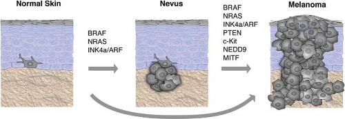
FIGURE 27.1 Validated genes mutated or deregulated in melanoma progression. Progression of normal melanocytes to metastatic melanoma is diagrammed with associated genetic events as a linear process, although in a majority of the cases, melanomas may not arise from a pre-existing nevus. The degree to which the association of each gene has been experimentally validated is shown in Table 27.1.


TABLE 27.1 SUMMARY OF VALIDATED GENES INVOLVED IN MELANOMA GENESIS AND PROGRESSION


Methods of Discovery


Melanoma Gene


Proposed Gene Product Behavior


Extent of Validation


H


E


I


O


K


X


P


M


Ref.


Linkage mapping


INK4A, ARF


Tumor suppressors


Reviewed in 17


Linkage mapping


NRAS, BRAF


Oncogenes


Reviewed in 17


Linkage mapping


PTEN


Tumor suppressor


Reviewed in 17


Copy number profiling


NEDD9


Metastasis enhancer


92


Copy number profiling


MITF


Oncogene


75


Expression profiling


WNT5A


Metastasis enhancer


86


Copy number profiling


GOLPH3


Oncogene


100


Copy number profiling


ETV1


Oncogene


79


DNA sequencing


ERBB4


Oncogene


72


RNAi screening


IGFBP7, GAS1


Tumor suppressor Metastasis suppressor


101, 102


H, (human gene aberrations) indicates known mutation, amplification, deletion, or focal loss of heterozygosity (LOH) in patients; E, (expression validation) is achieved by reverse transcription-polymerase chain reaction (RT-PCR), Northern, or Western blots; I, (immunohistocompatibility [IHC] or tissue microarrays) refers to histological protein analysis; O, overexpression; K, (knockdown/dominant negative); X, (xenograft) refers to manipulation of gene expression in cell lines; P, (pathway) indicates studies of interactions with putative pathway members; M, (mouse model of melanoma) indicates validation through genetic engineering in mice.


FIGURE 27.2 The unusual genomic structure and products of the CDKN2A locus. A: INK4A and ARF (p14ARF in human and p19ARF in mouse) initiate in different first exons and share the coding exon 2, but in an alterative reading frame, thus encoding two proteins with no amino acid similarity. The involvement of the neighboring and related CDKN2B locus in the pathology of melanoma is unclear, although it is often deleted in conjunction with CDKN2A. (From ref. 3.) B: ARF participates in the p53 pathway by binding to and sequestering the p53 antagonist, MDM2. The loss of ARF therefore results in the net degradation of p53 by MDM2-mediated ubiquitination. p16INK4A inhibits the action of the CDK4/CDK6-cyclin D1 complex in the retinoblastoma (RB) pathway. The loss of p16INK4A results in the phosphorylation and inactivation of pRB leading to its uncoupling from the transcription factor E2F, allowing for transcription of E2F target genes that are required for cell cycle progression from G1 to S phase.


CDK4

CDK4 is a direct target of inhibition by p16INK4A (Fig. 27.2) and is a primary regulator of RB activation. Rare germline mutations of CDK4 that render the protein insensitive to inhibition by INK4A (e.g., Arg24Cys) have been identified in a melanoma-prone kindred.9 These tumors retain wild type INK4A function, suggesting that INK4A is epistatic to CDK4 and that RB pathway deregulation is central to melanoma genesis. Somatic focal amplifications of CDK4 are also observed (albeit rarely) in sporadic melanomas.10 Carcinogen treatment induced melanomas in the animals without somatic Ink4a inactivation, similar to the mutual exclusivity observed in familial melanoma.11


RB1

Germline mutations in RB1 confer predisposition to melanoma in patients who have survived bilateral retinoblastoma.12 These melanomas exhibit loss of heterozygosity (LOH) of the remaining wild type RB1 allele. In such patients, estimates of increased lifetime risk of melanoma range from 4- to 80-fold. Interestingly, the RB1 gene locus is frequently deleted in primary cutaneous melanomas,13 and RB1 may be subject to genomic rearrangement in rare instances.14


THE p53 PATHWAY

The p53 pathway is critical for maintenance of the normal genome by regulating a multiplicity of mechanisms, including cell cycle checkpoints, DNA damage repair activation, and the appropriate induction of apoptosis. Mutations in the TP53 gene occur in over 50% of all tumors. Although the TP53 locus is rarely mutated in human melanomas,15 this region appears to undergo copy neutral LOH at enhanced frequency, at least in advanced tumors.4 Furthermore, loss of p53 in mice cooperates with activated H-Ras to induce melanomas.16 Thus, while TP53 is rarely deleted in human melanomas, inactivation of its pathway appears critical for melanoma genesis.


ARF

ARF-specific insertions, deletions, and splice donor mutations have been described in human melanomas.17 However, it remains ambiguous whether the genetic disruption of ARF alone is sufficient for tumorigenesis. In mouse models, Arf-specific deletion in conjunction with activated H-RAS leads to a similar melanoma phenotype as the Ink4a-specific deletion mouse mentioned above8; however, Arf-mutant melanomas were enriched for mutations in the RB pathway. Notably, upon UV radiation these mice developed
focal amplifications at the Cdk6 locus,18 which encodes an orthologue to CDK4. Furthermore, p53 heterozygous mouse melanomas retain Arf, demonstrating their epistatic relationship.16


THE MAP KINASE PATHWAY

Extensive genetic and mechanistic studies have unearthed a prevalence of activating mitogen-activated protein kinase (MAPK) pathway mutations across many tumor types. The focal point of MAPK activation is the ERK1/2 kinases, which mediate the transcription of many genes governing cell proliferation and survival (Fig. 27.3). In nontransformed cells, key MAPK effectors have also been shown to regulate differentiation and senescence.


FIGURE 27.3 The melanoma signaling cascades mitogen-activated protein kinase (MAPK) and phosphatidylinositol 3-kinase (PI3K). The MAPK pathway is hyperactivated in melanomas, mainly due to activating mutations in either the NRAS or BRAF genes. The sequential phosphorylations of the downstream MEK and ERK proteins ultimately results in the activation of a number of transcription factors, including MITF, which induce cell proliferation and survival. The PI3K pathway is mainly hyperactivated by loss of the PTEN tumor suppressor, which results in the phosphorylation and activation of the survival gene AKT and subsequent stimulation of the mitogenic mammalian target of rapamycin (mTOR) pathway. The relative independence of the MAPK and PI3K pathways is supported by the apparent need for mutations that affect both.


THE RAS FAMILY: H-, N-, AND K-RAS

Increasing evidence shows that the three different members of the classical RAS oncogene family exert functionally separable roles. N-RAS is the most frequently targeted in melanoma (33% of primary and 26% of metastatic samples19), followed by H-RAS (mainly in Spitz nevi).20 Despite their high incidence in other cancer types, K-RAS mutations are rarely observed in melanocytic lesions.21,22 Interestingly, although N-RAS mutations are found in 54% of congenital nevi, they are rare in dysplastic nevi,23 implying a distinct evolutionary path from dysplastic nevi to melanoma.

In mouse models, overexpression of activated H-RAS or N-RAS on an Ink4a/Arf-null background results in spontaneous melanoma formation.6,24 However, while H-RAS-induced
melanomas rarely, if ever, metastasize, N-RAS tumors frequently metastasize to draining lymph nodes and distal organs, in line with the apparent selection for N-RAS over H-RAS mutations in human melanomas. Knockdown of NRAS in human melanoma cell lines inhibits their viability, indicating dependency on this oncogene for tumorigenicity.25 Furthermore, shutting off transgene expression in an inducible H-RAS model caused regression of melanomas that arose following transgene induction, thereby confirming the RAS oncogene dependency in these tumors.26


BRAF

Somatic, activating BRAF mutations occur at high frequency in melanoma. These mutations are dominated by a single T→A transversion, resulting in a valine to glutamate amino acid substitution (V600E). Although the T→A transversion is not classically associated with UV-induced damage, BRAF mutations appear to be more common in melanomas arising at sites with intermittent exposure to UV.13,27 On the other hand, melanomas from chronically sun-damaged skin are typically wild type for BRAF.13 BRAF mutations may be an early preneoplastic event, given their high incidence in benign and dysplastic nevi (Fig. 27.1).28

BRAF is an immediate downstream target of RAS (Fig. 27.3) in the MAPK pathway. The BRAF(V600E) mutation confers more than 500-fold induction of kinase activity in vitro; this dramatic effect may explain why the V600E mutation dominates in BRAF-mutant melanoma. BRAF(V600E) mutations are often observed in conjunction with PTEN loss (see below), implying that dual modulation of MAPK and phosphatidylinositol 3-kinase (PI3K) pathways may promote a fully transformed phenotype.

Extensive data suggest that wild type BRAF operates on a senescence pathway in benign human nevi. Transgenic expression of BRAF(V600E) targeted to melanocytes in zebrafish produced benign nevuslike lesions, whereas invasive melanomas were produced (after extended latency) when crossed into p53 deficient zebrafish.29 Inducible expression of BRAF(V600E) alone in murine melanocytes resulted in excessive skin pigmentation and the appearance of nevi containing hallmarks of senescence.30 Human congenital nevi with activating BRAF mutations were shown to express senescence-associated acidic β-galactosidase (SA-β-Gal), the classical senescence-associated marker.2 This implied that activated BRAF alone is insufficient to induce tumor progression beyond the nevus stage in patients (Fig. 27.1). Interestingly, immunohistochemical staining of nevoid tissues found heterogeneous patterns of INK4A that only partially overlapped with SA-β-Gal, suggesting the presence of INK4A-independent pathway(s) operative in oncogene-induced senescence.

The high prevalence of activating BRAF and NRAS mutations implied that the MAPK cascade might confer a “drugable” melanoma tumor dependency when such mutations were present. This notion was supported by several lines of preclinical evidence, particularly in BRAF(V600E) melanomas. For example, constitutive ERK activation in BRAFV600E cells was required for their continued proliferation.31,32,33 RNAi knockdown of BRAF in this genetic context inhibited ERK activation, blunted cell growth, and in some cases induced apoptosis in vitro.31,32 BRAF silencing also suppressed anchorage-independent growth and tumor formation in BRAF(V600E) melanomas.34 As noted above, suppression of NRAS expression in NRAS-mutant melanomas resulted in similar growth inhibitory effects.25 Thus, BRAF or NRAS oncogenic mutations appeared to elaborate a stringent melanoma tumor dependency through aberrant MAPK pathway activation.

In BRAF(V600E) melanomas, the mechanistic basis for oncogene dependency appears to involve both proliferative and apoptotic regulation. Oncogenic BRAF inhibits BIM, a pro-apoptotic member of the BCL-2 protein family,35 and suppression of BRAF activity facilitates translocation of the pro-apoptotic protein BMF to the cytosol in cells harboring the mutation.36 Moreover, two downstream effectors of RAF signaling—ERK and RSK—were shown to phosphorylate and inhibit the LKB1 tumor suppressor. This results in an inability of LKB1 to activate AMP kinase, which normally down-regulates cell growth under various stress conditions.37 Thus, activated B-RAF and downstream MAPK effectors appear to augment multiple melanoma cell growth and survival pathways.

The emergence of selective RAF inhibitors has provided an opportunity to evaluate the “drugability” of BRAF and NRAS oncogene mutations in vitro and in the clinical setting. Toward this end, small molecule RAF inhibitors such as PLX4032 potently suppressed the growth of BRAF(V600E) melanoma cells, whereas this inhibitor had little effect on cancer cells lacking this mutation.38,39

Only gold members can continue reading. Log In or Register to continue

Stay updated, free articles. Join our Telegram channel

May 27, 2016 | Posted by in ONCOLOGY | Comments Off on Cutaneous Melanoma

Full access? Get Clinical Tree

Get Clinical Tree app for offline access