Current Management and Issues in Treatment of Endometrial Carcinoma


Current Management and Issues in Treatment of Endometrial Carcinoma


Tri A. Dinh MD

Reagan Street MD


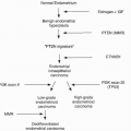
Endometrial cancer is the leading cause of gynecologic cancer in the United States, with approximately 40,000 cases diagnosed per year. Endometrial cancer follows only lung, breast, and colon cancers as a cause of cancer in women. It is the most common of the gynecologic cancers.1 Seventy-five percent of patients with endometrial cancer are postmenopausal, but 3% to 5% are younger than 40 years old.2 Management of endometrial cancer, whether surgical or medical, must take into consideration the histology of the cancer, the stage of the disease at presentation, and the patient’s comorbidities; in young women with early-stage endometrioid carcinoma, the patient’s desire for future fertility is also considered. Ninety percent of endometrial cancers arise in the endometrial glands as opposed to the endometrial stroma or myometrium.3 There are two types of endometrial cancers that are subdivided by histology. Type I accounts for 80% of endometrial cancer, is caused by hyperestrogenism, usually in the setting of obesity, and arises in a background of atypical hyperplasia. A European study reported that obesity is the cause of up to 39% of all endometrial cancers.4 Large amounts of adipose tissue aromatize estrogen, leading to increased estrogen effects on the uterus. Endometrial proliferation caused by estrogen may cause neoplastic change in the absence of progesterone, either endogenous or exogenous.4 Type II endometrial cancer develops in a population without a history of long-term unopposed estrogen, usually in nonobese women. Type II cancer occurs in the setting of an atrophic endometrium.4 Type I is usually low-grade endometrioid histology, whereas type II includes more aggressive clear cell and papillary serous cancers. Type I is almost always confined to the uterus, and outcomes of survival are considerably better than type II cancer, which has often metastasized prior to diagnosis even without evidence of extensive myometrial invasion.3

Endometrial cancer patients are typically obese and postmenopausal and are at high risk for cardiovascular disease and diabetes. Because the majority of patients diagnosed with endometrial cancer are older and have multiple associated comorbidities, surgical planning must consider issues of cardiac and pulmonary tolerance for major surgery, wound healing, and subsequent ability to tolerate adjuvant therapy, if needed.

HISTOLOGY AND CLINICAL CORRELATION


Endometrial Hyperplasia

Endometrial hyperplasia carries a reasonably high risk of progression to endometrial cancer if not adequately treated. Simple hyperplasia without cytologic atypia carries a less than 1% chance of progression to cancer, whereas complex hyperplasia without atypia has a 2% to 3% chance of progression to cancer. Thus, these two entities are usually treated medically using progesterone therapy. With cytologic atypia, simple hyperplasia and complex hyperplasia carry an 8% and 27% to 28% chance of progression to cancer, respectively. With cytologic atypia, hysterectomy is usually the treatment of choice unless future fertility is still desired.5

With a diagnosis of complex endometrial hyperplasia with atypia found on either in-office endometrial sampling or dilation and curettage, there is a significant risk of concurrent endometrial cancer at the time of hysterectomy. The Gynecologic Oncology Group (GOG), in a study with 302 patients diagnosed in the community with atypical endometrial hyperplasia (GOG Protocol 167A), found that there is poor reproducibility of the diagnosis when a panel of GOG pathologists reviewed the original biopsy slides. These patients, treated with hysterectomy within 12 weeks of study entry without other intervening treatments, had a 43% incidence of concurrent endometrial cancer.6 In a recent study using the extensive patient database of the Kaiser Permanente Group, Suh-Burgmann et al.7 found that there is a 48% incidence of endometrial cancer when the patient had a preoperative diagnosis of atypical endometrial hyperplasia.

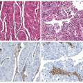
The high incidence of endometrial cancer when the patient undergoes hysterectomy for atypical endometrial hyperplasia underscores two dilemmas in diagnosis. First, it is often difficult to distinguish histologically between atypical endometrial hyperplasia and welldifferentiated endometrioid cancer. Second, typical office endometrial biopsy instruments sample only a small proportion of the entire uterine cavity, giving rise to the possibility of error due to sampling inadequacy8 Clinically, it is important to ascertain the diagnosis preoperatively because the majority of patients with atypical endometrial hyperplasia are treated surgically by general gynecologic surgeons who may not be trained to perform lymphadenectomy to complete surgical staging of an endometrial cancer.


Endometrioid Carcinoma

Preoperative grade of disease and depth of myometrial invasion are key factors in determination of extent of surgical staging (Tables 6-1 and 6-2). Patients with low-grade disease (grades 1 and 2) and minimal myometrial invasion have low risk (≤5%) of lymph nodal metastasis.9 Although pelvic and para-aortic lymphadenectomy to stage endometrial cancer is routinely performed by gynecologic oncologists, the procedure does have inherent risks of blood loss, infection associated with prolonging surgical time, injury to nearby nerves and vasculature, lymphocyst formation, and lymphedema. Surgery is more challenging due to the anatomic constraints of morbid obesity, a common characteristic of patients with endometrial cancer. Thus, the risk of complications due to the procedure must be weighed with the benefits of diagnosis of lymphadenopathy to diagnose and remove metastatic disease.

Although surgical staging has been advocated since 1988 with the inclusion of surgical criteria in the International Federation of Gynecology and Obstetrics (FIGO) staging guidelines, only 40% of patients in the United States are surgically staged. This dichotomy between
what is advocated and what is actually done is due to the previously stated fact that the majority of patients with endometrial cancer are treated by general gynecologic surgeons who are not trained in necessary techniques to complete surgical staging.10 In a multicenter study of 9,000 patients, only 30% of patients undergoing surgical treatment of endometrial cancer received lymph node assessment. In the same study, 28% of patients received postoperative radiation treatment despite lack of adequate staging information indicating the need for adjuvant radiation. Adequate surgical staging can identify as many as 20% of patients with extrauterine disease previously thought to have stage I disease based on clinical evaluation.11


Table 6-1 Histologic Grade and Depth of Invasion


Grade


Depth


G1 (%)


G2 (%)


G3 (%)


Total (%)


Endometrium only


44 (24)


31 (11)


11 (7)


86(14)


Superficial


96 (53)


131 (45)


54 (35)


281 (45)


Middle


22(12)


69 (24)


24 (16)


115 (19)


Deep


18 (10)


57 (20)


64 (42)


139 (22)


Total


180 (100)


288 (100)


153 (100)


621 (100)


Reprinted from Creasman WT, Morro CP, Bundy BN, et al. Surgical pathologic spread patterns of endometrial cancer. A Gynecologic Oncology Group study. Cancer. 1987;60(8 suppl):2035-2041.


Table 6-2 Frequency of Nodal Metastasis in Lymph Nodes Based on Uterine Risk Factors


Grade


Depth of Invasion


G1


G2


G3


(N = 180)


(N = 288)


(N = 153)


Pelvic Lymph Nodes


Endometrium only (N = 86)


0 (0%)


1 (3%)


0 (0%)


Inner (N = 281)


3 (3%)


7 (5%)


5 (9%)


Middle (N = 115)


0 (0%)


6 (9%)


1 (4%)


Deep (N = 139)


2 (11%)


11 (19%)


22 (34%)


Aortic Lymph Nodes


Endometrium only (N = 86)


0 (0%)


1 (3%)


0 (0%)


Inner (N = 281)


1 (1%)


5 (4%)


2 (4%)


Middle (N = 115)


1 (5%)


0 (0%)


0 (0%)


Deep (N =139)


1 (6%)


8(14%)


15 (23%)


Reprinted from Creasman WT, Morro CP, Bundy BN, et al. Surgical pathologic spread patterns of endometrial cancer. A Gynecologic Oncology Group study. Cancer. 1987;60(8 suppl):2035-2041.


Many gynecologic surgeons practice selective lymphadenectomy, believing that full lymph node dissection should be reserved for those at highest risk for metastatic disease. Data from the surgicopathologic study by the GOG (Protocol 33) suggest that uterine parameters including grade, depth of invasion, and cervical involvement may be used to determine the risk of lymph node involvement to decide whether or not to perform lymphadenectomy9 (see Table 6-2). Surgeons must decide for themselves their cutoff value for deciding to perform lymphadenectomy, taking into consideration the risk of positive lymph nodes, their own incidence and experience with complications, and the published risk of serious complications from the literature of less than 6%.12


High-Risk Histologies

Routine surgical staging for endometrial cancer, including total abdominal hysterectomy, bilateral salpingo-oophorectomy, and pelvic and para-aortic lymphadenectomy, may not be adequate for patients with high-risk histologies such as clear cell or serous carcinomas. Serous or clear cell histology carries a higher risk of positive lymph nodes.13 Chan et al.14 found that omentectomy may be needed to find the 25% of patients who present with stage IVB serous endometrial carcinoma. They advocate that patients with serous endometrial carcinoma undergo “extended” surgical staging similar to that done for patients with ovarian carcinoma, with the inclusion of omentectomy and peritoneal biopsies in addition to routine lymphadenectomy.


Carcinosarcoma

Carcinosarcoma (malignant mixed müllerian tumor) is histologically defined as having both malignant epithelial (carcinoma) as well as mesodermal (sarcoma) components. Many studies confirm that these cancers arise from a single clonal cell population and should be considered anaplastic carcinomas. Clinically, the behavior of the tumor is driven by the carcinomatous component of the cancer; most metastatic sites include only the carcinoma component. The carcinomatous components are more likely to appear more aggressive, invading into the myometrium or lymphovascular spaces.15 Clinical treatment for this cancer is typically geared toward treating the carcinomatous element.


FIGO STAGING FOR ENDOMETRIAL CANCER

In 2009, FIGO revised its staging criteria for endometrial cancer (Table 6-3). The new revision reflects improved understanding of the natural history of endometrial cancer and the survival and recurrence data obtained from decades of treating the disease. The changes may be summed up in several general statements. First, patients with minimally invasive cancer uniformly do well. Second, the prognosis of patients with cervical glandular involvement is dependent on other uterine factors (depth of invasion, grade, and the presence of lymphovascular invasion). Similarly, the presence of positive peritoneal cytology does not portend a bad prognosis without other histologic high-risk factors. Prior to 2009, uterine sarcoma (e.g., leiomyosarcoma), endometrial sarcoma, adenosarcomas, and carcinosarcomas have been staged using the same criteria as for endometrial carcinomas. In 2009, FIGO devised a separate staging guideline for sarcomas. Carcinosarcomas will continue to be staged using carcinoma guidelines.


Table 6-3 Revised 2009 FIGO Staging for Endometrial Carcinoma (changes from 1988 FIGO guidelines)


Only gold members can continue reading. Log In or Register to continue

Stay updated, free articles. Join our Telegram channel

May 22, 2016 | Posted by in ONCOLOGY | Comments Off on Current Management and Issues in Treatment of Endometrial Carcinoma

Full access? Get Clinical Tree

Get Clinical Tree app for offline access

Stage I*:Tumor confined to the corpus uteri


IA*: No or less than half myometrial invasion


IB*: Invasion equal to or more than half of the myometrium


(New guideline eliminates endometrium-confined cancer as a specific category.)


Stage II*: Tumor invades cervical stroma but does not extend beyond the uterus


(New guideline eliminates cervical glandular involvement as a specific category [stage IIA] in the 1988 guideline.)