CLINICAL SIGNS AND SYMPTOMS OF ABNORMAL SERUM PHOSPHATE LEVELS
A wide variety of diseases and syndromes with varying clinical manifestations have the characteristic biochemical abnormalities of hyperphosphatemia or hypophosphatemia. Further-more, a unique complex of disturbances often is directly related to the abnormal phosphate homeostasis. The recognition of these signs and symptoms may lead to appropriate biochemical testing, the diagnosis of an unsuspected disease, and initiation of lifesaving or curative treatment.
HYPERPHOSPHATEMIA
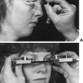
Hypocalcemia and consequent tetany are the most serious clinical sequelae of hyperphosphatemia.63 The decreased serum calcium concentration results from the deposition of calcium–phosphate salts in soft tissue, a process that may lead to symptomatic ectopic calcification. The dystrophic calcification is frequently seen in acute and chronic renal failure, hypoparathyroidism, pseudohypopara-thyroidism, and tumoral calcinosis. Indeed, deposition of calcium–phosphate complexes in the kidney may predispose a patient to acute renal failure. When the calcium–phosphate product exceeds 70, the probability that soft-tissue calcification will occur increases sharply. In addition, local factors, such as tissue pH and injury (e.g., necrotic or hypoxic tissue), may predispose the patient to precipitation of the calcium–phosphate salts. In chronic renal failure, calcification occurs in arteries, muscle tissue, periarticular spaces, myocardial conduction system, lungs, and kidney. Affected patients may also have ocular calcification, causing the “red eye” syndrome of uremia, and subcutaneous calcification, which also plays a role in uremic pruritus. Alternatively, a predisposition to calcification of periarticular surfaces of the hips, elbows, shoulders, and other large joints occurs in tumoral calcinosis.
In some disease states, hyperphosphatemia may also play an important role in the development of secondary hyperparathyroidism.27 A decrement in the serum calcium concentration secondary to hyperphosphatemia stimulates the release of PTH. Furthermore, hyperphosphatemia decreases the activity of renal 25(OH)D-1α-hydroxylase. The consequent diminished production of 1,25(OH)2D3 impairs the gastrointestinal absorption of calcium and induces skeletal resistance to PTH, which are influences that augment the development of hyperparathyroidism.
Thus, hyperphosphatemia triggers a cascade of events that have an impact on calcium homeostasis at multiple sites. The prevention of secondary hyperparathyroidism, metabolic bone disease, and soft-tissue and vascular calcification in affected patients, therefore, depends on ultimately controlling the serum phosphate concentration.
HYPOPHOSPHATEMIA
Related posts:

Stay updated, free articles. Join our Telegram channel

Full access? Get Clinical Tree


