Clinical and Pathologic Prognostic and Predictive Factors


Clinical and Pathologic Prognostic and Predictive Factors


Frederick L. Moffat


Prognostic and predictive factors and biomarkers are critical to clinical decision-making in oncology. A 1991 NIH Consensus Conference (1) stipulated that clinically useful prognostic and predictive factors in breast cancer must meet the following criteria:


  • They must provide significant, independent predictive value, validated by clinical testing.


  • Their identification and measurement must be feasible, reproducible, and widely available with quality control.


  • The results must be readily interpretable by clinicians and have therapeutic implications.


  • Measurement of biomarkers should not consume tissue needed for other tests, especially routine histopathological evaluation.

Prognostic markers provide information on the biological potential and most likely clinical course of a breast cancer irrespective of treatment (2, 3). Insight into the natural history of individual breast cancers may provide valuable information regarding the need for systemic adjuvant therapy, but is uninformative with respect to which specific treatment regimen is most likely to be effective.

Predictive factors inform on the likelihood of response of a breast cancer to specific therapies (3). Hormone receptor status predicts the responsiveness or lack of same of a breast cancer to endocrine therapy.

Some tumor biomarkers are of mixed significance. Estrogen receptor expression, while a strong predictor of response to endocrine therapy, is only weakly prognostic. HER2 expression has highly adverse prognostic implications but is predictive of tumor response to anti-HER2 therapy.

Conventional clinicopathological factors such as patient age, menopausal status, race/ethnicity, tumor size, nodal status, lymphovascular invasion, micrometastases or isolated tumor cells in regional lymph nodes, extracapsular extension of nodal metastases, tumor grade, tumor stage, presence or absence of the inflammatory phenotype, markers of tumor proliferation, and hormone receptor and HER2 status continue to be useful in estimating prognosis. The prognostic implications of the intrinsic breast cancer subtypes (4, 5), multigene tumor signature assays (6), and clinicopathological response to neoadjuvant systemic therapy (7) offer further opportunities for refinement of clinical decision making in breast cancer.

Numerous potential breast cancer biomarkers have been cited and characterized over the past several decades. Discerning their true magnitude of effect, reliability and clinical utility has been complicated by deficiencies in biomarker assays and measurement, the quality of evidence supporting the potential biomarker status of the factor(s) under study, and failures in clinical trials design and studied patient cohorts and populations to account for and control confounding variables. A number of expert panels have reviewed available information on breast cancer biomarkers and concluded that limitations in available data allow for only the most guarded recommendations (8). For these reasons, significant efforts have been directed toward standardizing the investigation and establishment of clinically relevant biomarkers.


GENERAL CONSIDERATIONS

For prognostic and predictive factors to be clinically useful, they must be detectable and reproducibly measurable by different laboratories at reasonable cost, yielding results promptly for clinical decision making. Their clinical correlations must be clearly defined in terms of their nature (prognostic, predictive, or both), and assay values, whether continuous or categorical, must be reliably associated with patient outcomes. The relevant clinical information being sought must not be available through another more readily accessible factor. The expected differences in outcomes must be significant and important from the patient’s perspective. Finally, useful prognostic and predictive factors must provide information upon which a choice among available treatment options can be based (9).


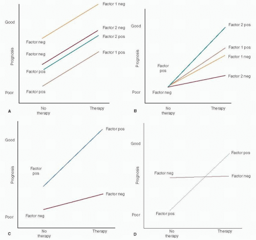
FIGURE 28-1 Schematic representation of prognostic and predictive factors: prognosis versus therapy as binomial variables. (A) Pure prognostic factor. (B) Pure predictive factor. (C) Mixed factor with weakly favorable prognostic effect and strong response to therapy. (D) Mixed factor with unfavorable prognosis and strong response to therapy. (Adapted from Henry NL, Hayes DF. Uses and abuses of tumor markers in the diagnosis, monitoring and treatment of primary and metastatic breast cancer. Oncologist 2006;11:541-552. Modified from Hayes DF, Trock B, Harris AL. Assessing the clinical impact of prognostic factors: when is “statistically significant” clinically useful? Breast Cancer Res Treat 1998;52:305-319, Springer Science and Business Media.)

Pure prognostic and predictive factors are schematically depicted in Figure 28-1, panels A and B, respectively.
Prognosis versus therapy are plotted as binomial variables (8, 10). A large incremental difference in prognosis related to positive and negative status is observed for factor 1 (a strong prognostic factor such as lymph node status) while that for factor 2 is much smaller (ER status). In panel B, factor 1 is a weak predictive factor while factor 2 is a much stronger one. Hayes et al. (10) proposed that prognostic factors in breast cancer be categorized quantitatively by their associated hazard ratios (HR), HR <1.5 denoting weak factors, 1.5 to 2.0 moderate factors, and >2.0 strong factors. They further proposed a similar rating of the strength of predictive factors by tumor response to and clinical benefit from a specific therapy. “Relative predictive value” (RPV), the ratio of the probability of response to treatment in a factor-positive patient as compared to that in a factor-negative patient, has been proposed as a means of quantifying the strength of predictive factors as weak (RPV = 1 — 2), moderate (RPV = 2 — 4), or strong (RPV > 4).

Panel C in Figure 28-1 represents a mixed prognostic and predictive factor such as ER status, the prognostic effect being weakly favorable and the predictive effect strongly so. Panel D depicts a mixed factor with an unfavorable prognosis but highly responsive to specific therapy, as exemplified by HER2 status.

Statistical significance in marker-positive or marker-negative patient outcomes is not infrequently mistaken for or conflated as evidence of clinical utility. This does not always hold and should never be assumed. Along with the magnitude of the effect and the relevance of the marker, technical reliability and reproducibility are critically important, as is the design and execution of clinical studies (10).

Technical shortcomings related to biomarker assay sensitivity, specificity, reproducibility, and reagent variability can be highly problematic. Standardization of assay methodologies has greatly improved, of late. For example, intra- and interobserver variation, well documented in immunohistochemical assays, has been controlled through automated and semiautomated processes (8).

Determination of cut-off points that distinguish positive from negative results is critical to the development of clinically useful assays. Cut-off points can be set arbitrarily or based on data. Arbitrary cut-off point selection has included the limits of detection of the assay, two standard deviations above the normal mean, the mean value in affected as compared to normal patients, or an arbitrarily defined appropriate percentage of positive cells (8).

Data-derived cut-off points have been based on plots of p-values versus outcomes, plots of the magnitude of marker effect versus patient outcome, construction of receiver operating characteristic curves (cut-off points established by determining the optimal trade-offs of sensitivity and specificity in an assay), or subpopulation treatment effect pattern plot (STEPP) analysis (11). The latter methodology evaluates outcomes to specific treatment regimens in subpopulations of patients within randomized trials or meta-analyses (8).

Once established in a test group of patients, cut-off points must be confirmed in a validation patient cohort similar to but completely independent of the initial test group. Having been identified and validated, the clinical value of a new tumor marker relative to well-established prognostic or predictive factors is then confirmed by multivariate analysis. This provides information on the potential clinical utility of the new marker in medical practice.

Study design is key to identifying and establishing new tumor biomarkers. The Tumor Marker Utility Grading System (TMUGS) (12) was developed as a frame of reference for grading the clinical utility of tumor markers based on published evidence. Putative markers are assigned a utility score according to degree of correlation with biological processes and end points (Table 28-1) and favorable clinical outcomes (Table 28-2), as determined by level of evidence (LOE; Table 28-3) (8) and grading of tumor marker studies (Table 28-4) (13).


TABLE 28-1 Scale to Evaluate Tumor Marker for Correlation with Biological Processes and End Points


Utility Scale


Explanation


0


Does not correlate with process or expected end point


NA


Data not available on marker correlation with process or end point for that use


+/-


Preliminary data suggestive, but substantially more definitive studies required


+


Assay probably associated with process or end point, but confirmatory studies required


++


Definitive studies show that assay reflects process or end point


From Hayes DF, Bast RC, Desch CE, et al. Tumor marker utility grading system: a framework to evaluate clinical utility of tumor markers. J Natl Cancer Inst 1996;88:1456-1466.


LOE levels I and II are the most robust and objective, the ideal level I clinical trial being prospective, randomized, appropriately powered, and designed specifically to evaluate the clinical utility of a putative tumor marker for a discrete, predetermined use. That noted, a clinical trial adequately powered to determine a clinical end point may be underpowered for analysis of tumor marker subgroups by as much as 25%, even when tissue samples are available for all participating patients (8).

Systematic overviews and pooled analyses of wellconducted LOE II studies are essentially equivalent to LOE I evidence. LOE III studies, with their greater variability in patient characteristics and therapies, are better suited to generating hypotheses than contributing clinically useful information (8).

Attention to detail is critical to clinical trials design and conduct. The appropriate patient population must be selected with particular attention to a similar profile among them in terms of known prognostic factors. Trials focused on predictive factors are ideally prospective, randomized, and controlled, comparing patients receiving the intervention in question to untreated controls (8).

The REMARK tool, a standardized reporting schema for tumor marker data, has been developed by the Working Group of the National Cancer Institute and the European Organization for Research and Treatment of Cancer (EORTC) (14). This project was undertaken to eliminate the highly variable and flawed approaches to tumor marker elucidation and to provide a standard template for investigation of potential markers in the future.


PROGNOSTIC FACTORS—CLINICAL


Age

Breast cancer patients aged 35 to 40 years or less at presentation have a significantly worse prognosis than older premenopausal patients or those over the age of 50. Chemotherapy is especially effective in premenopausal patients. However, those under 35 years of age receiving chemotherapy for endocrine-responsive breast cancer have a significantly higher risk of relapse than older premenopausal patients with such tumors. In contrast, outcomes among younger and older premenopausal patients receiving chemotherapy for endocrine nonresponsive disease are essentially equivalent (15).


TABLE 28-2 Scale to Evaluate Utility of Tumor Markers for Favorable Clinical Outcomes


Utility Scale


Explanation


0


Marker adequately evaluated for specific use; data definitively demonstrate no utility.


Marker should not be ordered for that clinical use.


NA


Data not available for the marker for that use because marker has not been studied for that use.


+/-


Data are suggestive that marker may correlate with biological processes and/or end points, and preliminary data suggest that use of the marker may contribute to favorable clinical outcome, but more definitive studies are required. Thus, marker is still considered highly investigational and should not be used for standard clinical practice.


+


Sufficient data available to demonstrate that marker correlates with the biological process and/or biological end point related to its use and that the marker might affect favorable clinical outcome for that use. However, marker still considered investigational and should not be used in standard clinical practice for one of three reasons:


  1. The marker correlates with another marker/test that has been established to have clinical utility, but the new marker has not been shown to clearly provide an advantage.


  2. The marker may contribute independent information but it is unclear whether the information provides clinical utility because treatment options have not been shown to change outcome.


  3. Preliminary data for the marker are quite encouraging, but the level of evidence is lacking to document clinical utility.


++


Marker supplies information not otherwise available from other measures that is helpful to the clinician in decision making for that use, but the marker cannot be used as the sole criterion for decision making. Thus, marker has clinical utility for that use, and it should be considered standard practice in selected situations.


+++


Marker can be used as the sole criterion for clinical decision making in that use. Thus, marker has clinical utility for that use, and it should be considered standard practice.


From Hayes DF, Bast RC, Desch CE, et al. Tumor marker utility grading system: a framework to evaluate clinical utility of tumor markers. J Natl Cancer Inst 1996;88:1456-1466.


TABLE 28-3 Levels of Evidence for Grading Clinical Utility of Tumor Markers


Level


Type of Evidence


I


Evidence from a single, high-powered, prospective controlled study specifically designed to test marker or evidence from meta-analysis and/or overview of level II and III studies. In the former case the study must be designed so that therapy and follow-up are dictated by protocol. Ideally, the study is a prospective controlled randomized trial in which diagnostic and/or therapeutic clinical decisions in one arm are determined at least in part on the basis of marker results, and diagnostic and/or therapeutic clinical decisions in the control arm are made independently of marker results. However, study design may also include prospective but not randomized trials with marker data and clinical outcome as primary objective.


II


Evidence from study in which marker data are determined in relationship to prospective therapeutic trial that is performed to test therapeutic hypothesis but not specifically designed to test marker utility (i.e., marker study is a secondary objective of the protocol).


III


Evidence from large studies from which variable numbers of samples are available or selected. Therapeutic aspects and follow-up of the patient population may or may not have been prospectively dictated. Statistical analysis for tumor marker was not dictated prospectively at the time of therapeutic trial design.


IV


Evidence from small retrospective studies which do have prospectively dictated therapy, follow-up, specimen selection, or statistical analysis. Study may use matched case-controls, etc.


V


Evidence from small pilot studies designed to determine or estimate distribution of marker levels in the sample population. Study design may include “correlations” with other known or investigational markers of outcome but is not designed to determine clinical utility.


From Hayes DF, Bast RC, Desch CE, et al. Tumor marker utility grading system: a framework to evaluate clinical utility of tumor markers. J Natl Cancer Inst 1996;88:1456-1466.


TABLE 28-4 Grade of Tumor Marker Studies for Level of Evidence


Grade


Study Description


A


Prospective


B


Prospective, using archived samples


C


Prospective, observational


D


Retrospective, observational


Level of Evidence


Grade


Validation Studies Available


I


A


None required


I


B


One or more with consistent results


II


B


None or inconsistent results


II


C


Two or more, consistent results


III


C


None or one, consistent or inconsistent results


IV-V


D


Not applicable: LOE IV and V unsatisfactory for determination of biomarker clinical utility


From Simon RM, Paik S, Hayes DF. Use of archived specimens in evaluation of prognostic and predictive biomarkers. J Natl Cancer Inst 2009;101:1446-1452.


In another meta-analysis (16), higher mortality (HR = 1.55; 95% confidence interval [CI] 1.20-2.00) and locoregional recurrence (HR = 2.34; 1.30-4.24) were observed in patients less than 35 years old as compared to those over 50. Mortality in patients aged 35 to 50 years as compared to those over 50 was no different (HR = 1.01; 0.87-1.16) but locoregional recurrence was more frequent (HR = 1.60; 1.14-2.25).

A SEER analysis of 243,012 breast cancer patients (17) reported that those less than 40 years of age (6.4% of the cohort) were more likely to be African American, single, to have presented with advanced disease, and to have undergone total mastectomy. Their tumors were of higher grade, larger size, and more often estrogen receptor-negative (-), progesterone receptor-negative (PR-), and lymph node-positive. The adjusted HR for mortality among younger women was significantly higher as compared to older patients overall (1.39; 1.34-1.45). For stage I disease, HR for mortality was 1.44 (1.27-1.64) and for stage II breast cancer, 1.09 (1.03-1.15). Among patients with stage IV disease, however, the mortality ratio for younger patients, at 0.85 (0.76-0.95), was significantly lower than for those over 40 years of age.

At 11 years median follow-up, a recent EORTC pooled analysis (18) reported that among patients less than 40 years of age, tumor size, nodal status, and intrinsic molecular subtype were independent prognostic factors for overall survival (OS). Among node-negative patients less than 40, only intrinsic subtype was significant. Ten-year survival among patients with luminal A tumors was 94% as compared to 72% for those with basaloid tumors. In a similar analysis of 315 patients less than 35 years of age (19), the excess risk of recurrence (HR = 1.65; 1.30-2.10) and mortality (HR = 1.78; 1.12-2.85) as compared to older patients was significant. Young patients with luminal B, triple-negative or HER2+ tumors were at particular disadvantage with respect to cancer recurrence and mortality.


Menopausal Status

Menopausal status may be a prognostic proxy for age as implied in the foregoing discussion. That noted, the time course of breast cancer recurrence varies as a function of menopausal status (20). Among node-positive premenopausal patients, the hazard function for relapse has two peaks, the first reaching its maximum 8 to 10 months postoperatively and the second at 28 to 30 months. In contrast, the hazard function in node-positive postmenopausal patients is significantly prolonged, peaking at 18 to 20 months. Primary tumor size correlates directly with the height of the hazard peaks in both pre- and postmenopausal patients, but has no effect on time to recurrence. In node-negative patients, the hazard function for recurrence increases to 18 to 24 months, decreasing somewhat thereafter but of much reduced amplitude at all time-points as compared to patients with positive nodes.


Race/Ethnicity

Five-year relative OS for African American breast cancer patients from 1988 to 2001 was 78% as compared to 90% for Caucasians (21). Stage distribution is less favorable among African Americans, but this factor alone does not explain the observed differences in outcomes. Treatment response rates are similar for African Americans and Caucasians, but African Americans are more likely to present with high-grade and triple-negative cancers and at a younger age. Moreover, there is an excess incidence of ER- inflammatory breast cancer in young African American patients (22).

In China, 20% of breast cancer patients are younger than 40 years of age as compared to only 6% of Caucasian patients in the United States. Moreover, the nonluminal HER2+ subtype with its earlier age of onset, poorer prognosis, and more advanced stage at presentation accounts for 26% to 31% of cases in China as compared to only 19% to 23% in Caucasian Americans (23).


Clinical Tumor Size

Clinical and radiographic estimates of primary tumor size tend to overstate the true dimensions of primary invasive breast cancers, especially small lesions, because of tumor-associated desmoplasia and in situ disease (24, 25).
The pathological dimensions of the invasive component are the accepted standard for determining primary tumor stage.


Clinical Stage

Clinical and pathological stage are critically important in treatment selection and outcomes. At present, the relevance of clinical staging relates primarily to locoregionally advanced disease presenting as a large primary breast tumor with or without one or more of the so-called grave signs (necrotic, fungating, and/or ulcerating tumor eroding through the breast skin with or without localized reactive cutaneous inflammation, peau d’orange, tumor invasion of the chest wall, and/or bulky nodal disease in axillary, internal mammary, and supraclavicular lymphatic basins). The most feared clinical presentation by far is inflammatory/T4d breast cancer with its sudden onset and rapid progression, often attended by bulky, fixed, or confluent disease in one or more nodal basins. Detectable distant metastases are present in 40% of these patients at the time of diagnosis (22, 26). The inflammatory phenotype, historically a harbinger of profoundly aggressive cancer biology and impending mortality, retains its grim prognostic implications even now, at least in relative terms.

Locoregionally advanced breast cancers are not infrequently unresectable or only marginally operable at presentation. These and inflammatory cancers remain the preeminent indications for neoadjuvant systemic therapy (27).


PROGNOSTIC FACTORS—PATHOLOGIC

In an overview of systematic reviews and meta-analyses published from 1999 through 2007 (28), the American Society of Clinical Oncology (ASCO) updated recommendations on breast cancer tumor markers. The data regarding DNA flow cytometric parameters were insufficient to impute any prognostic value to their routine use. Data on markers of tumor proliferation such as Ki-67, cyclin D, cyclin E (whole or fragments), p27, p21, thymidine kinase, topoisomerase IIα, and others were likewise inadequate to establish prognostic significance.

Only gold members can continue reading. Log In or Register to continue

Stay updated, free articles. Join our Telegram channel

Jul 9, 2016 | Posted by in ONCOLOGY | Comments Off on Clinical and Pathologic Prognostic and Predictive Factors

Full access? Get Clinical Tree

Get Clinical Tree app for offline access