Fig. 6.1
Multiple RICH with prenatal diagnosis on MRI. Two arrows are remarking the presence of 2 RICH on MRI
In some instances differential diagnosis with other congenital tumors is needed (Fig. 6.2).
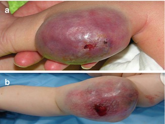
Fig. 6.2
Macroscopic similarity between RICH (a) and fibrosarcoma (b)
RICH differs from rapidly involuting congenital hemangiomas because they do not undergo a postnatal involuting phase. They are mainly located on the head or neck and limbs. In contrast to RICH, prenatal color Doppler ultrasound or MRI follow-up during pregnancy rarely leads to the detection of NICH [7].
PICH are congenital hemangiomas with a distinct behavior, evolving from RICH to persistent NICH-like lesions [8].
Tufted Angioma (TA)
TA is an uncommon vascular tumor that usually has its onset during infancy or early childhood but rarely can be congenital.
They had already been recognized half a century earlier by Japanese authors who referred to the tumors under the name of Nakagawa’s angioblastoma. Various presentations have been described, including solitary tumors, large infiltrated plaques sometimes having increased lanugo hair, and “port-wine stain-like” areas with a cobblestone surface. The appearance of the lesions changes with time and they are sensitive upon palpitation or even painful if subjected to trauma. Tufted angiomas may be associated with Kasabach-Merritt syndrome (KMS), in which thrombocytopenia can be life-threatening [9].
Kaposiform Hemangioendothelioma (KHE)
KHE is a rare and locally aggressive vascular tumor that can occur in the skin, pleura, and retroperitoneum and may be associated with severe thrombopenia with consumption coagulopathy (Kasabach-Merritt syndrome) in pediatric patients. The use of the term “kaposiform” relates to its unmistakable resemblance to Kaposi’s sarcoma, assumed by the compact spindled tumor cells characterized by the formation of slit-like lumen. KHE and TA have been viewed as two separate disease entities in past classifications of vascular tumors. However, the clinical similarities with TA, the association of both conditions with Kasabach-Merritt phenomenon, and the histological features of these syndromes have led to the suggestion that kaposiform hemangioendothelioma and tufted angioma are part of the same clinical spectrum of vascular tumors. In the largest reported case series of 107 patients, 71 % developed KMP (11 % of patients lacked cutaneous findings). Retroperitoneal and intrathoracic lesions, though less common, were complicated by KMP in 85 and 100 % of cases, respectively [10]. Current multidisciplinary management protocols have dramatically reduced mortality rates [11].
Spindle Cell Hemangioma (SCH)
SCH is a rare, benign vascular tumor of the dermis and subcutis. The lesions can be multifocal and are overrepresented in Maffucci syndrome, in which patients also have multiple enchondromas. Somatic mosaic R132C IDH1 hotspot mutations were recently identified in Maffucci syndrome (71 % SCHs harbored mutations in exon 4 of IDH1 or IDH2). Although originally spindle cell hemangioendothelioma was proposed as a specific clinicopathologic variant of hemangioendothelioma, currently, it is considered as an entirely benign lesion, and thus, the name spindle cell hemangioma seems to be the most accurate for this lesion [12].
Pyogenic Granuloma (PG)
Pyogenic granuloma is a condition usually occurring in the skin or mucosa and often related to prior local trauma or pregnancy. However, the etiopathogenesis of pyogenic granuloma is poorly understood, and whether pyogenic granuloma is a reactive process or a tumor is unknown. Pyogenic granuloma seems to be a reactive lesion resulting from tissue injury, followed by an impaired wound healing response, during which vascular growth is driven by FLT4 and the nitric oxide pathway.
Retiform Hemangioendothelioma (RH)
RH is a very rare tumor of the blood vessels that occurs mainly in the limbs of young adults.
RH mostly occurs as an asymptomatic, slow-growing single lesion. Most cases are present as exophytic, dermal, or subcutaneous nodules or plaques with a size range of 1–30 cm. Although metastasis or malignancy is rare, RH is known to recur in approximately 50 % of cases.
The blood vessels are arranged in an anastomosing pattern that resembles that of the rete testis and lined with hobnail endothelial cells with focal papillary projections. The endothelial cells reveal enlarged nuclei with vesicular chromatin and rare mitosis. Some lymphocytic infiltrate is observed. Immunohistochemically, the endothelial cells are diffusely positive for factor VIII-related antigen [12].
Due to the tendency for recurrence, surgical excision must be performed under histopathologically tumor-free margins (Fig. 6.3).
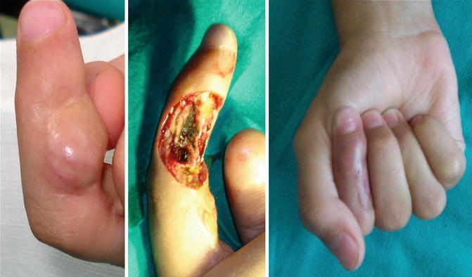
Fig. 6.3
Recurrence of retiform hemangioendothelioma needing radical excision
Papillary Intralymphatic Angioendothelioma (PILA, Dabska Tumor)
PILA is a locally aggressive, rarely metastasizing vascular lesion characterized by lymphatic and vascular-like channels and papillary endothelial proliferation. The tumor is extremely rare and often affects the skin and subcutaneous tissues of children. The clinical appearance is variable. However, in general, it presents as a slow-growing, usually intradermal nodule that grows up to 2–3 cm in diameter. Symptoms of pain can be present. Microscopically, the cuboidal or hobnail endothelial cells lining the vascular structures are characterized by a high nuclear cytoplasmic ratio and an apically placed nucleus that produces a surface bulge, accounting for the term “hobnail.” A growing number of cases described in the literature and the accumulating knowledge reveal that presence of highly specific lymphatic endothelial marker D2-40 and tumor marker vascular endothelial cell growth factor receptor type 3 (VEGFR3) suggests the tumor’s lymphatic origin.
Related posts:

Stay updated, free articles. Join our Telegram channel

Full access? Get Clinical Tree


