Chemotherapy of Multiple Myeloma


Chemotherapy of Multiple Myeloma


Robert A. Kyle

S. Vincent Rajkumar

Francis Buadi


Multiple myeloma (MM) is a plasma cell malignancy that accounts for slightly >10% of the hematologic malignancies. The historical overview of MM and the evolution of treatment for this disease has been reviewed recently.1 The annual incidence, age adjusted to the 2000 US population, is 4.3 cases per 100,000.2 The age-adjusted incidence has also remained stable in Malmo, Sweden, for the past 55 years, but the proportion of MM in persons ≥80 years of age has increased.3 The reported increased number of new cases during the past several decades is probably related to the increased availability of medical facilities for elderly patients and to improved diagnostic techniques rather than to an actual increased incidence. MM is twice as common in African-Americans as it is in the white population and is slightly more common in men than in women. The median age of diagnosis is 66 years and only 2% are younger than 40 years at diagnosis.4

MM evolves from monoclonal gammopathy of undetermined significance (MGUS) virtually in all patients. Ninety-five percent of MM patients had MGUS 5 years before diagnosis, whereas 82% had MGUS 8 years before the diagnosis of MM. MGUS was present in 100% of 71 patients 2 years before the diagnosis of MM.5


DIAGNOSTIC CRITERIA

The diagnosis of MM requires the presence of clonal bone marrow plasma cells, serum and/or urinary monoclonal (M protein), and evidence of end organ damage that is because of the plasma cell proliferative disorder (CRAB—hypercalcemia, renal insufficiency, anemia, and/or bone lesions).6 Anemia is present in 70% of patients at diagnosis and develops in almost all of the remainder during the course of the disease.

Hypercalcemia is found in one-fourth of patients at diagnosis, while the serum creatinine is elevated in almost one-half. Skeletal abnormalities are seen with conventional radiography in approximately 80% of patients at diagnosis. Approximately 80% of patients have a protein spike on serum protein electrophoresis, whereas immunofixation reveals a monoclonal protein in slightly >90%. In 15% to 20% of patients no heavy chain expression is found, and these patients are considered to have light chain MM. The M protein in these patients is almost always detected in the urine. Three percent of MM patients have no detectable M protein in serum or urine and are considered to have nonsecretory MM. The serum free light chain (FLC) assay is abnormal in about 70% of the so-called nonsecretory MM. Usually the bone marrow contains 10% or more clonal plasma cells.


DIFFERENTIAL DIAGNOSIS

MGUS, smoldering (asymptomatic) multiple myeloma (SMM), primary amyloidosis (AL) and solitary plasmacytoma are the most important entities in the differential diagnosis.

MGUS is found in 3% of persons ≥50 years of age and in 5% of those ≥70 years of age.7 It is characterized by the presence of an M protein <3 g per dl and a bone marrow containing <10% clonal plasma cells and no evidence of end organ damage. Approximately 1% of MGUS patients progress to MM, AL, Waldenstrom’s macroglobulinemia, or a related disorder each year.8 MGUS in the presence of postmenopausal osteoporosis, renal insufficiency from another cause such as diabetes or hypertension, or hypercalcemia because of hyperparathyroidism can complicate the differential diagnosis. Moreover, nearly one-half of women ≥ 60 years of age have osteopenia and a number of these have a vertebral compression fracture. In order to diagnose myeloma, the end organ damage must be felt related to the plasma cell proliferative process. The CT-PET scan of the spine may help in differentiating between postmenopausal osteoporosis and myelomatous bone disease. A renal biopsy may be needed in some patients in whom the etiology of renal dysfunction is not clear.

SMM must also be differentiated from MM. It is characterized by an M protein ≥3 g per dl and/or ≥10% bone marrow plasma cells but no evidence of end organ damage.9 In these patients, symptomatic MM develops at a rate of 10% per year for the first 5 years of follow-up. The risk of progression decreases to 3% per year for the next 5 years and then falls to 1% to 2% each year.

AL is a rare disorder that is characterized by the deposition of amyloid fibrils, which are composed of monoclonal immunoglobulin light chains. AL should be suspected in a patient with an M protein in the serum or urine and nephrotic range proteinuria with or without renal insufficiency, cardiomyopathy, hepatomegaly, or autonomic or peripheral neuropathy. Amyloid produces an apple-green birefringence under polarized light when utilizing a Congo red stain. The type of amyloid is most accurately determined by laser microdissection of Congo red staining tissue from the biopsy of an involved organ and then subjected to mass spectrometry.10


RISK STRATIFICATION OF NEWLY DIAGNOSED MM

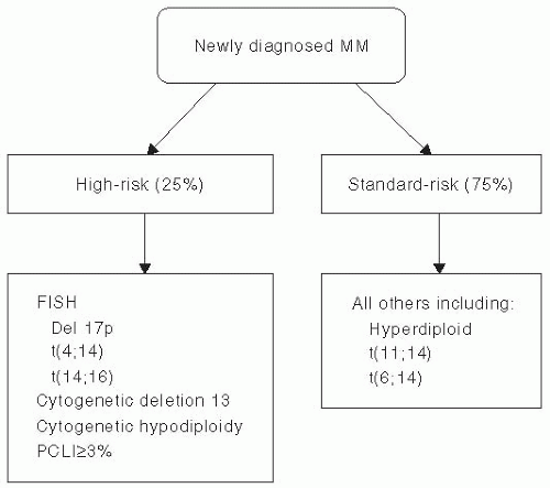
Patients with symptomatic MM may be classified into high-risk or standard-risk disease.11 We utilize a risk-stratification system termed mSMART (Mayo Stratification of Myeloma and
Risk-Adapted Therapy), which is based upon fluorescence in situ hybridization (FISH), metaphase cytogenetics, and the plasma cell labeling index (PCLI) (Fig 47.1). High-risk disease accounts for approximately 25% of MM patients and is characterized by del 17 p, t(4;14) or t(14;16) by FISH, deletion of chromosome 13, or hypodiploid by metaphase cytogenetics, or PCLI ≥3% (Table 47-1). Patients who have none of the high-risk features are classified as standard risk MM. Most standard risk MM patients have hyperdiploidy (which is found in 50%-60% of all myeloma patients), t(11;14) or t(6;14) by FISH studies. In addition to the risk factors listed above, lactate dehydrogenase and β2-microglobulin levels are additional important risk factors.

We strongly recommend clinical trials, but if the patient is not eligible or if clinical trials are not available, one may separate the patients into those who are autologous stem cell transplantation (ASCT) eligible or ineligible. Criteria for diagnosis, staging, risk stratification, and response assessment of MM have been described in detail.12 The treatment of newly diagnosed MM has been discussed in detail.13 Table 47-2 lists the most commonly used chemotherapy regimens for the treatment of myeloma.


INITIAL THERAPY FOR TRANSPLANT ELIGIBLE PATIENTS

Eligibility for ASCT varies from country to country. In general, ASCT is offered to patients <65 years of age, whereas in the United States decisions are made depending upon “physiologic age.” Patients aged >70 years, serum creatinine >2.5 mg per dl, and ECOG performance status grade 3 or 4 or New York Heart Association Functional Status class III or IV are generally ineligible for ASCT. Patients with renal failure may undergo transplantation, but the morbidity and mortality is higher.14


Figure 47.1 Myeloma Risk Stratification based on cytogenetic abnormalities and proliferative rate.

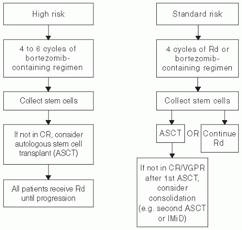
Those deemed to be eligible for ASCT receive four cycles of induction chemotherapy before ASCT (Fig 47.2). Alkylating agents such as melphalan (Alkeran) must be avoided before stem cell collection because they may impair mobilization of sufficient stem cells for a successful transplant. Most use granulocyte colony-stimulating factor (GCSF) with or without cyclophosphamide for stem cell collection. Plerixafor is also useful for mobilization of hematopoietic stem cells.15 One should collect enough stem cells for two transplants. Most of the currently accepted induction regimens contain an immunomodulatory drug (thalidomide or lenalidomide) and/or a proteasome inhibitor (bortezomib) in addition to corticosteroids.

We prefer lenalidomide 25 mg daily on days 1 to 21 plus dexamethasone 40 mg given weekly for each 28-day cycle as initial therapy for patients with standard risk disease.16 The addition of clarithromycin to lenalidomide and weekly dexamethasone is another option that produces an objective response in 90% of patients.17

Thalidomide plus dexamethasone is another active regimen. However, in a case-control retrospective study of 411 newly diagnosed patients with ≥partial response to lenalidomide plus dexamethasone (len/dex) or thalidomide plus dexamethasone (thal/dex), a partial response or better was achieved in 80% versus 61%, respectively. A very good partial response (VGPR), 34% versus 12%, was noted. Progression-free survival (PFS) (27 vs. 17 months) and overall survival (OS), not reached versus 57 months, favored len/dex.18


Table 47-1 Mayo Clinic Criteria for High-Risk Multiple Myeloma


High-risk Characteristic


Percentage of Newly Diagnosed Patients with the Abnormality (%)


Conventional cytogenetics


Deletion of chromosome 13 (monosomy)


14


Hypodiploidy


9


Either hypodiploidy or deletion 13


17


Fluorescent in situ hybridization (FISH)


t(4;14)


15


t(14;16)


5


17p-


10


Plasma cell labeling index (PCLI) studies: PCLI ≥3%


6


Any one of the above high-risk abnormalities


25-30


Reproduced with permission from Rajkumar SV, Kyle RA. Multiple myeloma: diagnosis and treatment. Mayo Clin Proc. 2005;80(10):1371-1382.


Table 47-2 Major Treatment Regimens in Multiple Myeloma


Regimen


Authors Preferred Dosing Schedulea


Melphalan-Prednisone (7-d schedule)


Melphalan 8-10 mg oral days 1-7


Prednisone 60 mg/d oral days 1-7


Repeated every 6 wk


Thalidomide-Dexamethasoneb


Thalidomide 200 mg oral days 1-28


Dexamethasone 40 mg oral days 1, 8, 15, 22


Repeated every 4 wk


Lenalidomide-Dexamethasone


Lenalidomide 25 mg oral days 1-21 every 28 d


Dexamethasone 40 mg oral days 1, 8, 15, 22 every 28 d


Repeated every 4 wk


Bortezomib-Dexb


Bortezomib 1.3 mg/m2 intravenous days 1, 8, 15, 22


Dexamethasone 20 mg on day of and day after bortezomib (or 40 mg d 1, 8, 15, 22)


Repeated every 4 wk


Melphalan-Prednisone-Thalidomide


Melphalan 0.25 mg/kg oral days 1-4 (use 0.20 mg/kg/d oral days 1-4 in patients over the age of 75)


Prednisone 2 mg/kg oral days 1-4


Thalidomide 100-200 mg oral days 1-28 (use 100 mg dose in patients >75)


Repeated every 6 wk


Bortezomib-Melphalan-Prednisoneb


Bortezomib 1.3 mg/m2 intravenous days 1, 8, 15, 22


Melphalan 9 mg/m2 oral days 1-4


Prednisone 60 mg/m2 oral days 1-4


Repeated every 35 d


Bortezomib-Thalidomide-Dexamethasoneb


Bortezomib 1.3 mg/m2 intravenous days 1, 8, 15, 22


Thalidomide 100-200 mg oral days 1-21


Dexamethasone 20 mg on day of and day after bortezomib (or 40 mg days 1, 8, 15, 22)


Repeated every 4 wk × 4 cycles as pre-transplant induction therapy


Cyclophosphamide-Bortezomib-Dexamethasoneb (CyBorD)


Cyclophosphamide 300 mg/m2 orally on days 1, 8, 15, and 22


Bortezomib 1.3 mg/m2 intravenously on days 1, 8, 15, 22


Dexamethasone 40 mg orally on days 1, 8, 15, 22


Repeated every 4 wkc


Bortezomib-Lenalidomide-Dexamethasoneb


Bortezomib 1.3 mg/m2 intravenous days 1, 8, 15


Lenalidomide 25 mg oral days 1-14


Dexamethasone 20 mg on day of and day after bortezomib (or 40 mg days 1, 8, 15, 22)


Repeated every 3 wkd


a All doses need to be adjusted for performance status, renal function, blood counts, and other toxicities.

b Doses of dexamethasone and/or bortezomib reduced based on subsequent data showing lower toxicity and similar efficacy with reduced doses.

c Omit day 22 dose if counts are low or when the regimen is used as maintenance therapy; When used as maintenance therapy for high-risk patients, delays can be instituted between cycles.

d Omit day 15 dose if counts are low or when the regimen is used as maintenance therapy; When used as maintenance therapy for high-risk patients, lenalidomide dose may be decreased to 10-15 mg per day, and delays can be instituted between cycles as done in total therapy protocols.


Reproduced from Rajkumar SV. Multiple myeloma: 2011 update on diagnosis, risk-stratification, and management. Am J Hematol. 2011;86(1):57-65.


Figure 47.2 Risk-adapted approach to the treatment of newly diagnosed multiple myeloma in patients who are candidates for stem cell transplantation.

Bortezomib plus dexamethasone is another option for induction therapy. This combination has resulted in an estimated survival of 67% at 4 years after transplant.19 Bortezomib appears to overcome the adverse effect of unfavorable cytogenetic abnormalities and is recommended for all patients with high-risk disease based on mSMART. Bortezomib plus dexamethasone is particularly useful in patients with acute renal failure. Bortezomib/dexamethasone can be combined with alkylating agents such as cyclophosphamide, as in the CyBorD regimen (bortezomib, cyclophosphamide, and dexamethasone),20 or with liposomal doxorubicin. Combinations of bortezomib with lenalidomide plus dexamethasone (VRD) or thalidomide plus dexamethasone (VTD) are also active in newly diagnosed myeloma. We favor bortezomib-containing regimens, especially CyBorD in patients with high-risk disease, in those in whom a rapid response to therapy is needed. The risk of severe peripheral neuropathy that is seen in approximately 15% of patients receiving bortezomib can be reduced using bortezomib in a once-weekly schedule rather than the usual twice-weekly schedule.

Only gold members can continue reading. Log In or Register to continue

Stay updated, free articles. Join our Telegram channel

Jun 19, 2016 | Posted by in ONCOLOGY | Comments Off on Chemotherapy of Multiple Myeloma

Full access? Get Clinical Tree

Get Clinical Tree app for offline access