Chapter 19 Charged Particle Radiotherapy
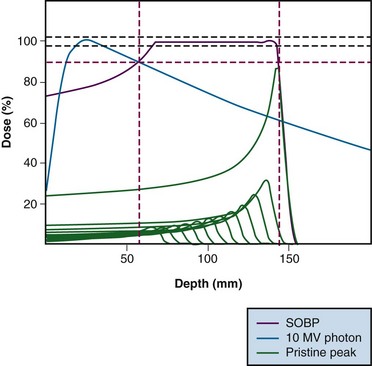
Interest in the use of charged particle radiotherapy has been primarily stimulated by the superior dose distributions that can be achieved with these particles compared with those produced by standard photon therapy techniques. Charged particles deposit energy in tissue through multiple interactions with electrons in the atoms of cells, although a small fraction of energy is also transferred to tissue through collisions with the nuclei of atoms. The energy loss per unit path length is relatively small and constant until near the end of the range where the residual energy is lost over a short distance, resulting in a steep rise in the absorbed dose (energy absorbed per unit mass). This portion of the particle track, where energy is rapidly lost over a short distance, is known as the Bragg peak (Fig. 19-1).
The initial low-dose region in the depth-dose curve, before the Bragg peak, is referred to as the plateau of the dose distribution and is about 30% of the Bragg peak maximum dose. The Bragg peak is too narrow for practical clinical applications. For the irradiation of most tumors, the beam energy is modulated in order to achieve a uniform dose over a significant volume. This is accomplished by superimposing several Bragg peaks of descending energies (ranges) and weights to create a region of uniform dose over the depth of the target; these extended regions of uniform dose are called spread-out Bragg peaks (SOBP) (see Fig. 19-1). Although the beam modulation used to spread out the Bragg peaks does increase the entrance dose, the proton dose distribution is still characterized by a lower-dose region in normal tissue proximal to the tumor, a uniform high-dose region in the tumor, and zero dose beyond the tumor.
Charged particles are generally characterized as having either high or low linear energy transfer (LET), the rate of energy loss by the particle in tissue. The LET influences the biologic impact of the energy deposited in tissue. X and gamma photons, protons, and helium ions are considered to be forms of low LET radiation. Heavier charged particles (e.g., neon ions, carbon ions) are considered to be forms of high LET radiation. There is an initial increase in the relative biologic effectiveness (RBE) with an increase in LET.1 Carbon ions have an RBE of about 3, whereas the recommended RBE of protons is 1.1.2,3 Higher-LET radiation is less influenced by tissue oxygenation and less sensitive to variations in the cell cycle and DNA repair. For particle radiation, the Gray equivalent dose is calculated by multiplying the physical dose administered by the RBE for that particle; the recommended nomenclature for expressing the dose is Gy(RBE) = physical dose in Gy × RBE.3
Development of Proton Beam Radiotherapy
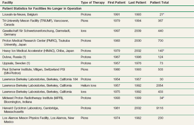
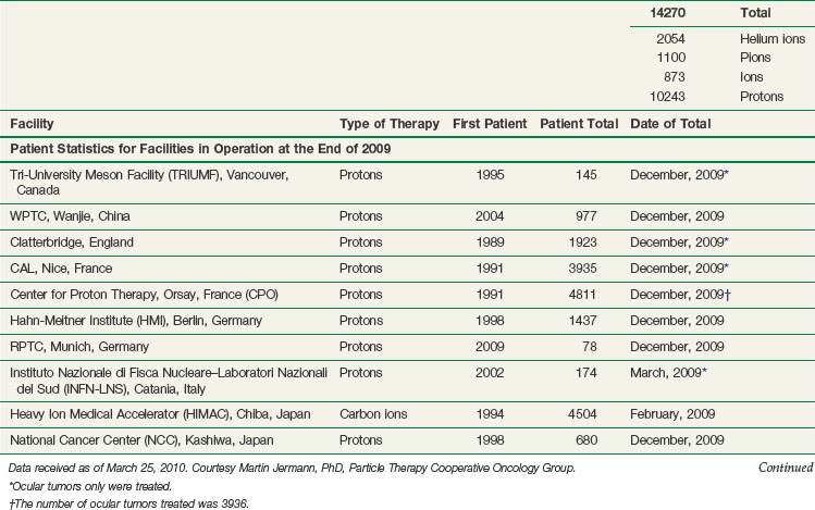
The vast majority of patients receiving charged particle therapy have been treated with protons. As of March 2010, over 67,000 patients had received part or all of their radiotherapy by proton beams.4 Table 19-1 lists currently operational proton beam treatment facilities worldwide. Several more sites are scheduled to begin using proton beam therapy over the next several years.
In 1946, Robert Wilson5 proposed that proton beams would provide superior dose distributions over photons and should be considered for clinical radiotherapy. Initially, patients were being treated at facilities designed and constructed for basic high-energy physics research, and this often meant that treatment delivery was cumbersome. The proton beams were limited to a fixed horizontal position, which meant that the patient had to be moved to align the tumor on the trajectory of the beam. This technique was in contrast to the isocentric capabilities of the modern linear accelerator, which rotates around a point in space and can effectively target any site in the body. In addition, for many of the proton machines, the energy of the beam (which defined the depth of the Bragg peak) was only sufficient to treat superficial lesions (such as those of the eye) or intermediate-depth lesions (such as those at the base of the skull). Because of these technical factors and the interests of the involved physicians, the tumors that initially received the most attention were uveal melanomas in the eye and sarcomas at the base of the skull. The major emphasis in proton therapy clinical research initially was on dose escalation for tumors for which local control with conventional radiotherapy was poor.
Treatment of Specific Cancers with Proton Beam Radiotherapy
Ocular (Uveal) Melanoma
As of December 2002, more than 3000 patients with uveal melanoma had been treated with protons at Massachusetts General Hospital in collaboration with the Massachusetts Eye and Ear Infirmary.6 The 5-year actuarial local control rate was 96% for all sites within the globe, with an 80% survival rate. The probability of eye retention at 5 years was estimated to be 90% for the entire group and 97%, 93%, and 78% for patients with small, intermediate, and large tumors, respectively. Independent risk factors for enucleation were involvement of the ciliary body, tumor height greater than 8 mm, and distance between the posterior tumor edge and the fovea. These results compare favorably with the 5-year local control rates of 93% reported with protons in Nice, France,7 98.9% after 1993 from the Paul Scherrer Institute in Villigen, Switzerland,8 and 96% from the Curie Institute in Orsay, France.9
Because some patients have experienced deteriorating vision after doses of 70 Gy(RBE), a randomized trial of 50 Gy(RBE) versus 70 Gy(RBE) for small and intermediate-sized lesions located within 6 mm of the optic disc or macula was conducted. Interim analysis of 188 patients, with a median follow-up of 60 months, suggested no reduction in either local control or survival rates. No significant improvement in visual outcome or complications has been observed. However, visual field analysis does show a smaller mean defect in the patients randomized to 50 Gy(RBE).10
Egger and colleagues11 reported long-term results of eye retention after treatment of uveal melanoma with proton beam therapy. A total of 2645 patients (2648 eyes) were treated at the Paul Scherrer Institute between 1984 and 1999. The overall eye retention rate at 5, 10, and 15 years after treatment was 89%, 86%, and 83%, respectively. Enucleation was related to large tumor size (mainly, tumor height), male gender, high intraocular pressure, and large degree of retinal detachment at treatment time.
Sarcomas of the Skull Base and Spine
At the Harvard Cyclotron Laboratory, Massachusetts General Hospital, physicians used a combination of protons and photons to treat patients with tumors of the skull base and cervical spine.12 A total of 169 patients with chordoma and 165 patients with chondrosarcoma were treated. The 10-year local control rate was highest for chondrosarcomas, intermediate for male chordomas, and lowest for female chordomas (94%, 65%, and 42%, respectively). For cervical spine tumors, 10-year local control rates were not significantly different for chordomas and chondrosarcomas (54% and 48%, respectively), nor was there any significant difference in local control rates between males and females. In a Cox multivariate analysis, predictors of local control included gender and equivalent uniform dose, or gender and target volume, or gender and minimum target dose.13 Five-year actuarial rates of endocrinopathy were as follows: 72% for hyperprolactinemia, 30% for hypothyroidism, 29% for hypogonadism, and 19% for hypoadrenalism. The minimum target dose (Dmin) to the pituitary gland was found to be predictive of endocrinopathy: Patients receiving 50 Gy(RBE) or more at Dmin to the pituitary gland had a higher incidence of and greater severity of endocrine dysfunction. Posterior pituitary dysfunction, represented by vasopressin activity with diabetes insipidus, was not observed.14
The French group at Orsay reported on the treatment of patients with skull base tumors, 34 with chordoma and 11 with chondrosarcoma.15 Irradiation was done with a combination of photons and protons, with protons used in one-third of the treatment regimens. The median total dose delivered was 67 Gy(RBE) (range, 60 to 70 Gy[RBE]). With a mean follow-up of 30.5 months (range, 2 to 56 months), the 3-year local control rate for chordomas was 83.1% and for chondrosarcomas was 90%. Three-year overall survival rates were 91% and 90%, respectively.
Between 1998 and 2005, 64 patients with skull base chordomas (42 patients) and chondrosarcomas (22 patients) were treated at the Paul Scherrer Institute with protons using a spot-scanning technique.16 Patients with chordoma received a mean dose of 73.5 Gy(RBE) (range, 67 to 74 Gy[RBE]), and patients with chondrosarcoma received a mean dose of 68.4 Gy(RBE) (range, 63 to 74 Gy[RBE]). With a mean follow-up of 38 months, actuarial 5-year local control rates were 81% and 94% for chordomas and chondrosarcomas, respectively. The actuarial 5-year rate for freedom from high-grade toxicity was 94%.
Torres and associates17 performed a planning study where they compared three-dimensional conformal proton (PR) therapy, intensity-modulated radiotherapy (IMRT) with photons (PH), and combined proton and IMRT photon (PP) irradiation of skull base chordomas to determine the optimal technique. For each of five patients, they generated four treatment plans: (1) an IMRT plan with a 1-mm planning target volume (PH1) for stereotactic treatment; (2) an IMRT plan with a 3-mm planning target volume (PH3) for routine treatment; (3) a PR plan with beam-specific expansion margins on the clinical target volume; and (4) a plan for PP treatment. The mean percentage of planning target volume (%PTV) receiving the prescription dose of 74 Gy(RBE) was highest in the PP plans and lowest in the PH3 plans. The PR plans were the least homogeneous and conformal. The PH3 plans had the highest mean percentage of volume (%V) of brain, brainstem, chiasm, and temporal lobes above the tolerance dose for those organs. The PH1 plans had the lowest brainstem mean %V receiving 67 Gy(RBE) and temporal lobe mean %V receiving 65 Gy(RBE). Global evaluation of the plans based on objective parameters revealed that the PP plans yielded the best target coverage and conformality. This study indicates that there may be dosimetric advantages to using a combination of IMRT and three-dimensional protons, to optimize conformality and minimize integral dose, which may be an important option until intensity-modulated proton therapy is more widely available.
Isacsson and colleagues18 compared conformal radiotherapy treatment plans with combination photon/proton plans for a patient with a cervical Ewing’s sarcoma. The comparison showed small but clear advantages of protons for the boost. At 1% normal tissue complication probability (NPTC) in the spinal cord, the calculated tumor control probability (TPC) was on average 5% higher for the photon/proton boost combination.
Hug and colleagues19 presented results on combined photon/proton treatment of 47 patients with osteogenic and chondrogenic tumors of the axial skeleton. Radiation was delivered postoperatively in 23 patients, preoperatively and postoperatively in 17 patients, and as the sole treatment in 7 patients. Mean radiation doses of 73.9 Gy(RBE), 69.8 Gy(RBE), and 61.8 Gy(RBE) were delivered to group 1 (20 patients with recurrent/primary chordoma or chondrosarcoma), group 2 (15 patients with osteogenic sarcoma), and group 3 (12 patients with giant cell tumors, osteoblastomas, or chondroblastomas), respectively. Five-year actuarial local control and survival rates for patients with chondrosarcoma were 100% and 100% and with chordoma, 53% and 50%. The actuarial 5-year local control rate for patients with osteosarcoma was 59%. The 5-year actuarial local control and survival rates for the group 3 patients were 76% and 87%. Overall, improved local control was noted for patients with primary versus recurrent tumors, those who underwent gross total resection, and those who received target doses of more than 77 Gy(RBE).
Weber and colleagues20 carried out a treatment planning comparison of intensity-modulated photon (IM) and proton therapy (IMPT) for paraspinal sarcomas. Plans for five patients were computed for IM photons (seven coplanar fields) and protons (three coplanar beams). The prescribed dose was 77.4 Gy(RBE) for protons to the gross tumor volume. Surface and center spinal cord dose constraints for all techniques were 64 and 53 Gy(RBE), respectively. Gross tumor volume coverage was optimal and equally homogeneous with both the IM photon and IM proton plans. The median heart, lung, kidney, stomach, and liver mean dose and the dose at the 50% volume level were consistently reduced by a factor of 1.3 to 25 with protons compared with photons. IMPT dose escalation (to 92.9 Gy(RBE) delivered to the gross tumor volume) was possible in all patients without exceeding the normal-tissue dose limits.
At the Francis H. Burr Proton Therapy Center at Massachusetts General Hospital, a phase II study was conducted of combined photon and proton beam radiation therapy, with or without surgical resection, for patients with spinal and paraspinal sarcomas.21 Doses of 77.4 Gy(RBE) at 1.8 Gy(RBE) per day were used for patients with gross residual disease and 70.2 Gy(RBE) for patients with microscopic residual disease. A total of 50 patients (29 with chordoma, 14 with chondrosarcoma, and 7 with other cancers) underwent gross total (25 patients) or subtotal (12 patients) resection or biopsy (13 patients). With a 48-month median follow-up, the 5-year actuarial local control, recurrence-free survival, and overall survival rates were 78%, 63%, and 87%, respectively. Two of 36 patients (5.6%) treated for primary tumors versus 7 of 14 patients (50%) treated for recurrent tumors developed local recurrence (p <.001). The spinal cord center dose was limited to 54 Gy(RBE) and the cord surface dose to 63 Gy(RBE) over a length of 5 cm or less. The cauda equina was constrained to 70.2 Gy(RBE), except for areas in direct contact with tumor, where the dose limit was 77.4 Gy(RBE). Five patients developed late radiation-associated complications; no myelopathy developed, but three grade 3 sacral neuropathies appeared after doses of 77.12 to 77.4 Gy(RBE) had been delivered.
Optic Pathway Glioma
At Loma Linda University, seven children with optic pathway gliomas were treated with proton radiation therapy.22 At a median follow-up of 37 months, all tumors were locally controlled. A reduction in tumor volume was seen in three patients, and tumor volume was stable in the other four. Visual acuity was stable in those that presented with useful vision. Proton plans were compared with photon plans for individual patients. With proton therapy radiation, there was a 47% reduction in the dose to the contralateral optic nerve. There was an 11% reduction in the dose to the chiasm and a 13% reduction in the dose to the pituitary gland. There was also a reduction in the dose to the temporal lobes and frontal lobes.
Astrocytoma
Between 1993 and 1998, 48 patients were treated for nonresectable grade II and III intracranial tumors at the Center for Proton Therapy in Orsay, France.23 Mean tumor doses ranged from 63 to 67 Gy at 1.8 Gy/fraction. With a median follow-up of 18 months, local control rates were 97% (33 of 34 patients) and 43% (6 of 14 patients) for nonparenchymal and parenchymal lesions, respectively.
At the Harvard Cyclotron Laboratory and Massachusetts General Hospital, a phase II study was undertaken by Massachusetts General Hospital researchers to assess whether dose escalation to 90 Gy(RBE) with conformal protons and photons in accelerated fractionation twice a day would improve local tumor control and survival rates.24 A total of 23 patients were enrolled, with ages of 18 to 70 years. Actuarial survival rates at 2 and 3 years were 34% and 18%, respectively. The median survival time was 20 months, with four patients alive 22 to 60 months after diagnosis. All patients developed new areas of gadolinium enhancement during the follow-up period. Histologic examination of tissues obtained at biopsy, resection, or autopsy was conducted in 15 patients. Radiation necrosis only was demonstrated in seven patients, and their survival was significantly longer than patients with recurrent tumor. Tumor regrowth occurred most commonly in areas that received doses of 60 to 70 Gy(RBE) or less; recurrent tumor was found in only one patient in the group that received a dose of 90 Gy(RBE). The authors concluded that attempts to extend local control by enlarging the volume would likely be complicated by a high incidence of radionecrosis.
Benign Meningioma
Between 1981 and 1996, 46 patients with partially resected, biopsied, or recurrent benign meningiomas were treated with combined proton/photon radiation at the Harvard Cyclotron Laboratory/Massachusetts General Hospital.25 The median dose for the tumor was 59 Gy(RBE). Overall survival rates at 5 and 10 years were 93% and 77%, respectively, and recurrence-free rates at 5 and 10 years were 100% and 88%, respectively. Three patients presented with local tumor recurrence at 61, 95, and 125 months. One patient died of focal brain necrosis at 22 months. Neurologic complications, including memory deficits and hearing loss, were also seen. Four patients developed ophthalmologic toxicity. In all of these cases, the maximum dose to the optic structures was more than 58 Gy(RBE). Endocrine abnormalities following treatment were also seen.
Investigators from the Paul Scherrer Institute reported on the treatment of 16 patients with recurrent, residual, or untreated intracranial meningiomas.26 The median prescribed dose was 56 Gy(RBE) (range, 52 to 64 Gy[RBE]) at 1.8 to 2 Gy(RBE) per fraction. Cumulative 3-year local control, progression-free survival, and overall survival rates were 91%, 91%, and 92%, respectively. No patient died of recurrent meningioma. Radiographic follow-up (median, 34 months) revealed an objective response in 3 patients and stable disease in 12 patients. The cumulative 3-year toxicity-free survival rate was 76%. One patient with an optic nerve sheath meningioma presented with sudden visual field deterioration of the ipsilateral eye 30 months after irradiation with 56 Gy(RBE). Another patient with optic nerve encasement by disease developed visual deterioration at 9 months. A third patient developed symptomatic brain necrosis 7 months after treatment. No radiation-induced hypothalamic/pituitary dysfunction was observed.
Paranasal Sinus, Nasal, and Nasopharyngeal Tumors
Mock and associates27 performed a planning comparison study of various photon and proton techniques for the treatment of paranasal sinus carcinoma. In five patients, proton plans were compared with conventional, conformal, and IMRT photon plans. The evaluations analyzed dose-volume histogram findings of the target volumes and organs at risk (i.e., the pituitary gland, optic pathway structures, and brain).
Between 1991 and 1996, 32 patients with carcinomas of the paranasal sinuses were treated with an accelerated photon/proton protocol.28 The stage distribution was T3 in two cases and T4 in 30 cases, and all were stages N0 and M0. Four patients had undergone a gross total resection, and the others had undergone only a biopsy or a subtotal resection. The median follow-up was 2.7 years. The actuarial disease-specific survival rate at 3 years was 62%. There have been 10 deaths, three with intercurrent disease and seven with metastatic disease. The 3-year actuarial local control rate was 89%. Late toxicity has included temporal lobe necrosis in three patients. Three patients have required surgical soft tissue repair.
Truong and colleagues29 performed a retrospective review of 20 patients with locally advanced primary sphenoid sinus malignant tumors treated between 1991 and 2005 with proton radiotherapy to a median dose of 76 Gy(RBE) to determine treatment outcome and prognostic factors. With a median follow-up of 27 months, the 2-year local, regional, and freedom from distant metastasis rates were 86%, 86%, and 50%, respectively. The disease-free and overall survival rates at 2 years were 31% and 53%, respectively. In multivariate analysis, oropharyngeal involvement (p = .005) and anterior cranial fossa invasion (p = .02) were predictive for poor disease-free survival rates. Brain invasion was predictive for decreased overall survival rates (p = .05). No grade 3 or 4 late visual toxicity was reported. Three patients developed chronic nasal symptoms after radiotherapy, consisting of common toxicity criteria (CTC) grade 2 to 3 nasal obstruction secondary to fibrous adhesions. One patient required surgical removal of adhesions to relieve chronic nasal congestion. One patient with symptomatic CTC grade 2 brain toxicity experienced seizures and short-term memory loss. The seizures were controlled with anticonvulsant medications and a short course of steroids. Two patients experienced cerebrospinal fluid leakage after surgery and irradiation. One patient developed a CTC grade 2 cerebrospinal fluid leak from the external auditory canal, secondary to tumor shrinkage and erosion of the petrous temporal bone 5 months after radiotherapy. The patient was offered surgery and declined treatment. One patient developed a CTC grade 5 cerebrospinal fluid leak without evidence of tumor recurrence at 2 months after completion of radiotherapy. The patient underwent four surgical repairs, including transethmoid packing of the ethmoid and sphenoid sinuses and placement of a lumboperitoneal shunt. The patient subsequently died from infectious meningitis. Two patients had endocrinopathies that were medically corrected. The authors concluded that proton radiation therapy results in excellent local control in patients with advanced primary sphenoid sinus malignant disease. Brain invasion and involvement of the oropharynx and anterior cranial fossa were important prognostic factors. Nasal symptoms, brain injury, endocrinopathies, and cerebrospinal fluid leaks, however, may complicate treatment.
Investigators at Massachusetts General Hospital performed a prospective study incorporating chemotherapy, surgery, and combined proton/photon therapy in the treatment of patients with neuroendocrine tumors of the sinonasal tract.30 Nineteen patients with olfactory neuroblastoma or neuroendocrine carcinoma were treated between 1992 and 1998. Patients received chemotherapy with two courses of cisplatin and etoposide followed by high-dose proton/photon radiotherapy to 69.2 Gy(RBE) using 1.6 to 1.8 Gy(RBE) per fraction twice daily in a concomitant boost schedule. Two further courses of chemotherapy were given to responders.
At Loma Linda University, 16 patients with recurrent nasopharyngeal carcinoma were treated with conformal proton irradiation.31 Patients had initially been treated with photon therapy at doses of 50 to 70 Gy. Conformal proton boost radiation was then delivered to bring the total dose to 59 to 70 Gy(RBE). With a mean follow-up of 23 months, the 24-month actuarial overall and locoregional progression-free survival rates were both 50%. No central nervous system complications were observed. An update now includes data on 39 patients Twenty-four-month actuarial overall and locoregional progression-free survival rates were 49% and 70%, respectively. When critical central nervous system structures were re-irradiated, doses were low (0 to 22 Gy[RBE]); one patient experienced grade IV central nervous system side effects. Grade III late head and neck toxicity occurred in seven patients and grade IV toxicity in two patients.
Acoustic Neuroma
Between 1991 and 1999, 30 patients with acoustic neuroma were treated with proton therapy at Loma Linda University.32 Patients with useful hearing before treatment received 54 Gy(RBE) in 30 fractions, and patients without useful hearing received 60 Gy(RBE). Follow-up ranged from 7 to 98 months (median, 34 months), during which no patients demonstrated disease progression on magnetic resonance imaging scans. Eleven patients demonstrated radiographic regression. Of the 13 patients with useful hearing before treatment, 4 (31%) maintained their hearing. No transient or permanent treatment-related trigeminal or facial nerve dysfunction was observed. Investigators are now interested in evaluating a reduction in tumor dose in an attempt to increase hearing preservation rates.
Carcinoma of the Prostate
Investigators at Massachusetts General Hospital completed a phase III trial comparing 67.2 Gy of photons with 75.6 Gy(RBE) of combined photons/protons using a conformal perineal proton boost.33 From 1982 to 1992, 202 patients with stage T3 or T4 prostate cancer received 50.4 Gy by four-field photons. Patients then received either 25.2 Gy(RBE) with conformal protons or a 16.8-Gy photon boost. No differences between the two groups were found in overall survival, total recurrence-free survival, or local recurrence-free survival rates. The local recurrence-free survival rate at 7 years for patients with poorly differentiated tumors (Gleason score of 9 or 10) was 85% for the proton arm and 37% for the photon arm. Rates of grade 1 and 2 rectal bleeding were higher in the proton arm (32% vs. 12%), as were those for urethral stricture (19% vs. 8%). Dose escalation to 75.6 Gy(RBE) by conformal proton boost led to increased late radiation sequelae but not to increased total survival rates in any subgroup. However, there was an improved local recurrence-free survival rate in patients with poorly differentiated tumors.
The Loma Linda University experience of conformal proton therapy for prostate cancer was reported34 for 1255 patients treated from 1991 to 1997. The overall biochemical disease-free survival rate was 73%, and it was 90% in patients with an initial prostate-specific antigen (PSA) level of 4 ng/mL or less; it was 87% in patients with posttreatment PSA nadirs of 0.50 ng/mL or less. Rates dropped with rises in initial and nadir PSA values.
Massachusetts General Hospital and Loma Linda University conducted a phase III randomized dose escalation trial in patients with early-stage prostate cancer.35
Stay updated, free articles. Join our Telegram channel

Full access? Get Clinical Tree


