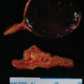
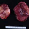
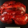
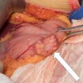
Carney triad (described in 1977) is a rare, nonfamilial, multitumoral syndrome, with three tumors in the initial description: gastrointestinal stromal tumor (GIST), pulmonary chondroma, and extraadrenal paraganglioma (PGL). , Subsequently, two other tumors, adrenal cortical adenoma and esophageal leiomyoma, were added as components—thus actually Carney triad is a “pentad.” Although it is rare, it is important for endocrinologists to be aware of this disorder because of the links to PGL and adrenocortical tumors.
Case Report
The patient was a 26-year-old woman referred for further management of Carney triad-related neoplasms. The first sign of the triad was at age 17 when she presented with microcytic anemia (hemoglobin 7.0 g/dL) that was treated with iron sulfate. The anemia proved refractory, and esophagogastroduodenoscopy (EGD) performed 1 year before consultation at Mayo Clinic found a large gastrointestinal stromal tumor (GIST), and she underwent an 80% gastrectomy for the removal of a 17-cm GIST. More recently it was noted that she had a mass in her left lingula, which was resected and proved to be a 3.3-cm chondroma. She was also noted to have a 4-cm mass near the aortic arch. She had labile hypertension controlled with a calcium channel blocker (amlodipine 10 mg daily). She had no signs or symptoms of adrenocortical or adrenomedullary hormone excess. On physical examination her body mass index was 28.7 kg/m 2 , blood pressure 138/84 mmHg, and heart rate 96 beats per minute.
INVESTIGATIONS
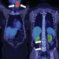
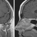
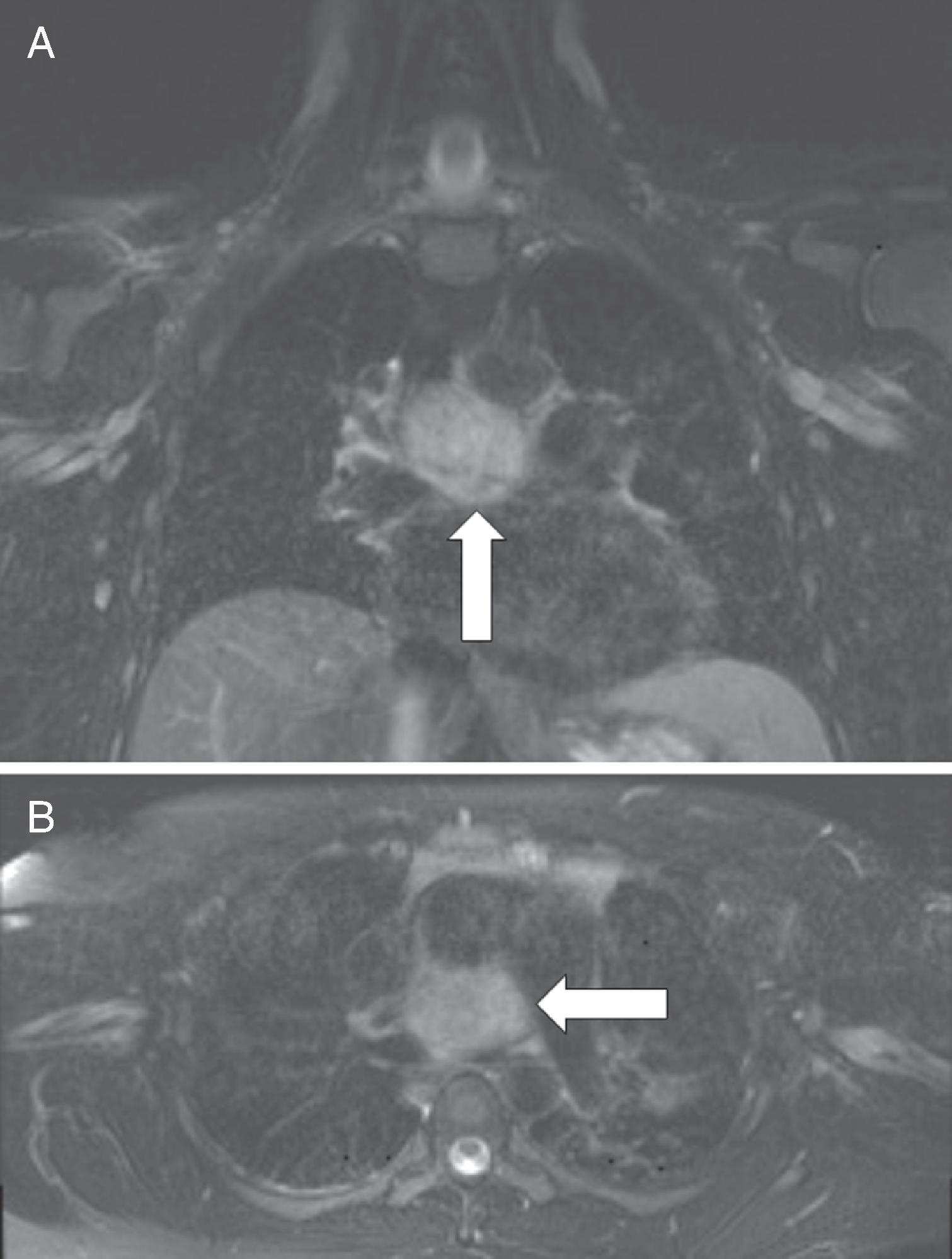
Chest magnetic resonance imaging (MRI) showed a 4.1 × 3.3–cm precarinal soft tissue mass ( Fig. 49.1 ). The plasma fractionated metanephrines and 24-hour urine for fractionated metanephrines and catecholamines, while not markedly elevated, were consistent with a catecholamine-secreting tumor ( Table 49.1 ). I-123 metaiodobenzylguanidine (MIBG) scintigraphy colocalized with the precarinal mass and two sites of abnormal uptake were seen in the upper abdomen just to the right and left of midline ( Fig. 49.2 ). MRI of the abdomen demonstrated two retroperitoneal lesions that colocalized with the findings on I-123 MIBG scintigraphy ( Fig. 49.3 ). In addition, the abdominal MRI detected a 2.5-cm right adrenal mass with imaging characteristics consistent with a cortical adenoma ( Fig. 49.4 ). The 1-mg overnight dexamethasone suppression test was normal (see Table 49.1 ). Finally, EGD showed no sign of residual GIST; however, two asymptomatic 1.5–2.0 cm esophageal leiomyomas were found.
| Biochemical Test | Result | Reference Range |
| Sodium, mmol/L | 137 | 135–145 |
Potassium, mmol/L | 3.9 | 3.6–5.2 |
Creatinine, mg/dL | 0.8 | 0.7–1.2 |
Plasma metanephrine, nmol/L | 0.32 | <0.5 |
Plasma normetanephrine, nmol/L | 2.23 | <0.9 |
1-mg overnight DST cortisol, mcg/dL | 1.5 | <1.8 |
24-hr urine: | ||
Metanephrine, mcg | 104 | <400 |
Normetanephrine, mcg | 979 | <900 |
Norepinephrine, mcg | 203 | <80 |
Epinephrine, mcg | 3.3 | <20 |
Dopamine, mcg | 305 | <400 |
Stay updated, free articles. Join our Telegram channel

Full access? Get Clinical Tree


