Neoplasms of the nasal cavity, the paranasal sinuses, the anterior skull base, and the orbit represent a heterogeneous group of disorders that are associated with multiple challenges and historically poor outcomes. Several factors have been implicated in the overall poor prognosis of these lesions including aggressive growth patterns, proximity to critical neurovascular structures, nonspecific clinical presentation, and initial presentation with locally advanced disease. A multidisciplinary approach is often invoked for these lesions with the goals of oncologic control and preservation of organ function. The relative rarity and heterogeneous variety of these lesions have to date precluded the ability to perform large, prospective, randomized studies. The current treatment algorithms are based largely on the collective experience of single-institutional experiences.
The past decade has witnessed an evolution in the management strategies and outcomes for sinonasal and anterior skull base malignancies. Distinction in the clinical behavior and treatment sensitivity of the various histopathologies has resulted in more tailored therapy. Additionally, the well-defined principles of open extirpative surgery, including extended skull base approaches and craniofacial surgery, have led to an increasing range of treatment options for advanced lesions. Finally, the increasing collective experience with the techniques and outcomes of endoscopic surgery is rapidly defining its role in the surgical treatment of select sinonasal and anterior skull base neoplasms.
ANATOMY—NASAL CAVITY AND PARANASAL SINUSES
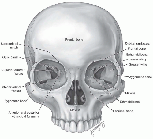
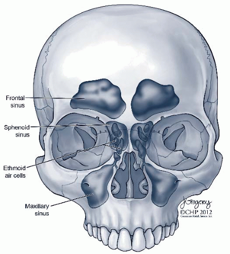
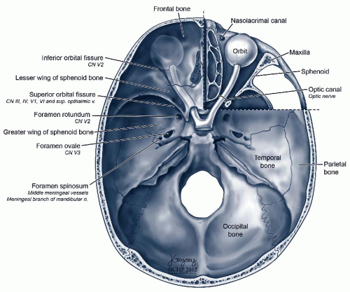
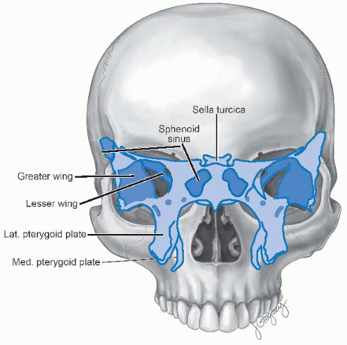
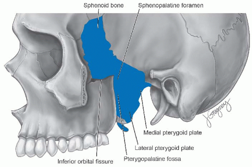
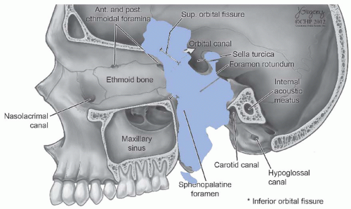
A thorough understanding of the anatomy of the sinonasal cavity, the anterior skull base, and the orbit is fundamental in the treatment of these lesions. An abridged review is presented below. The bony landmarks of the facial skeleton are presented in
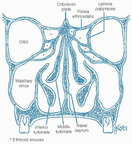
Figure 20-1. The nasal cavity and the paired paranasal sinuses are air-filled cavities, surrounded by a bony-cartilaginous framework and lined by pseudostratified, ciliated columnar (respiratory) epithelium interspersed with goblet cells and basal cells (
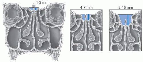
Fig. 20-2). Individual ciliated cells have approximately 50 to 200 cilia per cell, beating approximately 700 to 800 beats/minute and achieving a mucociliary clearance rate of 1 cm/minute. This physiology is easily impaired by the events that occur during cancer treatment including surgery and radiation therapy. The physiologic function of the sinonasal cavities include respiratory conductance of air flow, humidification and warming of inspired air, voice resonance, particle filtration, decreased skull weight, mediation of olfaction, absorption of midface and forehead trauma, and immune response to respiratory antigens. With regard to sinonasal neoplasms, the presence of compartmentalized, air-filled spaces allows for a period of unabated, local tumor growth with minimal, nonspecific symptoms and no significant findings on examination. This is one of the primary factors that predispose these lesions to a delay in diagnosis and advanced stage at initial presentation. Additionally, the intimate relationship of the sinonasal cavity to critical neurovascular structures, including the orbit, the internal carotid artery, and the intracranial cavity has significant implications on overall prognosis and treatment efficacy. In certain areas, the bony walls separating the sinonasal cavity from the orbit (lamina papyracea) and the intracranial cavity (anterior skull base) are thin and easily eroded by tumor (
Fig. 20-3). Additionally, the presence of multiple adjacent foramina and canals allow tumor spread along preformed perineural and perivascular channels (
Fig. 20-4). An understanding of these potential pathways is critical to defining the pathophysiology of tumor spread and is highlighted in the following paragraphs.
Nasal Cavity
The nose consists of paired nasal apertures and cavities supported by a bony and cartilaginous framework. It is divided by the midline nasal septum (
Fig. 20-5). The anterior most portion of the nasal cavity, termed the nasal vestibule, is lined by squamous epithelium and numerous hair follicles or vibrissiae. The remainder of the nasal cavity is lined by respiratory epithelium. The nasal cavity extends from the nasal vestibule anteriorly to the choana posteriorly. It is bounded by the columella and nasal septum medially and the lower lateral cartilages laterally. The floor of the nasal cavity corresponds to the hard and soft palate orally, whereas the roof corresponds to the cribriform plate intracranially. The paired choana open to the nasopharynx directly and are bordered by the medial pterygoid plates on each side. Olfactory epithelium is interspersed with respiratory epithelium along the superior aspect of the nasal septum, roof of the nasal cavity, and the superior and the middle turbinates. Olfactory nerve bundles pass through multiple perforations in the cribriform plate, providing a potential pathway for perineural spread of tumor intracranially.
The paired inferior, middle, and superior turbinates are bony projections lined by respiratory mucosa arising from the lateral
nasal wall. The inferior turbinates are independent osseous structures whereas the middle and superior turbinates are portions of the ethmoid bone. The area underneath each turbinate, termed meati, has important physiologic relationships. The lacrimal duct arises from the lacrimal sac in the inferomedial portion of the orbit, runs inferiorly along the medial-posterior surface of the maxilla, and empties through Hasner valve into the inferior meatus. The anterior paranasal sinuses composed of the maxillary, the anterior ethmoid, and the frontal sinuses physiologically clear their contents into the middle meatus. Similarly, the posterior ethmoid cells and the sphenoid sinus clear their contents into the superior meatus. The sphenopalatine foramen is located on the lateral nasal wall, inferior-posterior to the horizontal attachment of the middle turbinate, and transmits the sphenopalatine artery and vein from the pterygomaxillary space in addition to the nasopalatine and the superior nasal branches of the maxillary division of the trigeminal nerve.
The nasal septum is composed of a bony-cartilaginous wall sandwiched between two layers of tightly adherent mucopericondrium. The anterior quadrangular cartilage sits on the bony maxillary crest and articulates with the bony vomer posteriorly and the perpendicular plate of the ethmoid superiorly. In addition to providing structural support and normal nasal physiology, the nasal septum may act as a barrier for tumor transgression. Multiple neural and vascular sources supply the nasal septum. Arterial branches arise from both the external (sphenopalatine and facial arteries) and the internal carotid arteries (anterior ethmoid artery). Sensation is transmitted through both the ophthalmic (nasociliary nerve) and the maxillary (nasopalatine nerve) divisions of the trigeminal nerve.
Maxillary Sinus
The paired maxillary sinuses represent the largest of the paranasal sinuses and most common site of sinonasal malignancy. These inverted pyramidal structures have a total volume of approximately 15 mm. Dynamic mucociliary clearance transports sinus secretions toward the natural ostium in the superomedial aspect of the sinus into the middle meatus of the nasal cavity. Obstruction of the ostium results in stasis of secretions and potential bacterial superinfection. Accessory openings, termed fontanelles, may also exist in approximately 25% of the population. The maxillary sinuses are bordered medially by the lateral nasal wall and the nasal cavity (maxillary and palatine bones), superiorly by the orbital floor and the infraorbital canal, anteriorly by the anterior face of the maxilla, inferiorly by the alveolar process of the maxilla, posterolaterally by the infratemporal fossa, and posteromedially by the pterygopalatine fossa (PPF). The base of the pyramid is the nasal surface and the tip points toward the zygomatic process. The anatomic proximity of the maxillary sinus to these structures predicates the patterns of spread for advanced lesions and has significant treatment and prognostic implications. Anatomic variations of the maxillary sinus include the presence of septations and hypoplasia of the maxillary sinus. The floor of the maxillary sinus is intimately related to the upper gingiva and the hard palate and is typically 1 to 2 cm below the level of the nasal cavity floor in adults. Tumor extension inferiorly to the alveolus and medially to the lateral nasal wall is relatively more favorable and amenable to en bloc resection.
Ethmoid Sinus
The ethmoid sinus consists of multiple individual compartments and has been aptly termed the ethmoid labyrinth. Significant variability in the number, size, and pneumatization pattern of the ethmoid sinus cells has been described. Examples of this include aberrant pneumatization of ethmoid sinus cells within the inferior orbital wall (Haller or infraorbital cell), the frontal recess and sinus (frontal cell), the sphenoid sinus (Onodi or sphenoethmoid cell), and the middle turbinate (concha bullosa). The ethmoid sinuses are bordered by the middle and the superior turbinates medially, anterior face of the sphenoid sinus posteriorly, the lamina papyracea laterally, and the anterior skull base (fovea ethmoidalis) superiorly. The latter two boundaries are thin walled and easily breached by both neoplasm and surgical injury. In the normal configuration, there is an approximately 15-degree anterior-to-posterior down-sloping of the fovea ethmoidalis although variability in its height and slope exist. The periorbital perichondrium and dura of the anterior cranial fossa are tightly apposed to the respective bony walls and represent a natural barrier to tumor extension. The bony optic canal houses the optic nerve and may traverse immediately adjacent to the lateral border of the posterior ethmoid cells to a variable extent. The ethmoid sinuses are divided both anatomically and functionally into an anterior and posterior set of cells as demarcated by the ground (basal) lamella, the horizontal lateral bony attachment of the middle turbinate to the lamina papyracea. The paired anterior and posterior ethmoidal arteries traverse the roof of the ethmoid sinus. These vessels arise from the ophthalmic branch of the internal carotid artery. The anterior ethmoidal artery courses over the medial rectus muscle, penetrates the lamina papyracea, and courses the roof of the ethmoid sinus, posterior to the frontal. The posterior ethmoidal artery enters the orbit approximately 3 to 5 mm anterior to the optic foramen, emerges from the orbit approximately 12 mm posterior to the anterior ethmoidal artery courses across the roof of the posterior ethmoid cavity and terminates with the dural, the ethmoidal, and the nasal septum branches.
Sphenoid Sinus
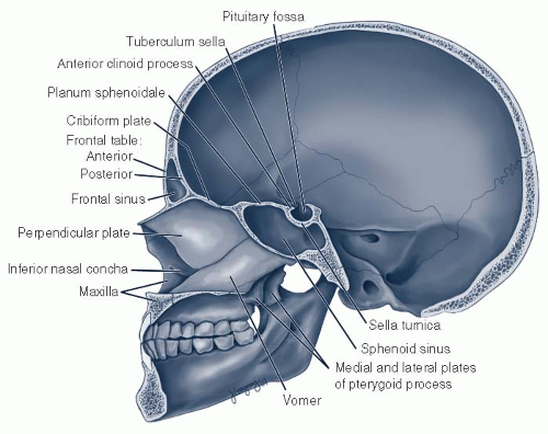
The sphenoid sinus is the posterior most extension of the sinonasal cavity and develops as a pneumatization of the sphenoid bone (
Fig. 20-6). It is bordered by the posterior ethmoid cells and the sphenoethmoid recess anteriorly, the sella and the clivus posteriorly, the internal carotid artery and the cavernous sinus laterally, and the planum sphenoidale superiorly. The natural ostium of the sphenoid sinus is located in the superior meatus, bounded by the superior turbinate laterally and posterior-most extension of the nasal septum medially. The ostium is located two-thirds up the vertical height of the sphenoid sinus cavity. The pneumatization of the sphenoid sinus is variable especially with regard to the sella medially and floor of the middle cranial fossa laterally. Variant numbers and insertions of the intersinus septation may also exist. The optic nerves run in the superolateral aspect of the sphenoid sinus, and the carotid artery canal is located at the junction of the lateral and posterior walls of the sinus. Dehiscences of the bony coverings of the optic nerve and the internal carotid artery within the sphenoid sinus may exist.
Frontal Sinus
The frontal sinus is the most anterior and superior extension of the sinonasal cavity. The majority of humans have two asymmetric frontal sinus cells separated by a thin septation. Variability exists in the degree of frontal sinus development with approximately 10% of the population lacking pneumatization of the frontal bone. The frontal sinuses empty into the inferomedial aspect of the sinus through the frontal recess into the middle meatus. The thicker anterior frontal table separates the frontal sinus from the soft tissue of the forehead. The thinner posterior frontal table separates the frontal sinus from the anterior cranial fossa.
Sinonasal Lymphatics
The lymphatic drainage of sinonasal malignancies is based on the location of the primary site. In general, the lymphatics are profuse and two distinct patterns are described for the sinonasal cavity. The lymphatics originating from the anterior nasal mucosa and nasal vestibule pass through the first echelon facial, submandibular, and parotid lymph nodes and eventually continue to the jugulodigastric nodes. The majority of the nasal cavity and paranasal sinuses drain posteriorly to the retropharyngeal lymph nodes and eventually to the superior deep cervical chain.
ANATOMY—ORBIT
Osteology
The bony orbit is pyramidal in shape with the anterior conjunctival surface (base) pointing to the posterior orbital apex (
Fig. 20-1). Direct neurovascular communication and bony foramina have the potential to act as a route of transmission among the orbit, the paranasal sinuses, and the intracranial cavity (
Fig. 20-9). The medial orbital wall extends from the anterior lacrimal crest to the orbital apex and separates the orbit from the ethmoid sinus. The thin-walled lamina papyracea comprises the anterior portion of the medial orbital wall, and the thicker sphenoid bone contributes to the posterior portion in the area of optic canal. The superior portion of the medial orbital wall transmits the anterior and posterior ethmoidal arteries through their respective foramina. The anterior ethmoidal artery pierces the medial orbital wall at the site of the frontoethmoid suture and then courses through the roof of the anterior ethmoid cells in the anterior ethmoidal sulcus, immediately posterior to the frontal recess. It enters the lateral lamella of the cribriform plate to supply the frontal sinus, the nasal septum, and the lateral nasal wall. A middle meningeal branch additionally enters the anterior cranial fossa. The floor of the orbit is composed of the orbital plate of the maxilla in addition to the zygoma and palatine bones and separates the orbit from the maxillary sinus. The infraorbital canal runs along the floor of the orbit and transmits the infraorbital artery and nerve to the cheek. The lateral wall of the orbit is formed by the zygoma anteriorly and the greater wing of the sphenoid posteriorly. The latter separates the orbit
from the middle cranial fossa. The superior and inferior orbital fissures in addition to foramina transmitting the zygomaticofacial and zygomaticotemporal arteries exist on the lateral orbital wall. The lateral orbital tubercle acts as an attachment point for multiple tendons and ligaments and is located on the lateral aspect of the lateral orbital rim, approximately 1 cm inferior to the frontozygomatic suture line. The orbital roof is composed of the orbital plate of the frontal bone and has variable pneumatization by the ethmoid and frontal sinuses. The trochlea exists in the anteromedial aspect of the orbital roof approximately 4 mm posterior to the orbital rim. The supraorbital notch or foramen transmitting the supraorbital neurovasculature exists at the boundary of the medial third of the orbital roof. A periosteal layer, termed the periorbita, lines the inner surface of the orbit and has complex attachments with various areas of the surrounding bone, the fascia layers of the orbit, and the dura surrounding the optic nerve.
Orbital Apex and Neurovascular Anatomy
The orbital apex provides direct intracranial communication and is marked by the posterior portion of the inferior orbital fissure, the superior orbital fissure, and the optic foramen. The optic foramen acts as an opening into the optic canal for the optic nerve and ophthalmic artery. The canal itself is housed within the lesser wing of the sphenoid bone, separating the canal contents from the posterior ethmoid cells and the sphenoid sinus medially. The superior orbital fissure lies between the greater and lesser wings of the sphenoid and transmits the oculomotor nerve (III), the trochlear nerve (IV), the lacrimal, the frontal, and the nasociliary branches of the ophthalmic nerve (V1), the abducens nerve (VI), branches of the ophthalmic vein and sympathetic fibers from the cavernous plexus. The oculomotor nerve divides into a superior and inferior branch within the cavernous sinus. The maxillary division of the trigeminal nerve from the PPF, the infraorbital branch of the maxillary artery, and the inferior ophthalmic vein pass through the inferior orbital fissure in the posterior-lateral portion of the orbital floor.
The optic nerve is approximately 5 cm in length and 4 mm in diameter. It consists of four anatomic segments in its length from the optic chiasm to the globe: intracranial, intracanalicular, intraorbital and intraocular. A dural layer surrounds the majority of the nerve course. The arterial supply to the optic nerve is primarily from the central retinal artery, a branch of the ophthalmic artery.
The ophthalmic division of the trigeminal nerve provides sensation to the orbit. It enters the orbit through the superior orbital fissure and divides into the frontal, the lacrimal, and the nasociliary nerves. The frontal branch divides into the supraorbital and supratrochlear nerves and supplies the upper eyelid and the forehead. The lacrimal nerve carries postganglionic parasympathetic fibers and somatic sensory innervation to the lacrimal gland. The nasociliary branch has multiple complex branches including the long and the short ciliary nerve (sympathetic and parasympathetic for pupil reactivity), anterior and posterior ethmoid nerves, the infratrochlear nerve (sensation to medial lower lid, conjunctiva, lacrimal sac, and side of the nose).
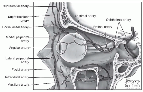
The primary vascular supply to the orbit is the ophthalmic artery (
Fig. 20-10). The artery arises from the internal carotid artery as it exits the cavernous sinus and enters the orbit through the optic canal. Multiple branches arise from the ophthalmic artery within the orbit. Branches supplying ocular function include the central retinal artery, the ciliary artery, and the collateral branches to the optic nerve. Nonocular orbital branches include the lacrimal artery, the muscular arteries, and the periosteal branches. Nonocular extraorbital branches include the anterior and the posterior ethmoid arteries, the supraorbital artery, the medial palpebral artery, the supratrochlear artery, and the dorsal nasal artery. Anastomoses between the ophthalmic branches and branches of the external carotid artery system exist, namely, the superficial temporal artery and the angular arteries. Additional anastomoses exist between the cavernous portion of the internal carotid artery and the ophthalmic artery via the inferolateral trunk. Anastomoses additionally exist with the internal maxillary artery via the middle meningeal artery. The infraorbital artery is a branch of the internal maxillary artery and enters the orbit
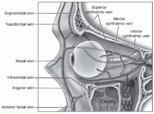
through the inferior orbital fissure. It travels across the floor of the orbit and exits the skull at the infraorbital foramen. The venous supply to the orbit is represented in
Figure 20-11.
Extraocular Muscles
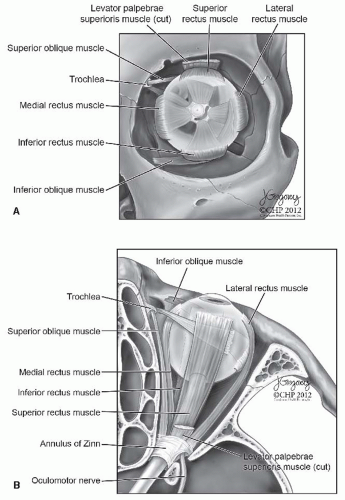
The annulus of Zinn is a ring of tight fibrous tissue at the orbital apex separating the superior orbital fissure into intra and extraconal spaces. The annulus acts as the origin site for the four rectus muscles (superior, medial, inferior, lateral) and the superior oblique muscle (
Fig. 20-12). Innervation of the extraoccular muscles are conferred by the oculomotor nerve with the exceptions of the trochlear nerve (superior oblique muscle) and the abducens nerve (lateral rectus muscle). The innervation courses in the intraconal space and enters the muscles at approximately the posterior one-third junction. The medial and superior recti originate adjacent to the insertion of the annulus to the body of the sphenoid bone adjacent to the optic foramen. The medial rectus courses adjacent to the lamina papyracea before inserting on the globe, making it prone to surgical and traumatic involvement from injury to the lamina as well as contiguous spread from ethmoid sinus neoplasms. The superior rectus runs inferior to the levator palpebrae superioris before attaching to the globe. The latter forms an aponeurotic band in the region of Whitnall ligament and inserts onto the tarsal plate of the eyelid skin and the medial and lateral canthal tendons. The inferior rectus originates from a tendinous portion of the annulus that spans the body and greater wing of the sphenoid bone. The muscle runs close to the orbital floor, especially in its posterior course, intimating a relationship with maxillary sinus pathology. The lateral rectus
originates from the body of the greater wing of the sphenoid near the lateral border of the superior orbital fissure and is separated from the optic nerve posteriorly by the ciliary ganglion. The insertion of the lateral rectus on the orbit is inferior to the lacrimal gland, the artery, and the nerve. The superior oblique muscle arises from the superomedial aspect of the annulus courses anteriorly through the trochlea before inserting in the posterolateral surface of the globe. The inferior oblique uniquely does not originate from the annulus of Zinn but rather arises from the periorbita of the inferomedial orbit lateral to the nasolacrimal duct. It courses lateral and posteriorly below the inferior rectus to insert on the inferolateral surface of the globe.
Lacrimal Gland
The oval-shaped, bilobed lacrimal gland is located adjacent to the globe in the superior-lateral aspect of the orbit in a depression of the frontal bone termed the lacrimal gland fossa (
Fig. 20-13). A pseudocapsule invests the gland. The orbital lobe of the lacrimal gland is approximately 2 to 3 times larger and is positioned posterior-superior to the smaller palpebral lobe. The two lobes are separated by the lateral horn of the levator aponeurosis, which along with Whitmall ligament and periostial fibers (Soemmering ligaments) provide support to the gland. Multiple excretory ducts exist for each lobe and ultimately pass through the palpebral lobe to reach the superior lateral conjunctival fornix, 5 mm above the superior tarsal margin. The boundaries of the lacrimal gland include the lateral rectus inferiorly, the orbital septum anteriorly, orbital fat posteriorly, and the globe medially. The serous function of the lacrimal gland is mediated by the columnar and myoepithelial cells that comprise the acini. The secretions formed in the acinar system drain into the intralobular space, followed by the interlobular space and ultimately into the main ducts. Arterial supply to the lacrimal gland is primarily from the lacrimal artery, a branch of the ophthalmic artery. Anastomoses between the lacrimal artery and the external
carotid artery system additionally exist including the middle meningeal artery, the anterior deep temporal artery, and, potentially, the infraorbital artery. Venous drainage is via the superior ophthalmic vein.
The complex pathway of the parasympathetic and the sympathetic innervations of the lacrimal gland initiates in the lacrimal nucleus in the pons and travels through the cerebellopontine angle as the nervus intermedius. The fibers course through the geniculate ganglion and exist as the greater superficial petrosal nerve. This merges with the deep petrosal nerve (sympathetic fibers from the internal carotid plexus) to form the Vidian nerve. Synapsing of the parasympathetic fibers occurs in the pterygopalatine ganglion. Postganglionic fibers travel via the zygomatic nerve and merge with the lacrimal nerve, a sensory branch of the ophthalmic division of the trigeminal nerve, prior to penetration of the lacrimal gland.
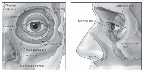
Lacrimal Drainage System
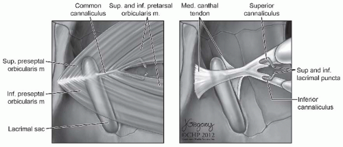
Flow of tears away from the nasal bulbar conjunctival surface through the lacrimal system begins at the inferior and superior lacrimal puncta (
Fig. 20-14). These openings into the lacrimal canaliculi face posteriorly toward the globe, are approximately 0.3 mm in diameter, and are separated by approximately 6 mm from the medical canthus. The puncta are positioned on an elevation termed the papilla lacrimalis. The paired upper and lower canaliculi traverse the distance from the puncta to the lacrimal sac. The canaliculi are lined by nonkeratinizing, stratified squamous epithelium surrounded by elastic tissue. The canaliculi have an initial vertical segment (2 mm) followed by an approximately 90-degree bend termed the ampulla and continue with the horizontal segment (8-10 mm). The common canaliculus is formed by the merger of the horizontal segments. The common
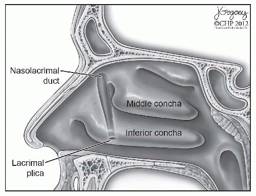
canaliculus pierces the lacrimal fascia to enter the lacrimal sac through the valve of Rosenmüller. The sinus of Maier is the dilation of the common canaliculus near the entrance to the lacrimal sac. The lacrimal fossa houses the lacrimal sac and is bounded by the anterior (frontal process of maxilla) and the posterior (lacrimal bone) lacrimal crests in the anterior-medial portion of the orbit. Visualization of the lacrimal sac through an intranasal approach, as is done in dacryocystorhinostomy, can be achieved by removal of the lacrimal bone in the superior aspect of the lateral nasal wall, anterior to the uncinate process. The lacrimal sac is divided into a superior portion termed the fundus and an inferior portion termed the body. The lacrimal fascia, an extension of the orbital periosteum, envelops the lacrimal sac and the nasolacrimal duct. Attaching fibers of the orbicularis oculi muscle are immediately deep to the fascia layer around the lacrimal sac. The nasolacrimal duct connects the lacrimal sac to the inferior meatus opening into the nasal cavity, termed Hasner valve. The nasolacrimal duct has a 12-mm superior intraosseous portion and a 5-mm inferior membranous portion and has a slight posterior-lateral trajectory as it enters the nose (
Fig. 20-15). The approximate diameter of the duct is 1 mm. The lacrimal sac and the nasolacrimal duct are lined by stratified columnar epithelium with foci of ciliated cells and interspersed goblet cells.
NASAL AND PARANASAL NEOPLASMS—NATURAL HISTORY, PATHOLOGY, PATHOGENESIS, AND PATTERNS OF SPREAD
Natural History
Sinonasal malignancies are rare, comprising fewer than 3% of upper aerodigestive neoplasms. The most common malignancy, squamous cell carcinoma (SCC), has an estimated incidence of <1:100,000.
2,3 The rarity of these lesions limits epidemiologic data and formation of prospective, randomized treatment protocols. Hence, the literature on sinonasal malignancies is largely composed of single institutional, retrospective case series. The origin of these cancers is from the maxillary sinus in 60% to 70%, from the nasal cavity in 20% to 25%, and the ethmoid sinus in 10% to 15% of cases. Malignancies arising from the frontal and sphenoid sinuses are extraordinarily unusual, making up fewer than 5% of all cases. At presentation, the majority of lesions extend beyond the site of origin.
4,5 Sinonasal malignancy most commonly occurs in patients older than 40 years of age and has a slight male preponderance.
2,3,4The majority of treatment failures are related to local recurrence. Lymphatic metastasis at initial presentation for sinonasal malignancy is uncommon. At the time of initial diagnosis, a 10% to 20% rate of lymphatic involvement is noted in patients with carcinoma of the maxillary sinus. The incidence increases up to 35% during the course of the disease.
6,7,8,9,10 Lymphatic spread for lesions originating from the ethmoid sinus is less common and ranges from approximately 5% to 10%.
11,12,13 The propensity for lymphatic and distant spread of disease additionally varies based on the tumor histology and grade. The retropharyngeal location of the majority of lymphatics implies inaccessibility during routine examination and is evaluated by imaging studies. Additionally, the surgical treatment implications for this pattern of spread are significant as the first echelon nodes are not routinely accessible at the time of resection and are better addressed with radiation therapy. The unusual presentation of a sinonasal cancer with a cervical metastasis may portend a locally advanced lesion and/or aggressive histology.
Benign Lesions
The differential diagnosis of sinonasal lesions is extensive and requires multimodality evaluation including endoscopic examination, radiography, and histopathology. Although this chapter primarily discusses malignancy, certain nonneoplastic processes can mimic the former and must be considered at the time of evaluation. One of the central challenges of successful detection of a sinonasal malignancy is differentiation from more common, inflammatory conditions including allergic rhinitis and sinusitis. Adding confusion to the issue is the frequent coexistence of sinusitis and neoplasm. Tumor growth may obstruct sinonasal outflow tracts and precipitate secondary infection. Older age at presentation, unilateral disease, symptoms, and examination findings consistent with aggressive behavior are more suggestive of malignancy and should increase clinical suspicion. These issues highlight the importance of close examination of preoperative imaging and collection of all sinus specimens for histology in patients undergoing surgery for presumed inflammatory sinusitis. An expanded discussion of two benign conditions, inverted papilloma and juvenile nasopharyngeal angiofibroma, is presented below given their clinical importance and the specific challenges associated with these lesions. The salient features of the other benign and inflammatory lesions that should be considered in the evaluation of a patient with a sinonasal lesion are presented in
Table 20.1.
Inverted Papilloma
Several different types of papilloma are identified in the sinonasal cavity. Squamous papilloma related to human papillomavirus (HPV) infection has a predilection for the nasal vestibule. Although local excision is considered curative, surveillance of the initial site and the remainder of the upper aerodigestive tract is required. Three different types of papilloma arising from the Schneiderian (respiratory) mucosa have been described. Although these lesions differ in their anatomic predilection and clinical behavior, the ultimate distinction requires histopathological analysis. Everted papilloma (fungiform) typically arise from the septum and are curable with local resection. Everted fronds of epithelium are noted on histology. Cylindric papillomas are rare and are similar in histology to everted papilloma. These latter two lesions are not felt to have significant malignant potential.
Inverted papilloma most commonly arise from the lateral nasal wall of the nasal cavity but may occur anywhere in the sinonasal tract. The overall incidence is estimated to be between 0.5 and 1.5 cases per 100,000 per year.
14,15 The etiology of inverted papilloma remains unclear and theories suggesting inflammatory and viral (HPV) origin have been described.
16,17 The name arises from the characteristic histological finding of infolded projections of the epithelium into the underlying stroma. Recurrence after treatment may occur in up to 20 % of lesions even with aggressive initial surgery. The potential for synchronous or metachronous transformation to squamous cell carcinoma has been described. A wide range of transformation rates has been described, with the majority of series reporting a rate of approximately 10%.
18 There is some uncertainty of the number, however, because carcinoma
in situ changes occur on the surface of the papillomas. To condemn such lesions as malignant papilloma may be an overstatement, especially since the percentage of them that go on to full-blown malignancies is unknown.
Although medial maxillectomy through a transfacial, lateral rhinotomy approach has historically been considered the treatment of choice for inverted papilloma, application of endoscopic techniques for resection of these lesions has demonstrated favorable results. Avoidance of an external incision is an obvious advantage over the more traditional procedures.
19,20,21 Additionally, the endoscopic approach can be tailored to the specific site and extent of lesion, allowing for complete tumor removal and preservation of functional sinus physiology (
Fig. 20-16). Although unsubstantiated by conclusive data, most experienced endoscopic surgeons believe the critical factor associated with successful surgical management of inverted papilloma lesions is identification of the site of origin and drilling of the underlying bone. This is greatly facilitated with the panoramic visualization
provided by endoscopy. In a large meta-analysis of surgical treatment for inverted papilloma, Hwang et al. reported a lower rate of recurrence (12% vs. 20%) in patients treated endoscopically versus conventional surgical means.
22
Juvenile Nasopharyngeal Angiofibroma
Juvenile nasopharyngeal angiofibroma (JNA) accounts for approximately 0.05% of all head and neck tumors and has an estimated incidence of 1:150,000.
23,24 These lesions occur exclusively in males and are most common in the second decade of life, likely secondary to androgen hormone responsiveness. The most common site of origin is the area around the sphenopalatine foramen and multiple potential patterns of local spread may exist. Medial spread occurs into the sinonasal cavity and nasopharynx with limited resistance. Lateral extension occurs into the pterygopalatine and infratemporal fossa. Posterior-superior growth may involve the sphenoid sinus and cavernous sinus. Erosion of the sphenoid bone may result in intraorbital and intracranial extension (middle cranial fossa). The most common presenting symptoms include nasal obstruction, epistaxis, facial pain, headache, and facial swelling. Symptoms related to orbital, sinonasal, and Eustachian tube mass effect may also occur. Endoscopic examination reveals a hypervascular appearing soft tissue mass.
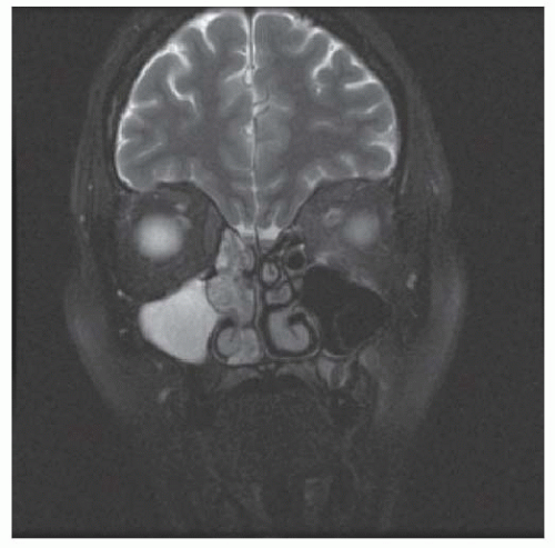
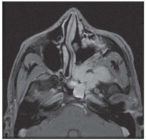
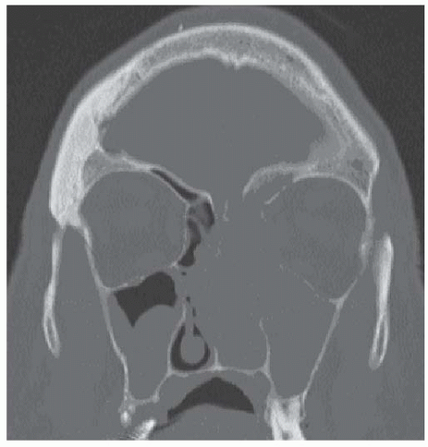
23A combination of CT and contrast-enhanced MRI are used to characterize JNA lesions including the soft tissue extension, bony erosion, anterior bowing of the posterior maxillary wall (Holman-Miller sign), and vascularity (
Fig. 20-17). The characteristic imaging studies combined with patient demographics is typically adequate for pretreatment diagnosis. Office-based biopsy is associated with the potential for extraordinary hemorrhage and should not be performed for suspected JNA. Histopathology from surgical specimens demonstrates a complex and heterogeneous proliferation of blood vessels with absent or thin elastic laminae and variably thick vascular walls. A background collagen matrix with spindle-shaped cells is noted around the blood vessels.
25 Extensive vascular arcades may be identified on angiography with the predominant vascular supply arising from the internal maxillary artery and a minor contribution possibly arising from the ascending pharyngeal artery, the internal carotid artery, and the contralateral external carotid artery system. Several staging systems have been proposed as described by Sessions, Chandler, and Fisch.
26,27,28 Staging is essential for reporting of treatment outcomes and possibly for predicting the prognosis for recurrence. For clinical purposes, the critical aspects of “advanced” disease that impact surgical planning include extension to the infratemporal fossa, the orbit, the cavernous sinus, and the middle cranial fossa.
Surgery represents the primary treatment modality for JNA. Although local control has been associated with radiation therapy, the potential long-term adverse effects in this young population (secondary malignancy, oribital and intracranial dysfunction) limit its use to unresectable and enlarging lesions.
29,30,31 Not surprising, given the complex anatomic location of JNA lesions, multiple surgical approaches have been described. The hypervascularity of these lesions is counteracted by presurgical angiography and emobilization. Choice of the available surgical approaches is based on the location and extent of the lesion. Open skull base approaches include midface degloving, facial translocation, lateral rhinotomy, transpalatal, craniofacial, and subcranial approaches. The application of expanded endoscopic techniques has rapidly emerged as a meaningful primary treatment modality for the majority of JNA lesions. Excellent tumor control rates have been reported from multiple centers with an overall favorable perioperative morbidity profile including decreased blood loss, shorter hospital stay, and avoidance of facial incisions. Lateral access to the infratemporal fossa and masticator space can be achieved with limited additional morbidity by the inclusion of a sublabial incision and a limited bony opening through the maxillary antrum.
32,33,34,35,36Regardless of the approach, a significant risk of recurrence is associated with JNA with reported rates ranging from 15% to 45%. The likelihood of recurrence correlates with the stage and location of the primary lesion especially if there is bony skull base involvement, prior history of recurrence, and an incomplete resection of both the soft tissue and bony components. Routine surveillance imaging with MRI scan is advocated to identify early recurrence of lesions.
37,38,39
