Cancer Biology


Cancer Biology


CANCER HALLMARKS & STEM CELLS

Alexander Drilon


Hallmarks of Cancer


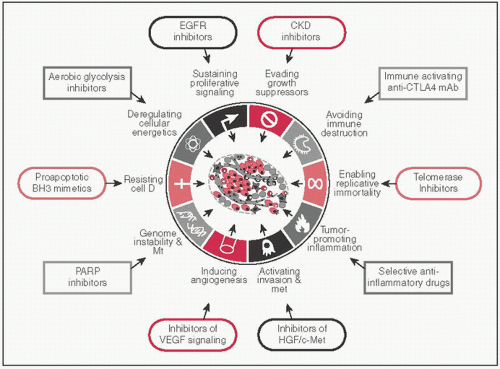
  • CAs share a set of common distinctive & complementary capabilities that enable tumor growth & met dissemination (Cell 2011;5:646)


Figure 4-1


Cancer Stem Cells


  • Characterized by self-renewal & ability to differentiate; found in a variety of CAs including AML, CML, gliomas, breast CA (NEJM 2006;355:1253)


  • Two pathways involved in regulation of pattern formation during ontogeny


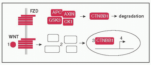
    • Wnt pathway: (1) WNT ligands bind to FZD (Frizzled) receptor, (2) disrupts assembly of a complex (AXIN, APC, CK1, GSK3) that tags CTNBB1 (β-catenin) for degradation, (3) CTNBB1 moves to nucleus → binds TCF/LEF freeing it from TLE (Groucho), (4) CTNBB1 & TCF/LEF activate target genes (eg, CCND1, MYC); FAP develops to germline inactivating Mt of APC (Nat Rev Cancer 2013;13:11)


      Figure 4-2


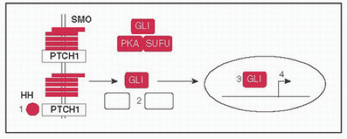
    • Hedgehog pathway: (1) HH ligands SHH (Sonic), DHH (Desert), & IHH (Indian) bind to PTCH1 (Patched) releasing inhibitory effect on SMO (Smoothened), (2) causes dissociation of protein complex including SUFU, PKA, & GLI, (3) GLI translocates to nucleus to (4) activate target genes; BCCs w/activating SMO Mt or inactivating PTCH1 Mt, Gorlin syndrome (BCCs, medulloblastomas, rhabdomyosarcomas) w/germline PTCH1 Mt (Nat Rev Cancer 2008;8:743)


      Figure 4-3


MECHANISMS OF ONCOGENESIS

Alexander Drilon

Carcinogenesis often proceeds in a multistep fashion w/the acquisition of several genomic aberrations via Mt, amp, rearrangements, & infxn


Mutations


  • Alterations in DNA sequence or structure to replication errors or carcinogens: Mt may be activating or inactivating


Type of Mutation


Examples


POINT Mt: Base substitution


transition G:C ⇔ A:T


transversion A:T ⇔ T:A, G:C ⇔ C:G


Can be silent (no change in AA) nonsense (early stop codon) missense (change in AA)


JAK2 V617F in polycythemia vera (PCV), ET


BRAF V600E in melanoma, hairy cell leukemia (HCL)


KRAS G12A in CRC, NSCLC


EGFR L858R in NSCLC


Note: 1st letter—original AA, number—AA position, 2nd letter—substituted AA, (EGFR L858R, ie, leucine at position 858 replaced w/arginine)*


DELETION: Deletion of one or more bases or larger stretches of material w/in chromosomes


EGFR exon 19 del in NSCLC


5q- syndrome in MDS


INSERTION: Insertion of one or more bases


ERBB2 (HER2) exon 20 ins in NSCLC


* Amino Acid Nomenclature: Arg (R), His (H), Lys (K), Asp (D), Glu (E), Ser (S), Thr (T), Asn (N), Gln (Q), Cys (C), Sec (U), Gly (G), Pro (P), Ala (A), Val (V), Ile (I), Leu (L), Met (M), Phe (F), Tyr (Y), Trp (W)


DNA Amplification


  • Selective ↑ in the number of copies of a gene (copy number alteration), mechanism by w/c the expression of a gene is enhanced above physiologic levels


  • Clinically relevant egs:


    • ERBB2 (HER2) Amp: BC (average of >6 gene copies/nucleus or HER2/CEP17 ratio >2.2 via FISH), gastric CA, predictive of response to HER2-targeted therapies (Nat Rev Clin Oncol 2011;9:16)


    • FGFR1 Amp: In squamous cell NSCLCs (Sci Transl Med 2010;2:62ra93)


Chromosomal Rearrangements


  • Result in creation of a gene fusion w/potent oncogenic activities: Novel sequences are juxtaposed to coding sequence of a TK domain or transcription factor → constitutive activation of TK or downstream target genes of transcription factor


Type


Examples


Translocation: Rearrangement of material between nonhomologous chromosomes


image


Reciprocal—even exchange of material


Robertsonian—2 acrocentric chromosomes fuse near centromere w/short arm loss


BCR-ABL1 t(9;22) in CML/Ph+ALL


PML-RARA t(15;17) in APL


NPM1-ALK t(2;5) in ALCL


RUNX1-RUNX1T1 t(8;21) in AML M2


EWSR1-FLI1 t(11;22) in Ewing sarcoma


Inversion: Chromosomal segment is reversed end-to-end (pericentric—includes centromere vs. paracentric—doesn’t include centromere)


image


EML4-ALK t(2;2) in NSCLC


CBFB-MYH11 inv(16) in AML M4 w/eosinophilia


Deletion: Genetic material lost w/in a chromosome (interstitial)


image


TMPRSS2-ERG del(21) in prost CA


FIP1L1-PDGFRA in hypereosinophilic syndrome


Infections


  • Play important roles as carcinogens in several diseases (see Cancer Epidemiology Chap. for more extensive list), carcinogenesis occurs through a variety of mechanisms eg, via the actions of viral/bacterial proteins, insertional mutagenesis


    • Viruses: HPV (cervical CA, oropharyngeal squamous cell CAs of the head & neck, viral proteins E6 & E7 inhibit TP53 & Rb1, respectively), EBV, KSHV


    • Bacteria: H. pylori (gastric CA), bacterial CagA protein → activates tyrosine phosphatase SHP2 affecting various pathways involved in cell migration, adhesion, & apoptosis


GROWTH FACTOR SIGNALING

Alexander Drilon


Oncogenes vs. Tumor Suppressor Genes (TSGs)


  • Oncogenes: Contribute to the development or maintenance of the neoplastic phenotype, converted from protooncogenes w/c have nl functions that are involved in cell differentiation, proliferation, & apoptosis → these may sustain gainof-function or activating Mt that result in constitutive activation (NEJM 2008;358:502)


    • Oncogene addiction: Process by w/c tumor cells become highly dependent on an oncogenic protein or pathway for sustained proliferation or survival, driver Mt in oncogenes are often mutually exclusive (Cancer Res 2008;68:3077)


    • Many oncogenes encode growth factor receptors or proteins involved in cell signaling


  • TSGs: Encode proteins responsible for inhibiting tumor formation or maintenance, involved in processes like DNA repair & apoptosis


Growth Factor Signaling


  • Initiated by ligand binding to growth factor receptors that activate downstream pathways; mechanism by w/c an extracellular signal is transduced through a cell, resulting in a change in gene expression, modifying specific cellular processes
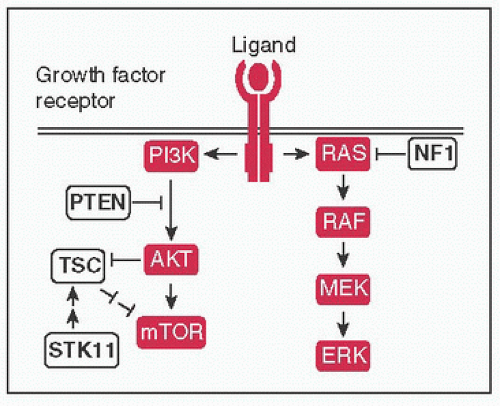


    Figure 4-4 Growth factor signaling: Ligand binding (eg, EGF, TGFα, epiregulin) → receptor dimerization & phosphorylation of intracellular TK domains → downstream pathway activation; ↑ pathway activity mediated by signal transduction proteins (protooncogenes) of MAPK and PI3K pathways, negative regulators (TSGs) shown in white → ultimately affect processes such as cell survival & proliferation, differentiation, angiogenesis, etc.


  • Can be targeted by mAbs (eg, bevacizumab for VEGF, cetuximab for EGFR, see Monoclonal Antibodies Chap.) or small molecules (eg, TKIs)


Growth Factor Receptor


Role in Carcinogenesis/Examples


ERBB family: Structurally related receptors, capable of forming homo or heterodimers, contain TK domains (except for ERBB3 or HER3) (Nat Rev Cancer 2005;5:341)


EGFR (ERBB1)


ERBB2 (HER2)


ERBB3 (HER3) & ERBB4 (HER4)


EGFR Mt in lung CA, EGFR overexpressed in lung adenoCA, glioblastoma, HNSCC


ERBB2 (HER2) Amp & ERBB2 (HER2) overexpression: breast CA & gastric CA


Dimerize w/other family members, overexpressed in a number of malignancies


FGFRs: FGFR1, FGFR2, FGFR3, FGFR4 (Nat Rev Drug Discov 2009;8:235)


FGFR1 Mt in glioblastomas & Amp in squamous NSCLC


FGFR3 Mt & FGFR3 overexpression in bladder CA


MET (HGFR)


MET Mt in sporadic/hereditary papillary RCCs


KIT


KIT Mt in GIST, melanoma, mast cell leukemia


PDGFRA


PDGFRA Mt in GIST, role in angiogenesis


VEGFR


Angiogenesis, overexpressed in a variety of CAs


IGF1R


Insulin signaling, potential predisposition to several CAs

Only gold members can continue reading. Log In or Register to continue

Stay updated, free articles. Join our Telegram channel

Jun 19, 2016 | Posted by in ONCOLOGY | Comments Off on Cancer Biology

Full access? Get Clinical Tree

Get Clinical Tree app for offline access