Skin Cancers and Sarcomas


Skin Cancers and Sarcomas


BASAL AND SQUAMOUS CELL CARCINOMAS

Parisa Momtaz

Allan C. Halpern


Epidemiology


  • Most common CA in US; collectively known as nonmelanoma skin CA


  • BCC is 5× more common than SCC


  • Rarely met, however, can be locally aggressive & disfiguring; SCC w/greater potential for met


Risk Factors/Pathogenesis


  • Sunlight: UVB strongly correlated w/SCC; correlation w/BCC is more complex


  • Fair-skinned individuals at greatest risk


  • Radiation exposure at a young age


  • Prolonged PUVA exposure


  • Immunosuppression (organ transplantation)


  • For SCC, other RFs include HPV (types 16, 18, 31, 33, 38), chemical carcinogens


  • SCC arise from keratinocytes


  • Most BCC arise from the epidermal cells differentiated in the primitive hair bulb


Genetics


  • Mts in BCC frequently involve the PATCH gene or members of the sonic hedgehog signaling pathway → overexpression of transcription factor Gli 1


  • Specific UV-induced Mts in the tumor suppressor p53 gene


  • Genetic syndrome predispose to BCC, SCC: Albinism, xeroderma pigmentosum, Nevoid BCC syndrome


  • Nevoid BCC syndrome (aka basal cell nevus syndrome, Gorlin-Goltz syndrome)


Nevoid BCC syndrome


Autosomal dominant; Mt in the PATCH gene at 9q22.3


Multiple BCC, starting in childhood


Palmoplantar pits, epidermoid cysts, lipomas


Developmental anomalies: Odontogenic cysts of the maxilla & mandible, dental defects, bifid ribs, scoliosis, hypertelorism, frontal bossing


Associated tumors: Medulloblastoma, ovarian fibromas, cardiac fibromas, mesenteric lymphatic cysts


Clinical Features


  • Most develop on head & neck (80%) & are more likely to recur compared to lesions developing on the trunk or extremities


  • SCC can arise on mucous membranes, BCC do not


  • SCCs appear as hyperkeratotic papules or plaques often w/crust or ulceration


  • SCC precursor lesions: Actinic keratoses, cutaneous horn, keratoacanthoma


  • Bowen Disease: SCC in situ on the skin


  • Basosquamous carcinomas: Mixed histology of both BCC & SCC; classified under SCC & risk of met determined by the squamous component


Types of BCC


Nodular BCC


Most common variant


Pearly, telangiectatic dome-shaped nodule often w/central


Ulceration


Superficial or multicentric BCC


Flat, tan/red patch usu on the trunk; often h/o radiation or arsenic exposure


Pigmented BCC


Both nodular & superficial BCCs can be pigmented, more common in darker-skinned individuals


Sclerosing BCC


Atrophic plaque, typically around nose or ears


Ulcus terebrans


Extremely aggressive BCC, invades underlying structures, frequently fatal but rare


Staging: BCC and SCC


Stage 0


Carcinoma in situ


Stage I


Tumor size ≤2 cm, <2 high-risk features


Stage II


Tumor size >2 cm, ≥2 high-risk features


Stage III


Tumor w/invasion of mandible, maxilla, orbit, or temporal bone & no nodal Involvement; OR tumor of any size & ipsilateral nodal involvement ≤3 cm


Stage IV


Ipsilateral nodal involvement >3 cm, bilateral or contralateral nodal involvement, multiple ipsilateral LN involvement, LN ≥6 cm, distant met


High-risk features: >2-mm thickness, Clark level IV, perineural invasion, primary site ear, primary site nonhair-bearing lip, poorly differentiated or undifferentiated


Treatment Localized Disease: BCC, SCC


  • Surgical excision w/histologic control of margins: Excision w/POMA, Mohs surgery


  • Electrodessication & curettage for low-risk lesions


  • Cryotherapy for pre-SCC lesions


  • LND if regional LN involvement


  • Adjuvant RT for pts who have undergone LND; o/w use of radiation Rx controversial; consider for positive margins, evidence of substantial perineural involvement


  • Superficial therapies: 5-FU, imiquimod, PDT


Treatment Metastatic Disease: BCC


  • Rare; however, systemic chemotherapy is indicated


  • Clinical trials


  • Vismodegib, inhibitor of the hedgehog pathway for residual or met disease (NEJM 2012;366:2171)


  • Platinum-based chemotherapy regimens


Treatment Metastatic Disease: SCC


  • Limited data; CIS-based regimens, cetuximab


  • Clinical trials


  • If transplant pt on immunosuppressive Rx, can consider adjusting dose of immunosuppressive Rx if appropriate


Prognosis and Follow-up


  • Localized disease has good prognosis


  • Worse prognosis for SCC of the genitalia, mucous membranes


  • Perineural involvement increases the risk of recurrence for BCC & SCC & increases met risk for SCC


  • Close surveillance for high-risk pts (immunosuppression, organ transplantation)


  • Encourage sun protection strategies & self skin checks


  • Pts w/h/o BCC or SCC are likely to develop more lesions; perform annual or bi-annual skin exams


Figure 16-1 SCC courtesy of Dr. Allan Halpern (MSKCC)


Figure 16-2 BCC courtesy of Dr. Allan Halpern (MSKCC)


MELANOMA

James J. Harding

Paul B. Chapman


Epidemiology


  • Incidence: ˜76000 new cases/y & ˜10000 death/y in US


  • Only a small % of all skin CA (˜4%) but has the highest morbidity & mortality


  • ↑ Incidence, affects younger individuals


  • Acquired RFs: Higher number of moles, atypical nevi, hx of prior melanoma, 1st-degree relative w/melanoma, prior nonmelanoma skin CA, freckles, sunburns easily, UVA/UVB exposure (esp before age 15), hx of indoor tanning, red-hair, immunosuppression


  • Hereditary RFs: Rarely inherited, some syndrome include: Familial atypical multiple mole melanoma syndrome (CDKN2A, p16), Xeroderma pigmentosum (Mts in NER), LFS (p53), RB1, BRCA2, PTEN (Cowden syndrome), WRN (Werner syndrome), BAP1 (syndrome of ocular & cutaneous melanoma & mesothelioma)


Pathology


  • Histologic Subtypes


    • Cutaneous (most common)


      • Superficial spreading: Most common, radial growth


      • Nodular: Vertical growth into dermis, worse prognosis


      • Lentigo maligna: Sun-damaged skin in elderly or middle-aged pts, usu on face


      • Acral lentiginous: Most common in Asians or African Americans, usu on palms or soles


      • Desmoplastic/Neurotropic: Locally invasive, less likely to metastasize, CN involvement


    • Uveal: Arise from melanocytes in iris, ciliary body or choroid


    • Mucosal: Arise from melanocytes in mucosal surfaces (ie, nasopharynx, anus, vagina)


  • Molecular Subtypes


    • The majority of melanomas are driven by overactivation of the MAPK pathway


      • BRAF V600 (˜50-60%): V600E >V600K, commonly observed in younger pts w/nodular/superficial spreading melanoma of the trunk. Sensitive to vemurafenib or dabrafenib


      • NRAS (15-20%): No specific targeted Rx available (MEK inhibition in clinical trials)


      • KIT: ↑ In mucosal, chronically sun-damaged skin, acral sites (˜20%); similar Mts as in GIST


      • GNAQ/GNA11: Not observed in cutaneous melanomas, ↑ in uveal melanomas (>80%), activates heterotrimeric G-protein coupled receptors → activates MAPK pathway


      • Other Mts: PIK3CA/AKT, PTEN loss, NF-1, & BAP1


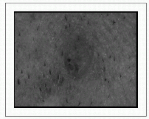
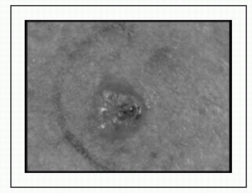
Clinical Manifestations


  • Sx: Cutaneous: Most melanomas found by pt at early stage; adv disease → enlarged LN, skin nodules, sx related to lung, liver, bowel/mesenteric, or brain mets; uveal: Incidental finding or visual complaints; mucosal: Mucosal bleeding


  • PEx: Evaluate skin & mucosal surfaces; ABCDEs: Asymmetry, Borders irregular, Color variegated, Diameter >5 mm. Evolution; check for LAN or subcutaneous met, sequela of widespread met disease, melanosis → rare, blackening of the skin & urine due to ↑ melanin production, poor prognosis


  • Labs: Microcytic anemia, suspect chronic blood loss from bowel mets; ↑ LDH


Diagnostic Evaluations and Staging


  • Punch bx preferred over shave bx; shave bx can transect the tumor & prevent proper depth assessment (T-staging)


  • EOD w/CT-CAP or CT-Chest w/PET (especially if lesions is in the distal extremity), if distant disease, FNA or Core bx


  • Baseline MRI-brain w/gadolinium for Stage III disease or higher to r/o intracranial met.


  • Evaluation/staging/tx of uveal melanoma is different


AJCC Staging of Cutaneous Melanoma (JCO 2009;27:6199)


Stage (˜% at DX)


Tumor


Node


Mets


5-y OS


IA/B


≤1 mm or 1.01-2 mm w/o ulceration


None


None


90-100%


IIA-C (I/II = 80-85%)


1.01-2 mm w/ulceration, or any ≥2.01 mm


None


None


˜50-80%


IIIA-C (10-15%)


Any T


Regional LN or in transit mets


None


˜20-70%


IV(1A-1C) (5%)


Any T


Any N


Distant Mets


<10-40%*


* Long-term OS includes pts w/indolent biology or eligible for metastasectomy


Treatment of Localized Cutaneous Melanoma (Stages I-III)


  • Wide local excision of : Margin of 1 cm if tumor ≤1 mm deep, margin of 2 cm for all other lesions


  • Sentinel LN mapping & bx: Lymphoscintigraphy w/Tc99 colloid, usu used for lesions >1 mm, if <1 mm consider if high-risk features


  • Completion LND: Performed if sentinel LN positive, whether this procedure improves OS is subject of ongoing phase III study


  • Adjuvant Rx & surveillance: IFN-α ↑ RFS; radiotherapy ↑ local regional control, no RFS/OS advantage; clinical trial participation; or observation (serial exam/imaging in IIB-IV NED)


Metastatic Cutaneous Melanoma


  • Metastatectomy: In properly selected pts, long-term OS 20-40%


  • Immunotherapy:


    • Ipilimumab (CTLA-4 blocking Ab):OS in 2 RCT (NEJM 2010;363:711 & NEJM 2011;364:2517), slow acting, monitor for immune-mediated tox (ie, colitis, dermatitis, hepatitis, etc.)


    • Programmed D-1 receptor: RR ˜30% (NEJM 2012;366:2443)


    • IL-2: Given in ICU setting, 2-6% pts w/durable disease control/cure


    • Adoptive cellular Rx/TILs: Experimental, high RR


  • Targeted Rx:


    • RAF inhibitors (vemurafenib, dabrafenib): Rapid RR 50-60%, ↑ PFS compare to DTIC, ↑ OS for Vem (NEJM 2011;364:2507); causes keratoacanthomas & SCC of skin


    • MEK inhibitor (trametinib):OS for trametinib (NEJM 2012;367:107); combo BRAFi + MEKi may be superior to BRAFi


    • c-KIT inhibitors (imatinib/dasatinib/nilotinib): May be particularly effective for exon 11 & exon 13 Mts


  • Chemotherapy: Dacarbazine (DTIC)/Temozolomide, RR 7-19%; Combination chemotherapy: Cisplatin, vinblastine, temozolomide (CVT) RR 30-40%, Carboplatin/Taxol RR 20-30%, no OS advantage; Biochemotherapy = chemo + IL-2 + IFN-α, has ↑ RR, no OS advantage over chemo


GASTROINTESTINAL STROMAL TUMOR

James J. Harding

William D. Tap


Epidemiology

Aug 17, 2016 | Posted by in ONCOLOGY | Comments Off on Skin Cancers and Sarcomas

Full access? Get Clinical Tree

Get Clinical Tree app for offline access