Breast: Stage Tis, T1, and T2 Tumors


Breast: Stage Tis, T1, and T2 Tumors


ANATOMY


  • The mammary gland lies over the pectoralis major muscle and extends from the second to the sixth rib craniocaudally, and from the sternum to near the midaxillary line. The breast itself consists of glandular tissue arranged in multiple lobes composed of lobules connected in ducts, areolar tissue, blood vessels, and the overlying skin (32, 87).


  • A network of lymphatics is formed over the entire surface of the chest, neck, and abdomen and becomes dense under the areola.


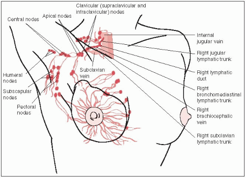
  • The following lymphatic pathways originate mostly in the base of the breast: the axillary or principal pathway passes from the upper and lower halves of the breast to the chain of nodes situated between the second and third intercostal space; the trans pectoral pathway passes through the pectoralis major muscle to the supraclavicular lymph nodes; and the internal mammary pathway passes through the ipsilateral pectoralis major and intercostal muscles (usually close to the sternum) to the nodes of the internal mammary chain (Fig. 24-1).


NATURAL HISTORY


  • As breast cancer grows, it travels along the ducts, eventually breaking through the basement membrane of the duct to invade adjacent lobules, ducts, fascial strands, mammary fat, and skin. It then spreads through the breast lymphatics and into the peripheral lymphatics; tumor can invade blood vessels.


  • About 10% to 40% of newly diagnosed stage T1 and T2 breast cancers, respectively, have pathologic evidence of axillary nodal metastases; the incidence is strongly correlated with tumor size (32).


  • Metastases to the internal mammary nodes are more frequent from inner quadrant and central lesions; they occur more often when there is axillary node involvement. Supraclavicular nodes are occasionally involved.


  • Vascular invasion by tumor and hematogenous metastases to the lungs, pleura, bone, brain, eyes, liver, ovaries, and adrenal and pituitary glands occur, even with small tumors.


  • Metachronous bilateral carcinoma of the breast occurs in 5% to 8% of patients and is simultaneous in approximately 1%.


CLINICAL PRESENTATION


  • Most patients with carcinoma in situ, T1, or T2 breast cancers present with an abnormal screening mammogram or have a painless or slightly tender breast mass.


  • Approximately 40% to 50% of these lesions are detected by mammography only; approximately 35% of tumors detected by mammography and physical examination are invasive carcinomas smaller than 1 cm (60).


FIGURE 24-1 Anatomy of the breast, with lymphatic drainage highlighted in red. (From Agur AMR, Dalley AF., eds. Grant’s atlas of anatomy, 12th ed. Philadelphia, PA: Lippincott Williams & Wilkins, 2009:740-741; with permission.)


DIAGNOSTIC WORKUP


  • The workup of a patient with an abnormal mammogram or breast mass, including complete clinical and family history, is summarized in Table 24-1. A pelvic examination should be done, if one has not recently been performed.


  • The consistency, tenderness, mobility or fixation, and size of both a breast mass and lymph nodes should be noted, including the number of the latter. The incidence of metastatic axillary lymph nodes is shown in Table 24-2. No tumor is found in 25% to 30% of patients with clinically palpable axillary nodes.


  • Mammography is invaluable in the detection of over 90% of breast cancers (17).


  • Ultrasonography, which has a sensitivity of 73% and specificity of 95%, is helpful in differentiating cysts from solid tumors (60).


  • The use of magnetic resonance imaging is rapidly increasing, and has been shown to have a role in complementing mammography for screening of high-risk women, for diagnosis of questionable findings seen on physical examination, mammography, or ultrasound, and for assessing response following neoadjuvant treatment for certain breast cancers (39).


  • In patients with stage 0, I, or II disease, the incidence of abnormal bone scan is approximately 2%; it is not routinely obtained in these patients.


  • Positron emission tomography using F-18 2-deoxyglucose is more frequently used for detection of regional lymph nodes or distant metastases (2).


PATHOLOGIC STUDIES


  • Histopathologic diagnosis is obtained by fine-needle aspiration, stereotactic core biopsy, or excisional biopsy of solid masses (59).


  • In nonpalpable lesions, needle localization with radiographic techniques is necessary to identify the tissue to be removed.


  • Estrogen (ER) and progesterone receptor (PR) assays are routinely performed on tissue obtained from patients with both invasive and noninvasive breast cancer in the United States. These parameters correlate with prognosis and tumor response to chemotherapeutic and hormonal agents.


  • The her-2-neu (c-erbB-2) proto-oncogene encodes a transmembrane protein tyrosine kinase receptor, 185 kDa; an assay to assess overexpression is routinely performed in invasive carcinomas. Overexpression is correlated with poorer prognosis (12).


TABLE 24-1 Diagnostic Work-up for Carcinoma of the Breast, Stages T1 and T2


General


History with emphasis on presenting symptoms, menstrual status, parity, family history of cancer, other risk factors


Physical examination with emphasis on breast, axilla, supraclavicular area, abdomen


Special Tests


Biopsy (core biopsy directed by physical examination, ultrasound, or mammography as indicated, or needle localization)


Radiologic Studies


Before biopsy


Mammography/ultrasonography


Chest radiographs


Magnetic resonance imaging of breast (selected cases)


After positive biopsy


Bone scan (when clinically indicated, for stage II or III disease or elevated serum alkaline phosphatase levels)


CT of chest, abdomen and pelvis for stage II or III disease and/or abnormal liver function tests


Laboratory Studies


Complete blood cell count, blood chemistry


Urinalysis


Other Studies


Hormone receptor status (ER, PR)


HER2/neu status


Consider genetic counseling/BRCA testing in selected cases


ER, estrogen receptor; PR, progesterone receptor


Source: Taken from Table 53.8 Haffty B, Buchholz TA, Perez CA. Early stage breast cancer. In: Halperin EC, Perez CA, Brady LW., eds. Principles and practice of radiation oncology, 5th ed. Philadelphia, PA: Lippincott Williams & Wilkins, 2008, with permission.


TABLE 24-2 Incidence of Metastatic Axillary Lymph Nodes in Carcinoma of Breast Correlated with Primary Tumor Size


Study (Reference)


Tumor Size° (cm)


≤0.5


0.6-1.0


1.0-2.0


2.1-3.0


3.1-5.0


Washington Universityb


3/55 (5%)


25/203 (12%)


59/294 (20%)


38/113 (34%)


9/31 (29%)


Tinnemans et al. (81)


1/13 (7.7%)


3/24 (12.5%)


13/44 (29.5%)


Silverstein et al. (72)


3/96 (3%)


27/156 (17%)


115/357 (32%)


145/330 (44%)c


Kambouris (40)


13/357 (32%)


15/25 (60%)


1/7 (14%)


Greco et al. (29)


40/306 (13%)


49/69 (71%)


Fein et al. (19)


6/68 (9%)


7/48 (15%)


16/50 (32%)


aNumber of patients with axillary metastasis/total number of patients with tumor size indicated.


bUnpublished data.


cT2 tumors (2 to 5 cm).


Source: Taken from Table 53.4 by Haffty B, Buchholz TA, Perez CA. Early stage breast cancer. In: Halperin EC, Perez, CA, Brady LW, eds. Principles and practice of radiation oncology, 5th ed. Philadelphia, PA: Lippincott Williams & Wilkins, 2008, with permission.


STAGING SYSTEMS


  • The American Joint Committee on Cancer staging system (3) is the most widely used system in the United States (Table 24-3).


TABLE 24-3 American Joint Committee on Cancer Staging System for Breast Cancer


Primary Tumor (T)


TX


Primary tumor cannot be assessed


T0


No evidence of primary tumor


Tis


Carcinoma in situ.


Tis (DCIS)


DCISa


Tis (LCIS)


LCISa


Tis (Paget’s)


Paget’s disease of the nipple is NOT associated with invasive carcinoma and/or carcinoma in situ (DCIS and/or LCIS) in the underlying breast parenchyma. Carcinomas in the breast parenchyma associated with Paget’s disease are categorized based on the size and characteristics of the parenchymal disease, although the presence of Paget’s disease should still be noted


T1


Tumor ≤20 mm in greatest dimension


T1mi


Tumor ≤1 m in greatest dimension


T1a


Tumor >1 mm but ≤5 m in greatest dimension


T1b


Tumor >5 mm but ≤10 mm in greatest dimension


T1c


Tumor >10 mm but ≤20 mm in greatest dimension


T2


Tumor >20 mm but ≤50 mm in greatest dimension


T3


Tumor >50 mm in greatest dimension


T4


Tumor of any size with direct extension to the chest wall and/or to the skin (ulceration or skin nodules)b


T4a


Extension to the chest wall, not including only pectoralis muscle adherence/invasion


T4b


Ulceration and/or ipsilateral satellite nodules and/or edema (including peau d’orange) of the skin which do not meet the criteria for inflammatory carcinoma


T4c


Both T4a and T4b


T4d


Inflammatory carcinomac


Regional Lymph Nodes (N)


NX


Regional lymph nodes cannot be assessed (e.g., previously removed)


N0


No regional lymph node metastasis


N1


Metastases to movable ipsilateral level I, II axillary lymph node(s)


N2


Metastases in ipsilateral level I, II axillary lymph nodes that are clinically fixed or matted; or in clinically detectedd ipsilateral internal mammary nodes in the absence of clinically evident axillary lymph node metastases


N2a


Metastases in ipsilateral axillary lymph nodes fixed to one another (matted) or to other structures


N2b


Metastases only in clinically detectede ipsilateral internal mammary nodes and in the absence of clinically evident axillary lymph node metastases


N3


Metastases in ipsilateral infraclavicular (level III axillary) lymph node(s) with or without level I, II axillary lymph node involvement; or in clinically detectedd ipsilateral internal mammary lymph node(s) with clinically evident level I, II axillary lymph node metastases; or metastases in ipsilateral supraclavicular lymph node(s) with or without axillary or internal mammary lymph node involvement


N3a


Metastases in ipsilateral infraclavicular lymph node(s)


N3b


Metastases in ipsilateral internal mammary lymph node(s) and axillary lymph node(s)


N3c


Metastases in ipsilateral supraclavicular lymph node(s)


Pathologic Classification (pN)


pNX


Regional lymph nodes cannot be assessed (e.g., previously removed or not removed for pathologic study)


pN0


No regional lymph node metastasis identified histologically


pN0(i-)


No regional lymph node metastases histologically, negative IHC


pN0(i+)


Malignant cells in regional lymph node(s) no >0.2 mm (detected by H&E or IHC including ITC)


pN0(mol-)


No regional lymph node metastases histologically, negative molecular findings (RT-PCR)


pN0(mol+)


Positive molecular findings (RT-PCR), but no regional lymph node metastases detected by histology or IHC


pN1


Micrometastases; or metastases in one to three axillary lymph nodes; and/or in internal mammary nodes with metastases detected by sentinel lymph node biopsy but not clinically detectedf


pN1mi


Micrometastases (>0.2 mm and/or more than 200 cells, but none >2.0 mm)


pN1a


Metastases in one to three axillary lymph nodes, at least one metastasis >2.0 mm


pN1b


Metastases in internal mammary nodes with micrometastases or macrometastases detected by sentinel lymph node biopsy but not clinically detectedf


pN1c


Metastases in one to three axillary lymph nodes and in internal mammary lymph nodes with micrometastases or macrometastases detected by sentinel lymph node biopsy but not clinically detectedf


pN2


Metastasis in four to nine axillary lymph nodes; or in clinically detected(f) internal mammary lymph nodes in the absence of axillary lymph node metastases


pN2a


Metastases in four to nine axillary lymph nodes (at least one tumor deposit >2.0 mm)


pN2b


Metastases in clinically detectede internal mammary lymph nodes in the absence of axillary lymph node metastases


pN3


Metastases in ten or more axillary lymph nodes; or in infraclavicular (level III axillary) lymph nodes; or in clinically detectede ipsilateral internal mammary lymph nodes in the presence of 1 or more positive level I, II axillary lymph nodes; or in more than three axillary lymph nodes and in internal mammary lymph nodes with micrometastases or macrometastases detected by sentinel lymph node biopsy but not clinically detectedf; or in ipsilateral supraclavicular lymph nodes


pN3a


Metastases in ten or more axillary lymph nodes (at least one tumor deposit >2.0 mm); or metastases to the infraclavicular (level III axillary lymph) nodes


pN3b


Metastases in clinically detectede ipsilateral internal mammary lymph nodes in the presence of one or more positive axillary lymph nodes; or in more than three axillary lymph nodes and in internal mammary lymph nodes with micrometastases or macrometastases detected by sentinel lymph node biopsy but not clinically detectedf


pN3c


Metastases in ipsilateral supraclavicular lymph nodes


Distant Metastasis (M)


M0


No clinical or radiographic evidence of distant metastases (no pathologic M0; use clinical M to complete stage group)


cM0(i+)


No clinical or radiographic evidence of distant metastases, but deposits of molecularly or microscopically detected tumor cells in circulating blood, bone marrow or other nonregional nodal tissue that are no larger than 0.2 mm in a patient without symptoms or signs of metastases


M1


Distant detectable metastases as determined by classic clinical and radiographic means and/or histologically proven larger than 0.2 mm


Note:


1. Definitions for classifying the primary tumor (T) are the same for clinical and pathologic classification. If the measurement is made by physical examination, the examiner will use the major headings (T1, T2, orT3). If other measurements, such as mammographic or pathologic, are used, the telescoped subsets of T1 can be used. Tumors should be measured to the nearest 1-mm increment


2. Isolated tumor cell (ITC) clusters are defined as small clusters of cells not greater than 0.2 mm, or single tumor cells, or a cluster of fewer than 200 cells in a single histologic cross-section. ITCs may be detected by routine histology or by immunohistochemical (IHC) methods. Nodes containing only ITCs are excluded from the total positive node count for purposes of N classification but should be included in the total number of nodes evaluated aDCIS, ductal carcinoma in situ; LCIS, lobular carcinoma in situ


bInvasion of the dermis alone does not qualify as T4.


cInflammatory carcinoma is restricted to cases with typical skin changes involving a third or more of the skin of the breast. While the histologic presence of invasive carcinoma invading dermal lymphatics is supportive of the diagnosis, it is not required, nor is dermal lymphatic invasion without typical clinical findings sufficient for a diagnosis of inflammatory breast cancer.


dClassification is based on axillary lymph node dissection with or without sentinel lymph node biopsy. Classification based solely on sentinel lymph node biopsy without subsequent axillary lymph node dissection is designated (sn) for “sentinel node,” for example, pN0(sn).


eClinically detected is defined as detected by imaging studies (excluding lymphoscintigraphy) or by clinical examination and having characteristics highly suspicious for malignancy or a presumed pathologic macrometastasis based on fine-needle aspiration biopsy with cytologic examination. Confirmation of clinically detected metastatic disease by fine-needle aspiration without excision biopsy is designated with an (f) suffix, for example, cN3a(f). Excisional biopsy of a lymph node or biopsy of a sentinel node, in the absence of assignment of a pT, is classified as a clinical N, for example, cN1. Information regarding the confirmation of the nodal status will be designated in site-specific factors as clinical, fine-needle aspiration, core biopsy, or sentinel lymph node biopsy. Pathologic classification (pN) is used for excision or sentinel lymph node biopsy only in conjunction with a pathologic T assignment.


fNot-clinically detected is defined as not detected by imaging studies (excluding lymphoscintigraphy) or not detected by clinical examination.


Source: TNM Staging from Edge SB, Byrd DR, Compton CC, eds. AJCC cancer staging manual, 7th ed. New York, NY: Springer Verlag, 2009; with permission.


PATHOLOGIC CLASSIFICATION


  • The World Health Organization has classified proliferative conditions and tumors of the breast into the following categories: benign mammary dysplasias, benign or apparently benign tumors, carcinoma, sarcoma, carcinosarcoma, and unclassified tumors (69). The American Joint Committee on Cancer has developed an alternative system.


  • Intraductal carcinoma or ductal carcinoma in situ (DCIS) is a noninvasive lesion with five histologic subtypes: comedo, solid, cribriform, papillary, and micropapillary.


  • Lobular carcinoma in situ (LCIS) is a noninvasive proliferation of abnormal epithelial cells in the lobules of the breast that confers an increased risk of subsequent invasive breast cancer in either breast.


  • Microinvasive carcinoma is defined as “an invasive carcinoma with no focus measuring greater than 1 mm.”Lesions that fulfill this definition are staged as T1mi, a subset of T1 breast cancer and are “nearly always encountered in a setting of DCIS (or, less often, LCIS) where small foci of tumor cells have invaded through the basement membrane into the surrounding stroma…”


  • Invasive (infiltrating) ductal carcinoma, the most common type of breast cancer, accounts for more than 80% of all cases.


  • Tubular carcinoma has a nonaggressive growth pattern; axillary lymph node involvement is reported in approximately 10% of patients.


  • Medullary carcinoma is well circumscribed, with infrequent lymph node metastases.


  • Invasive lobular carcinoma (ILC) may be interspersed with LCIS; it is often “mammographically silent” and is much more commonly ER-positive than invasive ductal carcinoma.


  • Mucinous carcinoma, also called mucoid or colloid carcinoma, is most often observed in older women. Survival is appreciably better than with infiltrating ductal carcinoma.


  • Primary neuroendocrine small cell carcinoma has occasionally been reported.


  • Paget’s disease describes involvement of the nipple by tumor that extends from subjacent ducts in the nipple or metastases from an underlying carcinoma.


  • Cystosarcoma phyllodes is generally benign; these tumors are large and usually encapsulated, without invasion of the adjacent breast. Initially, they have slow growth, followed by a sudden, rapid increase in size. A few cases of metastases to the contralateral breast, axillary lymph nodes, mediastinum, and lungs have been reported.


  • Primary mammary lymphomas are rare; most are non-Hodgkin’s lymphoma. Immunohistochemistry studies of 13 such tumors showed that 12 were B-cell in origin (60).


  • Other unusual tumors occasionally described in the breast include sarcoma, squamous cell carcinoma, basal cell carcinoma, and adenocystic carcinoma.


PROGNOSTIC FACTORS


Intrinsic Factors


  • Tumor size and clinical stage are strong prognostic factors influencing local recurrence, nodal and distant metastases, and survival.


  • Results of tumor excision and breast irradiation are equivalent in patients with infiltrating lobular or infiltrating ductal carcinoma.


  • The incidence of local recurrence is greater and survival decreased with higher nuclear grade, vascular invasion, inflammatory infiltrate, and undifferentiation or necrosis of the tumor.


  • Tumor location in the breast does not affect prognosis.


Extensive Intraductal Component


  • According to the Harvard University definition of an extensive intraductal component (EIC), 25% or more of the primary tumor comprises intraductal carcinoma. Intraductal carcinoma must also be seen outside (adjacent to) the infiltrating border (60).


  • Some groups have reported that EIC is associated with a higher incidence of in-breast tumor recurrence, but Clarke et al. (11), Fisher et al. (21), van Limbergen et al. (84), and an analysis of the experience at Washington University, St. Louis, MO, found no significant correlation between local tumor control and the presence of EIC. This difference may be related to several factors, including the pathologic criteria used to define EIC, adequacy of tumor excision, careful assessment of surgical margins, and doses of irradiation delivered to the boost volume.


Involvement of Axillary Nodes by Tumor


  • In patients treated with radical or modified radical mastectomy, there is a direct relationship between tumor involvement of axillary nodes and chest wall recurrence and an inverse correlation with survival.


  • Overall survival (OS) is lower in node-positive patients than in node-negative patients (20, 32).


S-Phase Thymidine Labeling and DNA Index


  • A significant correlation between high S-phase (greater than 6%) and a tendency to develop distant metastases and lower survival has been reported (51).


  • Diploid tumors have a better prognosis than those with an aneuploid chromosomal distribution.


Oncogenes


  • The most frequently mutated gene known to date in sporadic breast cancer is the tumor suppressor gene p53.


  • In 316 primary breast tumors, the 5-year relapse-free survival rate was significantly lower for patients with p53 variant tumors (73).


  • p53 status does not predict response to adjuvant chemotherapy, although there is a trend for patients with p53 variant tumors to benefit from chemotherapy.


  • p53 overexpression is an unfavorable prognostic factor in node-positive patients and is an indication for aggressive adjuvant chemotherapy (12). Its prognostic value in node-negative patients is less clear, and its predictive value for response to chemotherapy in these patients remains controversial (9, 12, 85).


BRCA1 and BRCA2 Genes


  • About 45% of families with site-specific breast cancer have a mutation in BRCA1, a gene that maps to chromosome 17q21.


  • The cumulative breast cancer risk among women carrying a mutant BRCA1 is approximately 50% at 50 years of age and 85% at 70 years of age. The risk of ovarian cancer is 29% by 50 years of age and 44% by 70 years of age (60).


  • BRCA2 mutations may account for 3% of breast cancers overall. Approximately 10% to 20% of families at high risk for breast cancer have no linkage to either BRCA1 or BRCA2.


  • A specific BRCA1 mutation is associated with breast cancer (21%) in Jewish women younger than 40 years, particularly in Ashkenazi families (22).


  • Of 61 women classified as probable BRCA1 carriers, breast cancer was diagnosed in 35; 13 of these had bilateral disease. Lifetime disease penetrance of the BRCA1 gene was 88% (60).


  • Marcus et al. (48) concluded that BRCA1-related hereditary breast cancers have a higher tumor-cell proliferation rate and are more frequently aneuploid than other hereditary breast cancers. Paradoxically, patients with BRCA1-related hereditary breast cancer have lower recurrence rates than other hereditary breast cancer patients.


  • Genetic studies in patients with breast cancer place a second susceptibility BRCA2 locus in chromosome 13q12-13.


  • Like BRCA1, BRCA2 appears to confer high risk of early-onset breast cancer; but unlike BRCA1, it does not carry a substantial elevated risk of ovarian cancer.


  • The risk of breast cancer in men carrying BRCA2 mutations, although small, is probably greater than in men carrying BRCA1 mutations.


  • Approximately 45% of hereditary breast cancer cases may be explained by BRCA1, and approximately 70% of the remaining hereditary breast cancers may be explained by BRCA2.


Cathepsin D Assay


  • Elevated cathepsin D levels are correlated with shorter disease-free survival and a trend toward shorter overall survival (80). In a multivariate analysis, a high level of cathepsin D was the most important independent prognostic factor in node-negative breast cancer (78).


Extrinsic (Host) Factors


  • Young age may be a risk factor for breast recurrence in conservation surgery and irradiation; it may be correlated with EIC, high tumor grade, and a major mononuclear cell reaction. de la Rochefordiere et al. (15) noted that younger patients had significantly lower survival rates and higher local and distant relapse rates than older patients.


  • Black women are commonly diagnosed with more advanced stages of breast cancer than white women. Black women, in general, have lower 5-year breast cancer survival rates; however, when adjusted for income, in addition to stage and age, the effect of race on survival is reduced (5).


  • Although it was believed in the past that pregnancy after the diagnosis of breast cancer was associated with a worse prognosis, recent evidence suggests the opposite.


GENERAL MANAGEMENT


Ductal Carcinoma In Situ and Lobular Carcinoma In Situ


  • Several reports document a 30% to 50% risk of developing an invasive breast cancer after a diagnosis of DCIS goes untreated within 10 years (60).


  • Patients with LCIS also have a propensity to develop invasive lesions (35% to 45% in 10 to 20 years) (43, 58).


  • DCIS that presents as a large mass (greater than 2.5 cm) has a significantly higher potential for occult invasion, multicentricity, axillary lymph node metastases, and local recurrence than nonpalpable lesions, as well as worse overall survival.


  • Multicentricity (occult malignancies located outside the quadrant of the primary tumor) should be differentiated from multifocality (malignant foci within the same quadrant as the primary tumor or residual disease). Multicentricity rates range from 15% to 78% (average, 35%) (37).


  • Schwartz et al. (70) noted that solid and cribriform patterns of DCIS are rarely multicentric or microinvasive, whereas papillary and micropapillary patterns are often multicentric and more diffuse but rarely microinvasive.


  • Comedo carcinomas have a high incidence of ber-2-neu-protein overexpression and a high proliferative index on thymidine-labeling studies.


  • Analysis of the pathologic data from the National Surgical Adjuvant Breast Project (55) B-17 protocol showed that the only predictors for ipsilateral breast recurrence were comedo-type necrosis and involved or uncertain excision margins (20).


Treatment of Ductal Carcinoma In Situ


  • Optimally, treatment of patients with DCIS must be individualized based on the natural history of the disease, tumor extent, histologic features, and patient preference.


  • The usual therapeutic options are total mastectomy or breast-conserving surgery, with or (in selected patients) without irradiation.


  • Important prognostic factors include tumor size, pathologic subtype, nuclear grade, necrosis, extent of microscopic tumor, and status of surgical margins (20, 71, 77).


  • Routine axillary lymph node dissection has been eliminated for DCIS because very few patients have positive nodes. A possible exception is the large or extensive intraductal cancer (≥3 cm), which is known to have a small incidence (1%) of axillary spread.


Treatment of Lobular Carcinoma In Situ


  • Haagensen et al. (31) recommended close follow-up for patients with LCIS because of the equal increased risk of cancer in both breasts and the long interval to the development of invasive cancer.


  • Treatment options for LCIS include close follow-up, bilateral mastectomies, and hormonal manipulation (reference prevention trial).


  • Prophylactic bilateral total mastectomies with breast reconstruction may be indicated in highly anxious patients or in women who have a strong family history of breast cancer.


  • Currently, there is no information regarding the use of breast irradiation in LCIS.


Management of Invasive Breast Cancer

Jun 1, 2016 | Posted by in ONCOLOGY | Comments Off on Breast: Stage Tis, T1, and T2 Tumors

Full access? Get Clinical Tree

Get Clinical Tree app for offline access