Approach to the Microscopic Evaluation of Blood and Bone Marrow


Approach to the Microscopic Evaluation of Blood and Bone Marrow


Kseniya Petrova-Drus, MD, PhD

Babette B. Weksler, MD


EXAMINATION OF THE PERIPHERAL BLOOD


Slide Preparation and Staining

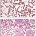
A hematologic evaluation begins with laboratory assessment of complete blood count parameters performed by automated hematology analyzers. Blood component indices are obtained from instruments that utilize various combinations of spectrophotometric, chemical, electrical impedance, and electro-optical analyzers. When the various cell parameters fall within a predefined normal range, the results are automatically validated and reported by the instrument. However, when suspect values outside the predetermined range are encountered, the instrument flags the sample to alert the technologist that additional investigation is required before the results can be released. In most laboratories, user-defined parameters prompt repeat or further automated testing, including automated slide making and staining for the evaluation of a peripheral blood smear. Despite increasing use of automation, manual preparation and staining of peripheral blood smears continues to be a common and important practice in many hematology laboratories.

Manual preparation of peripheral blood smears employs either the wedge or coverslip method. The wedge technique uses a spreader slide held at a 30- to 45-degree angle above a drop of blood placed near one end of a second glass slide so that the end of the spreader slide contacts the blood which extends along the entire edge between the two slides. The angled spreader slide is then pushed rapidly to generate the blood smear on the second slide, which is quickly air-dried and stained. An optimal peripheral blood smear prepared by this method contains a thick area that gradually transitions to a thin area with an even separation of red cells. The coverslip method utilizes a small drop of blood placed between two coverslips positioned so that the corners form an octagonal star. After the drop is spread, the coverslips are pulled apart in the same plane, followed by air-drying, staining, and mounting on a slide. Automated staining methods may employ similar mechanics or utilize centrifugal force to generate an even blood smear.

Aniline dyes, containing basic dyes such as methylene blue and acidic dyes such as eosin, are used to stain the smears. The most common methods used today include Wright, Giemsa, or May-Grünwald stains, or a combination of variants, based on the original method developed by Dmitri Romanowsky, a Russian protozoologist, who first used it 1890 to see malaria parasites. Eosin gives a red to orange color to the alkaline components of cells, such as hemoglobin and the granules of eosinophils, that contain an alkaline spermine derivative. Alkaline methylene blue dye gives a bluish-purple color to the acidic cellular elements, including nucleic acids (DNA, RNA), nucleoproteins, and the granules of basophils, which contain the acid heparin.

Other stains and methods may be utilized to better visualize unique morphologic features, such as supravital brilliant cresyl blue that stains residual RNA in young red cells (reticulocytes), giving the appearance of strands of dark blue net-like material in these cells.


EXAMINATION OF THE BONE MARROW ASPIRATE SMEAR AND TOUCH PREPARATION


Slide Preparation and Staining

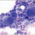
As with peripheral blood smears, optimal preservation of morphology requires either air-drying of aspirates without the use of anticoagulants or the use of EDTA as the anticoagulant of choice. Heparin introduces a staining artifact that alters the morphology of the cells. Bone marrow aspirate smears are prepared by a method similar to that used for making peripheral blood smears. A drop of the aspirate containing grossly visible marrow particles, which look like gray specks to the naked eye, is placed at one end of the slide and the marrow particles are gently dragged with a spreader slide leaving behind a trail of cells. A crush/squash preparation entails placing marrow particles between two slides and applying slight pressure and rapidly pulling apart the two slides. When a bone marrow sampling fails to yield any aspirated marrow particles (dry tap), the fresh unfixed core biopsy specimen can be picked up with forceps and touched several times to a slide producing touch preparations. Smear, crush, or touch preparations should be rapidly air-dried and stained with a Romanowsky’s stain, such as May-Grünwald Giemsa or Wright-Giemsa. An optimal specimen of the marrow aspirate provides adequate morphology for a thorough evaluation of the cytologic features of the marrow components and for a manual differential count. Although touch preparations may be the only specimen available for a cytologic examination of the individual marrow elements, they are prone to artifacts, making them suboptimal for an accurate morphologic review as they may not be representative. Nonetheless, touch preparations can sometimes provide useful information and should always be attempted in a dry tap.

The presence of iron is examined by staining the aspirate smear with Prussian blue (Perl’s reaction produces a blue-green color when hemosiderin or ferritin is present) and counter-staining with safranin-O or Kernecht Red (nuclear fast red). This stain allows evaluation of storage iron present in the macrophages in the marrow particles. The iron stain to assess storage iron is most reliable when it is positive; absence of iron staining may be due to uneven distribution of iron-laden macrophages in the particles. Additionally, the iron stain is used to visualize sideroblasts, which are normoblasts containing one or more particles of iron. The presence of abnormal sideroblasts and ring sideroblasts should be noted as they are seen in various hereditary and acquired hematologic diseases.

Special cytochemical stains can be performed on the aspirate smears that aid in identifying various cell lineages in the marrow. This is especially important when evaluating an excess of immature cells. The differential reactivity with these chemical reagents among the marrow precursors served as the basis for the early neoplastic classification systems. The most common stains performed include myeloperoxidase for the myeloid lineage, and nonspecific esterases for the monocytic cells. Rarely other stains may be utilized, such as Sudan black for the myeloid lineage, periodic acid-Schiff (PAS) for the erythroid and T-cell precursors, and toluidine blue to highlight the granules of mast cells and basophils. Flow cytometry of the aspirated cells and immunohistochemical stains of the biopsy have mostly replaced the need for cytochemical stains.

In rare instances, immunohistochemical stains can be performed on the aspirate smears if flow cytometry or a biopsy/clot section is not available; however, these are technically challenging and are difficult to interpret because of artifacts. Aspirate smears can also be used for fluorescence in situ hybridization (FISH) analysis or molecular tests, though this is not done routinely.


EXAMINATION OF THE BONE MARROW BIOPSY AND CLOT SECTION


Processing for Histologic Sections

The trephine biopsy specimen should be taken at right angle to the cortex and be at least 1.5 cm in length. A long core biopsy increases the likelihood of finding focal lesions (i.e., lymphoma, granuloma, and metastatic disease). After the touch preparations are made, the trephine core specimen is placed in a container with appropriate fixative. Methods for fixation vary and can significantly affect morphologic detail and immunohistology. Commonly used fixatives include B5, Bouin’s, zinc formaldehyde, isotonic buffered saline, or formaldehyde and glutaraldehyde. Typically, neutral buffered formalin is used with a 6-hour fixation period, but the fixation times can also range from 1 hour to a maximum of over 24 hours, depending on the method. Decalcification is a necessary step to soften the bone, allowing cutting of thin sections without tearing them. However, decalcification chelates the storage iron, affects the morphologic detail and immunohistochemistry, and interferes with molecular assays.

A bone marrow clot preparation is processed in a similar manner as the trephine core biopsy with the exception of the decalcification step. A clot section can provide additional information, especially if the trephine core biopsy is inadequate. It is generated at the time of the bone marrow aspiration, when a portion of the aspirate containing marrow particles is allowed to form a clot. Various agents may be used to facilitate clotting. The clot is then placed into an appropriate fixative without further decalcification. An important advantage of the clot preparation is that it may be used for FISH analysis or extraction of nucleic acid material suitable for molecular-based testing. Furthermore, lack of decalcification produces more reliable immunoreactivity, although interpreting these stains may be more challenging than on the trephine biopsy section.

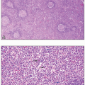
Automated tissue processors are utilized for the bone marrow clot and trephine biopsy specimens, similar to other routine surgical biopsy material. Specimens are then embedded in paraffin, sectioned at 3 to 5 µm in thickness, and mounted on glass slides. The prepared sections are routinely stained with hematoxylin and eosin (H&E), although other stains may be used in addition. For instance, PAS helps to highlight the megakaryocytes, whereas Giemsa is helpful to identify mast cells, eosinophils, plasma cells, and to differentiate between proerythroblasts and myeloblasts. Evaluation for the presence of fibrosis is done routinely on sections by staining for reticulin by using silver impregnation, which reacts with type III collagen, whereas Masson’s trichrome is used to detect type I collagen. Other useful histochemical stains include Ziehl-Neelsen stain for acid-fast organisms, Gomori’s methenamine silver for fungi, and Congo red for amyloid. Iron stores can also be evaluated by Prussian blue on the histologic sections; however, decalcification chelates sideroblast and storage iron, so that iron evaluation is unreliable on the trephine biopsy sections.

Immunohistology is used routinely on histologic sections and aids in lineage and subtype identification of cells by probing for specific lineage-associated antigens. Testing for expression of certain proteins may also provide information regarding mutational status or proliferation, which can impact prognosis in neoplastic conditions. Although flow cytometry provides information on protein expression of individual cells and allows assessment of cell populations, immunohistology provides correlation with direct morphologic assessment. Furthermore, immunohistology is especially important if the aspirate specimen obtained for flow cytometric analysis is hemodiluted or if the cells of interest are not adequately represented in the aspirate.

Nov 20, 2018 | Posted by in HEMATOLOGY | Comments Off on Approach to the Microscopic Evaluation of Blood and Bone Marrow

Full access? Get Clinical Tree

Get Clinical Tree app for offline access