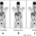
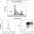
Fig. 8.1
Mini Deluxe Phantom tested by UIH PET/CT scanner
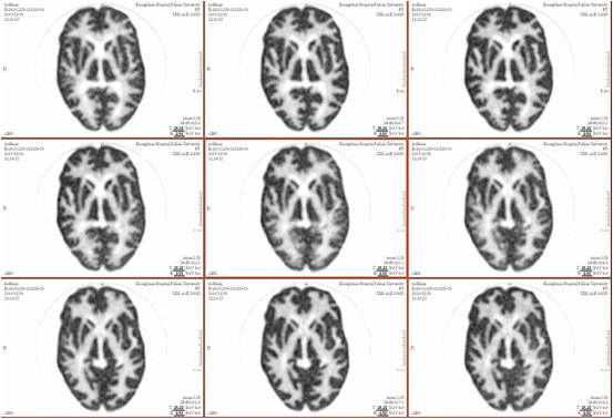
Fig. 8.2
Hoffman brain phantom tested by UIH PET/CT scanner
8.2 Animal Study
Animal studies were applied to focus on using high-resolution detector with appropriate physics corrections and advanced image reconstruction methods to get high-quality images of great practical importance. For instance, a normal mouse scanned by uMI S-96R PET/CT with 0.5 mCi 18F–FDG and 10 min of acquisition time was demonstrated good anatomic details and FDG uptake distributions (Fig. 8.3a–d). With the same imaging parameters, a tumor-bearing nude mouse with hepatocellular carcinoma showed a large, well-circumscribed soft tissue mass with heterogeneous FDG uptake (Fig. 8.4a–b). The high-resolution modality was also used to scan dog with 18F–FDG whole body PET/CT imaging (Fig. 8.5a–d). In particular, the non-gating heart of dog with rest or stress was displayed via a cardiac scan with brilliant metabolic PET imaging (18F–FDG, 5 mCi; scanning time, 3 min; LYSO, 2.4 × 2.4 mm; and matrix, 256 × 256) and high-resolution anatomical CT imaging (kV,120 and mAs, 70). The fusion tools are helpful for displaying powerful PET/CT fusion images, which can locate papillary muscles of the heart of a dog with metabolic profile (Figs. 8.6a–b and 8.6c–e). F-18 NaF whole body bone imaging can display the entire skeletal system with PET, CT multiplanar reformation (MPR), PET/CT fusion, and CT 3D reconstruction modes (Fig. 8.7a–i). In a comparative study, the image quality of whole body bone imaging of a dog scanned by UIH F-18 NaF PET/CT was superior to that of the same dog by the same type of PET/CT from another manufacturer (Fig. 8.8a–f). In addition, UIH F-18 NaF PET/CT imaging can easily detect fractures of the bilateral distal femoral bones of a dog prior to and after operation (Fig. 8.9a–d).
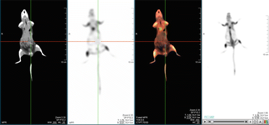
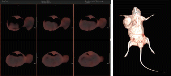
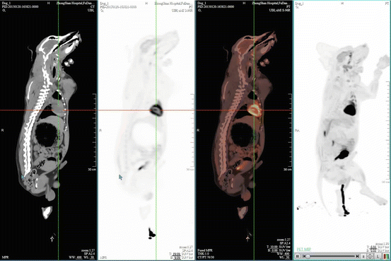
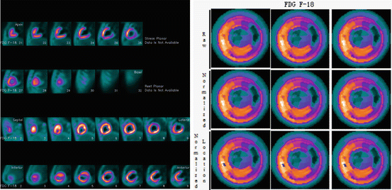
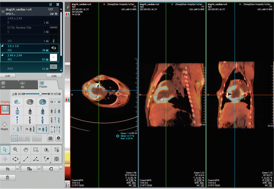
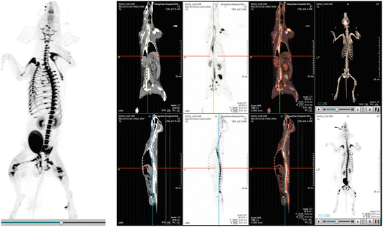
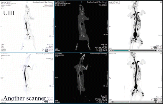
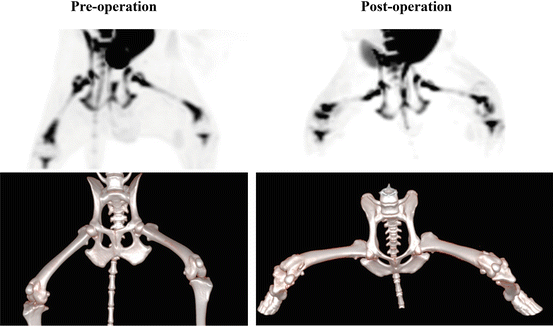

Fig. 8.3
(a–d) Normal mouse 18F–FDG PET/CT imaging

Fig. 8.4
(a–b) Tumor-bearing nude mouse 18F–FDG PET/CT imaging

Fig. 8.5
(a–d) 18F–FDG dog whole body imaging

Fig. 8.6a–b
18F–FDG dog heart imaging

Fig. 8.6c–e
Fusion tools for 18F–FDG PET/CT – display with dog heart

Fig. 8.7
(a–i) 18F–NaF dog whole body bone imaging

Fig. 8.8
(a–f) 18F–NaF dog whole body bone imaging

Fig. 8.9
(a–d) 18F–NaF dog bone imaging with the bilateral distal femoral fractures
8.3 Clinical Evaluation
The UIH high-resolution time-of-flight PET/CT provided more doctor-friendly information in oncology, infection, and cardiovascular diseases. The clinical oncology applied PET/CT to diagnose tumor, to search for unknown primary malignant tumor, to perform tumor staging and restaging, and to evaluate therapy response.
- (1)
There are several cases (including lung cancers, gastric cancer, and pleural mesothelioma) to show the clinical implication of the high-resolution time-of-flight PET/CT.
Case 1:
A 51-year-old woman presents with no fever and cough (Figs. 8.10a–b and 8.10c–f).
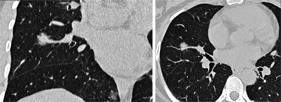
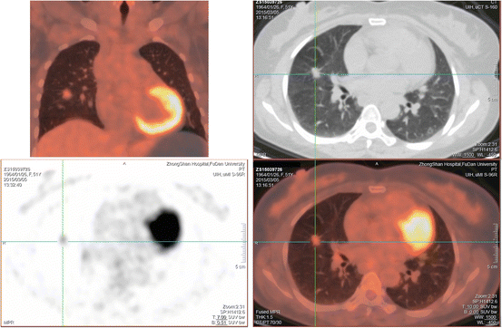

Fig. 8.10a–b
CT images on lung window showed an oval, spiculated, mixed ground-glass opacity nodule at the middle lobe of the right lung

Fig. 8.10c–f
18F–FDG PET/CT was performed for differential diagnosis of a solitary pulmonary nodule. PET/CT images with MPR demonstrated that the solitary pulmonary nodule was of marked FDG uptake with 2.37 of SUVmax and 0.58 of SUVmean. The nodule was pathologically confirmed to be a Grade II alveolar type lung carcinoma
Case 2:
A 74-year-old man presents with stimulating dry cough, chest tightness, and shortness of breath for a week (Figs. 8.11a–c and 8.11d–l).

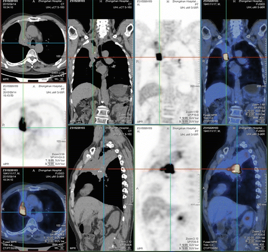

Fig. 8.11a–c
Axial non-enhanced chest CT images on lung window (2A) and mediastinal window (2B) showed marked tapering and obstruction in the initial segment of the bronchus of the right upper lobe. Axial contrast-enhanced chest CT scan (2C) showed a segmental atelectasis of the right upper lobe caused by a mass in the right hilum. 18F–FDG PET/CT was performed for differential diagnosis of the mass

Fig. 8.11d–l


PET/CT images with MPR demonstrated that the right hilar mass was of marked FDG uptake. Lung cancer was confirmed by fiber-optic bronchoscopy with biopsy. Then, a right upper lobectomy was performed, and the mass was pathologically verified to be squamous cell carcinoma. Two lymph node metastases were found in a regional lymph node dissection
Stay updated, free articles. Join our Telegram channel

Full access? Get Clinical Tree


