Fig. 9.1
Types of dedicated breast PET scanners. (a) Positron emission mammography (PEM). (b) Fully tomographic dedicated breast PET with dual-plate detector rotating around the breast. (c) Fully tomographic dedicated breast PET with ring-shaped detector
Table 9.1
Examples of dedicated breast (db) PET systems
PEM | Fully tomographic dedicated breast PET | |
|---|---|---|
Rotating planar head type | Ring-shaped detector type | |
PEM Flex Solo II (Naviscan) High-resolution PEM (Stanford U) | Clear-PEM (Hospital of Portuguese Institute of Oncology) PEM/PET (West Virginia U) dbPET/CT (UC Davis) | MAMMI (Oncovision) Elmammo (Shimadzu) Partial ring dbPET (Pennsylvania U) |
PEM has two detector heads integrated with planar or curved breast compression paddles, which acquire limited-angle tomographic images from incomplete 3D data obtained with a mildly compressed breast (Fig. 9.1a). The positioning of the breast is similar to that in mammography, and two projections (craniocaudal and mediolateral oblique) are usually obtained. The Flex Solo II scanner (Naviscan, San Diego, USA) is a PEM device that was cleared for marketing by the US Food and Drug Administration (FDA) in 2009 [24] and is currently the most common and commercially available dedicated breast PET system in the United States. Usually, a scanning time for one projection is 7–10 min [25, 34]. The in-plane spatial resolution was reported to be 2.4 ± 0.3 mm full width at half maximum (FWHM) for images reconstructed with a three-dimensional (3D) list-mode maximum likelihood expectation maximization (MLEM) algorithm. On the other hand, the cross plane spatial resolution is low, with a reported FWHM of 8.0–8.2 mm [24, 25], but attenuation and scatter corrections are available. This system is equipped with quantitative metrics to measure quantitative values, called PEM uptake values (PUV) [36]. A biopsy capability is included in this model.
The fully tomographic dedicated PET systems are newer generations of PET devices that acquire complete 3D data from an uncompressed breast, and there are several variations in the detector design. One is a dual- or multi-plate detector rotating around the breast (Fig. 9.1b), such as Clear-PEM developed by the Portuguese consortium under the framework of the Crystal Clear Collaboration at CERN [1] and PEM/PET system developed at West Virginia University [32]. Another type of the fully tomographic dedicated PET systems is a ring-shaped detector encircling the breast (Fig. 9.1c), which includes MAMMI (Oncovision, Valencia, Spain) and Elmammo (Shimadzu, Kyoto, Japan).
In Kyoto University, authors examined a scanner performance of the Elmammo prototype (Shimadzu, Kyoto, Japan) and have been performing human imaging with this system since 2009 [26]. Elmammo has a complete ring-shaped detector, consisting of 36 detector blocks arranged in three contiguous rings with 12 detector modules, which have a transaxial diameter of 185 mm and an axial FOV of 155.5 mm (Fig. 9.2). Each detector block has four layers with a 32 × 32 array of 1.4 × 1.4 × 4.5 mm3 lutetium gadolinium oxyorthosilicate (LGSO) crystals, coupled to a 64-channel position-sensitive photomultiplier tubes (PSPMT). This system has also DOI measurement capability. Elmammo is one of those systems that can achieve the highest spatial resolution among PET systems for human applications. With the Elmammo prototype, minimal FWHMs in the radial, tangential, and axial directions are 1.6, 1.7, and 2.0 mm and 0.8, 0.8, and 0.8 mm, for filtered back projection (FBP) and 3D dynamic row-action maximum likelihood algorithm (DRAMA) reconstructions, respectively [26]. These values are much smaller than the FWHM obtained with whole-body PET or PET/CT systems of approximately 5–7 mm [7, 12]. Elmammo has capabilities of attenuation and scatter corrections and holds a quantitative metrics to obtain standard uptake value (SUV). A breast is usually scanned for 5 min with a patient lying in a prone position. Because the axial FOV of the ring-shaped scanner is designed to be large enough to cover the entire depth of a breast, the whole image is obtained at once without changing the position of the detector. Elmammo is commercially available since 2014 in Japan.
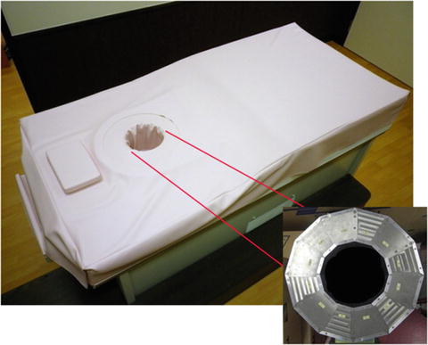

Fig. 9.2
Elmammo prototype (Shimadzu, Kyoto, Japan). This is a fully tomographic dedicated breast PET system with ring-shaped detectors. A patient lies in the prone position with her breast in the aperture of the ring-shaped detector (Cited from Miyake et al. [26])
9.3 Clinical Values of Focused Breast PET Systems
In comparison to conventional whole-body PET, the potential advantages of the high-resolution PET systems may include (1) improvement of detection of breast cancer uptake, (2) visualization of detailed distribution of PET tracers accumulated in breast lesions, and (3) quantification of tumor uptake with less biased interpretation, resulting from the partial volume effect. Although available published data are still limited, there have been several published studies that addressed the diagnostic values of dedicated breast PET with 18F-FDG in the detection of breast cancers in patients with suspicious lesions or established cancers. However, no or very few studies have addressed the diagnostic values of dedicated PET systems in the screening of asymptomatic women or assessment of treatment efficacy. In the following section, we summarize results of reported studies and describe potential advantages and disadvantages for the clinical use of dedicated breast PET.
9.3.1 Diagnostic value in Patients with Confirmed Breast Cancers
Several studies have addressed the diagnostic values of dedicated breast PET with 18F-FDG in the detection of breast cancers in patients with known or suspected cancer lesions. As summarized in Table 9.2, most of these studies were performed with PEM scanners, which showed sensitivities of 80–95% and varied specificities that ranged from 33 to 100% in the detection of breast cancers [4, 14, 23, 28, 33]. There has been one study for a ring-shaped dedicated PET device (Elmammo prototype) that showed a sensitivity of 82% and a specificity of 50% [21]. Recently, a meta-analysis was performed for a total of eight PEM articles published between 2000 and 2012, which showed pooled sensitivities and specificities on lesion-basis analysis of 85% (95% CI, 83–88%) and 79% (95% CI, 74–83%), respectively, with area under the curve (AUC) of 0.88 [11]. These results are highly similar or slightly better than those of whole-body PET, with a sensitivity of 83% (95% CI, 73–89%) and a specificity of 74% (95% CI, 58–86%) [10].
Table 9.2
Overall diagnostic performance of 18F-FDG dbPET in detection of cancers in patients with suspected or known breast cancer
Authors | Year | dbPET type | No. of patients | Sensitivitya | Specificitya |
|---|---|---|---|---|---|
Murthy et al. | 2000 | PEM | 16 | 80% | 100% |
Levine et al. | 2003 | PEM | 16 | 86% | 91% |
Rosen et al. | 2005 | PEM | 23 | 86% | 33% |
Berg et al. | 2006 | PEM | 94 | 90% | 86% |
Eo et al. | 2012 | PEM | 101 | 95% | NA |
Iima et al. | 2012 | Ring shaped | 69 | 82% | 50% |
However, these studies enrolled patients with confirmed breast cancers or suspicious lesions diagnosed based on conventional breast examinations, such as clinical diagnosis and mammography. Hence, overall sensitivities could have been elevated due to the high ratio of relatively large cancers. From a clinical point of view, in patients with recently diagnosed breast cancers, the main role of the dedicated breast PET may be the detection of additional lesions, because the new findings may change the treatment strategy, particularly in cases in which a conservative therapy is envisioned. Thus, some studies provided data with stratifying lesions to known index cancers and additional lesions in the ipsilateral or contralateral breasts.
For known index breast cancers, reported sensitivities of 18F-FDG PEM are uniformly high, ranging from 92 to 95% (Table 9.3) [4, 5, 22, 34]. Two studies showed the sensitivities of PEM for index cancers were significantly higher than those of whole-body PET (56–68%) [22, 34]. In addition, Kalinyak et al. showed PEM (95%) had higher sensitivity than whole-body PET/CT (87%, p = 0.03) [22].
Table 9.3
Sensitivities of 18F-FDG PET systems for index breast cancer
Authors | Year | No. of patients | Analysis | PEM | wbPET | wbPET/CT |
|---|---|---|---|---|---|---|
Berg et al. | 2006 | 77 | Lesion basis | 93% | ||
Kalinyak et al. | 2014 | 69 | Breast basis | 92%a | 56% | |
109 | Breast basis | 95%b | 87% | |||
Schilling et al. | 2011 | 208 | Lesion basis | 93%a | 68% | |
Berg et al. | 2011 | 388 | Lesion basis | 93% |
For additional ipsilateral breast cancer, PEM had sensitivity of 41–85% and high specificity ranging from 74 to 91% (Table 9.4) [5, 22, 34]. Although the sensitivity of PEM was still limited, Kalinyak and colleagues demonstrated that PEM (47–57%) was more sensitive than either whole-body PET (6.7%, p < 0.05) or whole-body PET/CT (13%, p < 0.01) [22] (Table 9.4).
Table 9.4
Diagnostic performance of 18F-FDG PET systems in detection of additional ipsilateral breast cancer
Authors | Year | No. of patientss | Analysis | Sensitivity | Specificity | ||||
|---|---|---|---|---|---|---|---|---|---|
PEM | wbPET | wbPET/CT | PEM | wbPET | wbPET/CT | ||||
Kalinyak et al. | 2014 | 69 | Breast basis | 47%a | 6.7% | 91% | 96% | ||
109 | Breast basis | 57%a | 13% | 91% | 95% | ||||
Schilling et al. | 2011 | 208 | Lesion basis | 85% | NA | 74% | NA | ||
Berg et al. | 2011 | 388 | Breast basis | 51% | 91% | ||||
Lesion basis | 41% | 80% | |||||||
In the detection of additional breast cancers in the contralateral breasts, Berg et al. showed sensitivities of 18F-FDG PEM were only 20% (3 out of 15) in the prospective reading session and 73% (11 out of 15) in the retrospective reading sessions [6].
Detection and visualization of sub-centimeter breast cancers (≤10 mm) have been challenging issues for conventional PET imaging. Whole-body PET has a limited sensitivity for sub-centimeter cancers, with reported sensitivities of 0% for T1a (>1 mm and ≤5 mm) invasive cancers and 13–39% for T1b (>5 mm and ≤ 10 mm) invasive cancers [3, 22]. However integrated whole-body PET/CT could provide improved sensitivities of 0–40% for T1a and 71–83% for T1b [21, 22]. In dedicated breast PET systems, according to data from small subpopulations, sensitivities of dedicated breast PET range 25–100% (average 46% for 28 reported lesions) for T1a and 46–86% (average 81% for 86 reported lesions) for T1b (Table 9.5) [4, 5, 21, 22, 34]. Collectively, these data suggest that dedicated breast PET scanners are more sensitive than conventional PET scanners in the detection of sub-centimeter tumors. Kalinyak and colleagues demonstrated that in T1b cancers of index tumors, PEM has a significantly higher sensitivity than whole-body PET in the same population (95% versus 37%, n = 19, p = 0.002) [22].
Table 9.5


Sensitivities of 18F-FDG dbPET for sub-centimeter invasive cancer
Stay updated, free articles. Join our Telegram channel

Full access? Get Clinical Tree


