AND ORAL CAVITY (ICD‐O C00, C02–06)


LIP AND ORAL CAVITY (ICD‐O C00, C02–06)


Rules for Classification


The classification applies only to carcinomas of the vermilion surfaces of the lips and of the oral cavity, including those of minor salivary glands. There should be histological confirmation of the disease.


Anatomical Sites and Subsites


Lip* (Fig. 19)
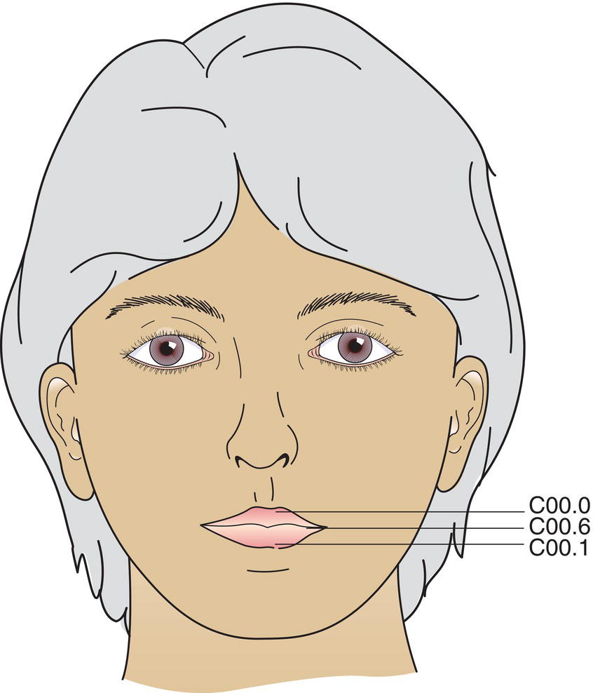


  1. External upper lip (vermilion border) (C00.0)
  2. External lower lip (vermilion border) (C00.1)
  3. Commissures (C00.6)
Schematic illustration of External upper lip, External lower lip, and Commissures.

Fig. 19


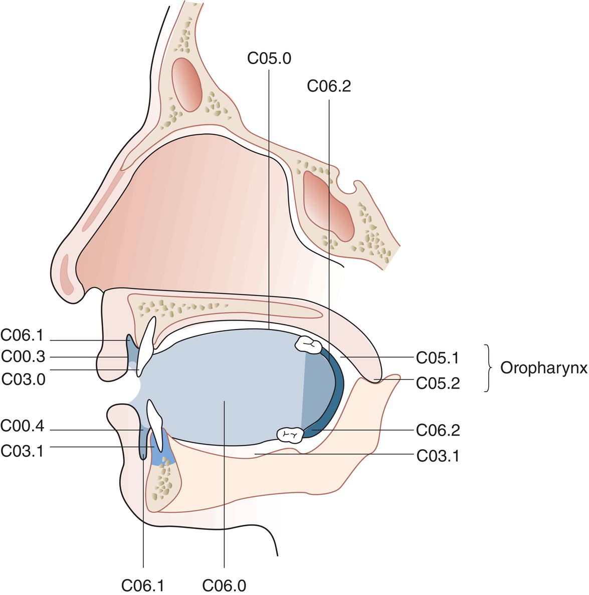
Oral Cavity (Figs. 20, 21, 22)


  1. Buccal mucosa

    1. Mucosa of upper and lower lips (C00.3, 4)
    2. Cheek mucosa (C06.0)
    3. Retromolar areas (C06.2)
    4. Bucco‐alveolar sulci, upper and lower (vestibule of mouth) (C06.1)

  2. Upper alveolus and gingiva (upper gum) (C03.0)
  3. Lower alveolus and gingiva (lower gum) (C03.1)
  4. Hard palate (C05.0)
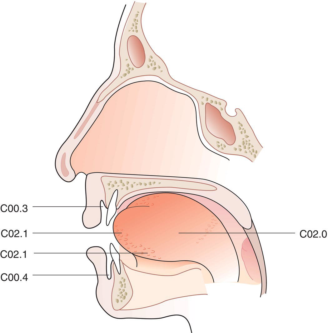
  5. Tongue*

    1. Dorsal surface and lateral borders anterior to vallate papillae (anterior two‐thirds) (C02.0, 1)
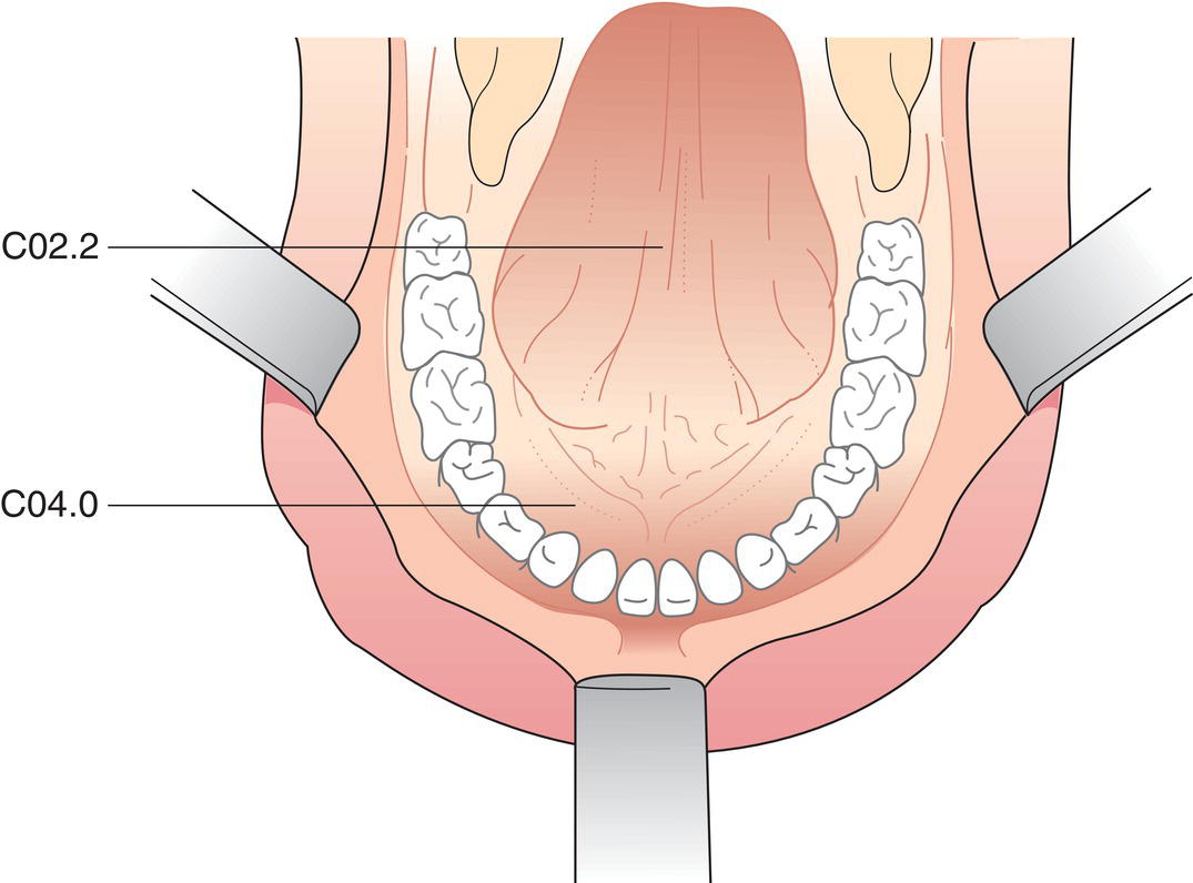
    2. Inferior (ventral) surface (C02.2)
Schematic illustration of the indications of Buccal mucosa, Upper alveolus and gingiva, Lower alveolus and gingiva, Hard palate, and tongue.

Fig. 20

Schematic illustration of the indications of Buccal mucosa, Upper alveolus and gingiva, Lower alveolus and gingiva, Hard palate, and tongue.

Fig. 21

Schematic illustration of the indications of Buccal mucosa, Upper alveolus and gingiva, Lower alveolus and gingiva, Hard palate, and tongue.

Fig. 22


TN Clinical Classification


T – Primary Tumour


Only gold members can continue reading. Log In or Register to continue

Stay updated, free articles. Join our Telegram channel

Tags:
Dec 15, 2022 | Posted by in ONCOLOGY | Comments Off on AND ORAL CAVITY (ICD‐O C00, C02–06)

Full access? Get Clinical Tree

Get Clinical Tree app for offline access
TX Primary tumour cannot be assessed
T0 No evidence of primary tumour
Tis Carcinoma in situ
T1 Tumour 2 cm or less in greatest dimension and 5 mm or less depth of invasion (Figs. 23, 24, 25, 26, 27)
T2 Tumour 2 cm or less in greatest dimension and more than 5 mm depth of invasion (Figs 28, 29), or