ANATOMY AND EMBRYOLOGY OF THE OPTIC CHIASM
The optic chiasm may be considered the “Grand Central Station” of the vision sensory system, containing some 2.4 million afferent axons. Many of the disease processes that involve the intracranial optic nerves likewise involve the chiasm. Because of the relationship of the nerves and chiasm to the basal structures of the anterior and middle cranial fossae, pituitary adenomas and parasellar meningiomas frequently encroach on the anterior vision pathways. Failure of early diagnosis of chiasmal disorders may endanger the life of the patient and lessen the likelihood of reversal of visual deficits.
The chiasm is situated in the anteroinferior recess of the third ventricle. The inferior aspect of the chiasm usually is 8 to 13 mm above the nasotuberculum line (i.e., the plane of the diaphragma sellae or clinoid processes). The intracranial portion of the optic nerves is inclined as much as 45 degrees from the horizontal and measures 17 ± 2.5 mm in length (Fig. 19-1). The chiasm measures ˜8 mm from anterior to posterior notch, 12 mm across, and 4 mm in height. The inferior surface of the chiasm projects more or less directly above the bony dorsum sellae (79%). When the chiasm lies more anteriorly over the diaphragm sellae, it is said to be prefixed (17%), and behind the dorsum, postfixed (4%).1, 2 and 3 The lateral aspect of the chiasm is embraced by the supraclinoid portion of the internal carotid artery. The anterior cerebral arteries of the circle of Willis (Fig. 19-2) pass over the dorsal surface of the optic nerves as they converge. The optic nerves are fixed at the intracranial entrance of the optic canals, the dorsal aspect of which is formed by an unyielding falciform fold of dura.
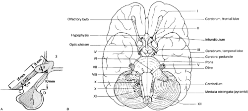 FIGURE 19-1. A, The relationships of the optic nerves (ON) and chiasm (X) to sellar structures and the third ventricle (3). (C, anterior clinoid; D, dorsum sellae; P, pituitary gland in sella.) B, Base of the brain, showing some of the pertinent anatomic structures. The cranial nerves are numbered. (A, From Glaser JS. Topical diagnosis: the optic chiasm. In: Glaser JS. Neuro-ophthalmology, 3rd ed. Philadelphia: Lippincott, 1999:81; B, From Akesson EJ, Loeb JA, Wilson-Pauwels L. Thompson’s core textbook of anatomy, 2nd ed. Philadelphia: JB Lippincott, 1990:81.)
Related posts:Stay updated, free articles. Join our Telegram channel
Full access? Get Clinical Tree
 Get Clinical Tree app for offline access

|


