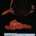
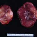
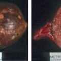
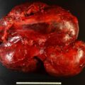
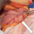
Adrenal myelolipoma is a benign adrenocortical tumor composed of adipose tissue and bone marrow elements; it is reported in one out of 500–1250 autopsy cases. Adrenal myelolipoma is typically diagnosed in the fifth and sixth decades of life with no sex predilection. Although large myelolipomas can lead to mass effect symptoms, most are asymptomatic and detected incidentally on cross-sectional imaging performed for other reasons. There are a few types of adrenal masses in which the imaging phenotype is diagnostic. Herein we share such a case.
Case Report
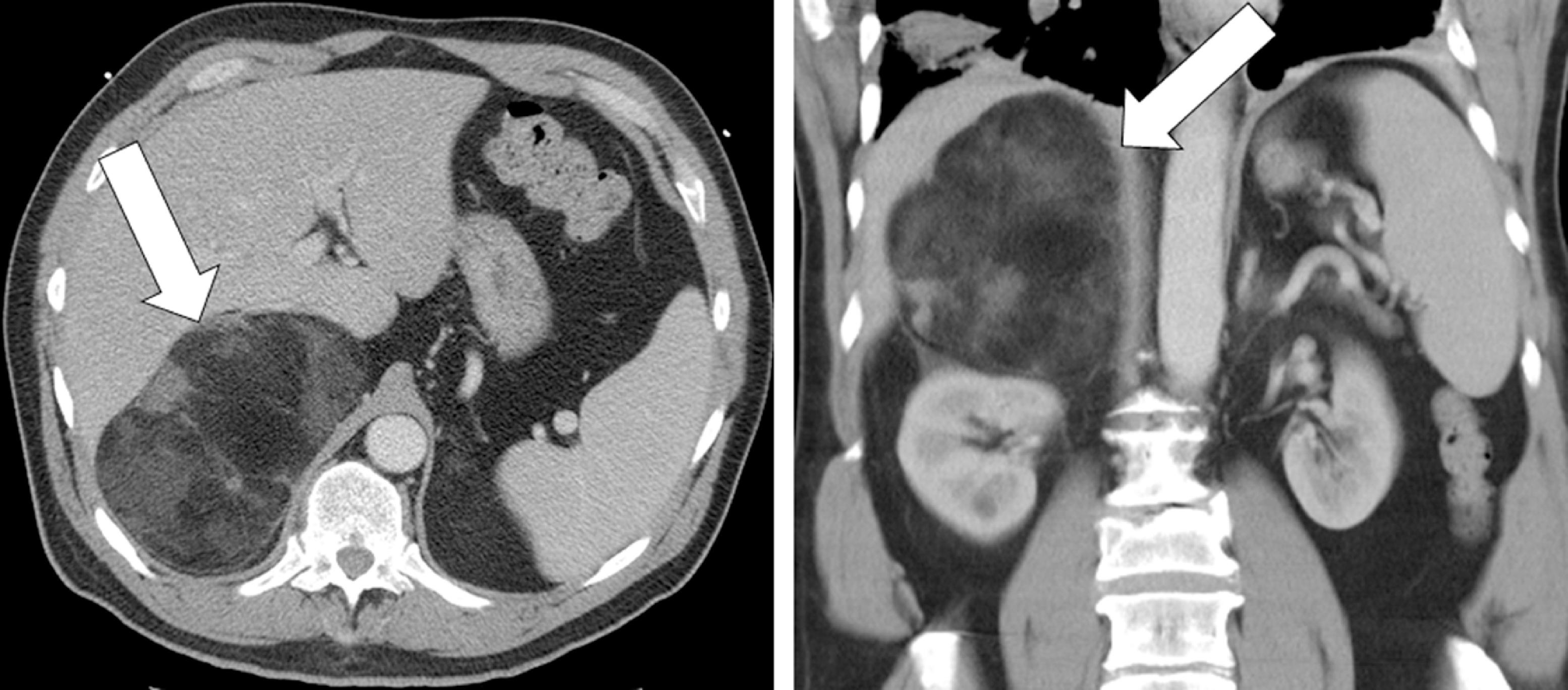
The patient was a 52-year-old man referred for evaluation of an incidentally discovered right adrenal mass. This patient was well until 1 month previously when he had signs and symptoms of pulmonary embolism that was confirmed on a chest computed tomography (CT) study. The chest CT scan incidentally discovered a large right adrenal mass (14 × 9 × 13 cm) that was predominately fat density ( Fig. 65.1 ). He had a history of hypertension controlled on a calcium channel blocker (amlodipine 5 mg daily) and a diuretic (hydrochlorothiazide 12.5 mg daily). The question was whether compression of the inferior vena cava by the right adrenal mass predisposed the patient to pulmonary embolism. Doppler ultrasound of the lower extremities was normal. His only other medication at the time of our consultation was warfarin. On physical examination his body mass index was 31.9 kg/m 2 , blood pressure was 133/85 mmHg, and heart rate 56 beats per minute. There was no right upper quadrant abdominal discomfort.

Stay updated, free articles. Join our Telegram channel

Full access? Get Clinical Tree


