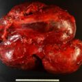
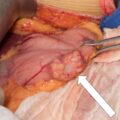
Lipid-poor adrenal masses raise concerns about the underlying pathology, especially in young people. Herein we present a case of a lipid-poor adrenal mass in a 22-year-old woman that proved to be a benign adrenal hemangioma.
Case Report
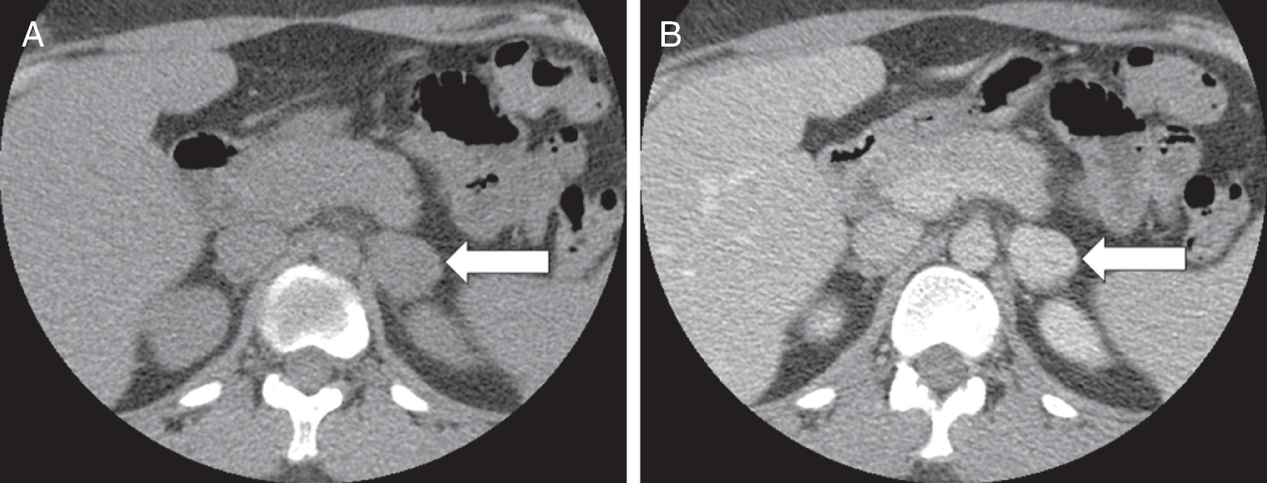
The patient was a 22-year-old woman who was found to have a 2.5-cm left adrenal mass incidentally discovered on a chest computed tomography (CT) that was obtained to evaluate pneumonitis. On a follow-up abdominal CT scan 1 year later it measured 2.5 × 2.5 cm with an unenhanced CT attenuation of 29 Hounsfield units (HU) and 61% contrast washout at 15 minutes ( Fig. 73.1 ). She had no paroxysmal symptoms and there was no history of hypertension. Her weight had been stable. She had no signs or symptoms of Cushing syndrome. There was no history of hypokalemia. Her only regular medication was an oral contraceptive pill. On physical examination her body mass index was 33.5 kg/m 2 , blood pressure 122/91 mmHg, and heart rate 94 beats per minute. She had no stigmata of an adrenal disorder. Heart and lung examinations were normal.
INVESTIGATIONS
The laboratory studies were normal ( Table 73.1 ). There was no biochemical evidence for functioning pheochromocytoma or cortisol secretory autonomy.
| Biochemical Test | Result | Reference range |
| Sodium, mmol/L Potassium, mmol/L Fasting plasma glucose, mg/dL Creatinine, mg/dL 1-mg overnight DST cortisol, mcg/dL Plasma metanephrine, nmol/L Plasma normetanephrine, nmol/L | 140 4.3 91 0.9 0.7 <0.2 0.5 | 135–145 3.6–5.2 70–100 0.6–1.1 <1.8 <0.5 <0.9 |
Stay updated, free articles. Join our Telegram channel

Full access? Get Clinical Tree


