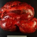
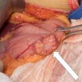
Adrenal ganglioneuroma is a rare benign tumor that is characterized by ganglion cells individually distributed in Schwannian stroma. Adrenal ganglioneuroma is found in approximately 2% of adrenalectomies. The unique imaging characteristics of this tumor can lead to a presumptive diagnosis, which is potentially important because when small, adrenal ganglioneuromas may not need to be resected. Herein we present a case of a young patient with a 5-cm adrenal mass that was suspected to be an adrenal ganglioneuroma based on imaging.
Case Report
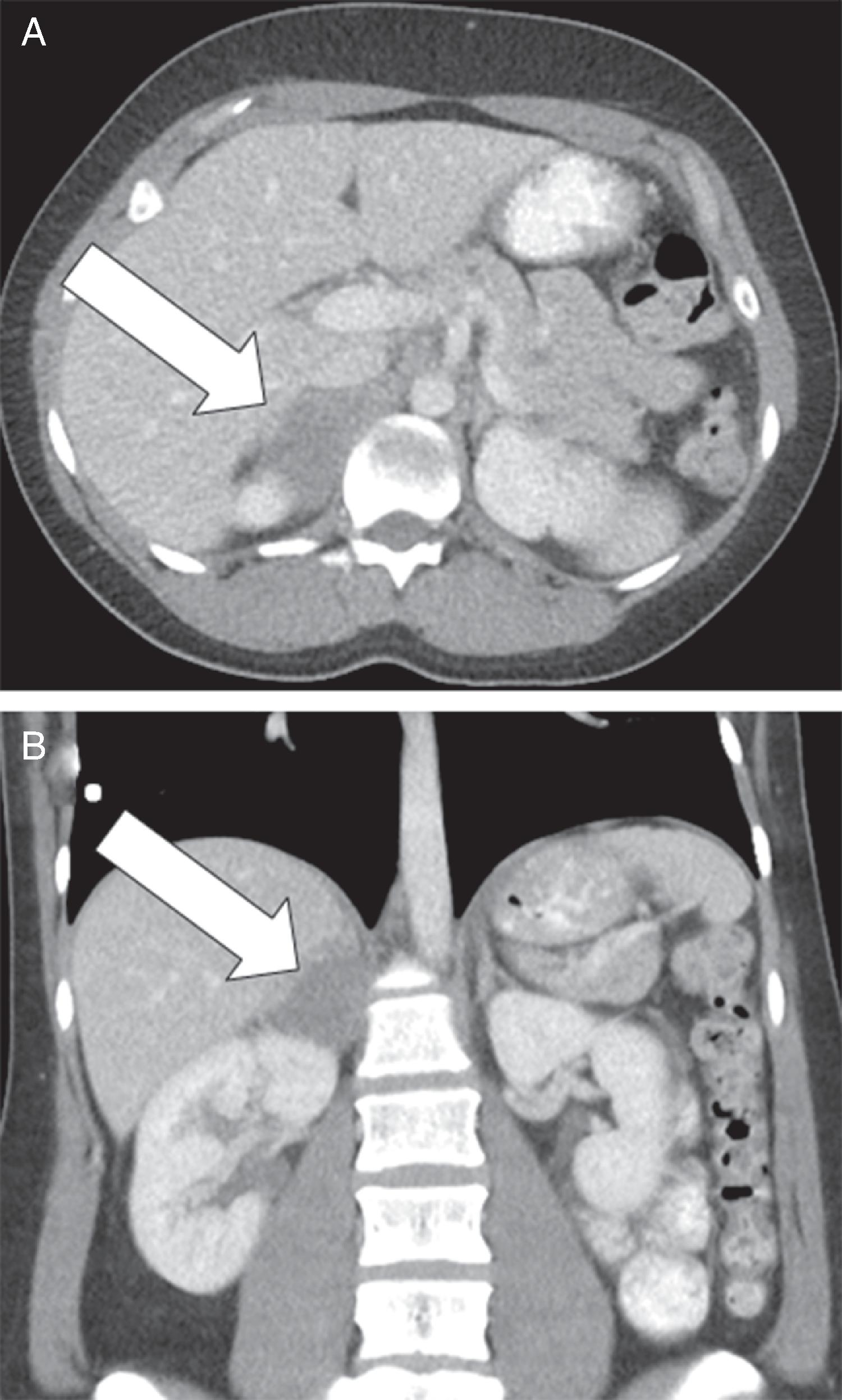
This 28-year-old woman was seen in endocrine consultation for the incidental discovery of a 5-cm right adrenal mass on computed tomography (CT) performed for pelvic pain. The CT findings were unique, with the mass having a triangular shape, uniform ground-glass density, well-defined tumor edges, indeterminate unenhanced CT attenuation, and relative lack of vascularity with contrast administration ( Fig. 74.1 ). She had no signs or symptoms of an adrenal disorder. She had normal blood pressure and stable body weight. She had been taking no regular medications. She had never been treated with anticoagulants and did not take aspirin.
INVESTIGATIONS
The laboratory studies were normal with the exception of the mild elevation in plasma normetanephrine ( Table 74.1 ). The false-positive rate of plasma normetanephrine is 15%, and because of the rarity of pheochromocytoma, 97% of patients with increased plasma normetanephrine on the Mayo Clinic campus do not have a pheochromocytoma. Mild elevations in plasma normetanephrine always need to be interpreted with the clinical context in mind. In this case, the uniform density and lack of vascularity on CT made pheochromocytoma extremely unlikely.
| Biochemical Test | Result | Reference Range |
| Sodium, mmol/L Potassium, mmol/L Fasting plasma glucose, mg/dL Creatinine, mg/dL Plasma metanephrine, nmol/L Plasma normetanephrine, nmol/L 1-mg overnight DST cortisol, mcg/dL 24-Hour urine: Metanephrine, mcg Normetanephrine, mcg Norepinephrine, mcg Epinephrine, mcg Dopamine, mcg | 137 4.1 66 0.7 0.38 1.2 1.1 194 472 69 14 313 | 135–145 3.6–5.2 70–100 0.6–1.1 <0.5 <0.9 <1.8 <400 <900 <80 <20 <400 |
Stay updated, free articles. Join our Telegram channel

Full access? Get Clinical Tree


