Acute Myeloid Leukemia and Myelodysplastic Syndromes


Acute Myeloid Leukemia and Myelodysplastic Syndromes


Robert J. Arceci

Soheil Meshinchi


HISTORICAL BACKGROUND AND DEFINITIONS

The myeloid malignancies represent a heterogeneous group of acute and chronic clonal disorders that arise from bone marrow precursor or progenitor cells. They include acute myeloid leukemia (AML), myelodysplastic syndromes (MDS), and subacute myeloproliferative disorders (MPD) such as juvenile myelomonocytic leukemia (JMML) and chronic myelogenous leukemia (CML). This chapter focuses on AML and MDS. Combined, they represent about 20% of childhood leukemias.

The origin of the term “myeloid” derives from the Greek “myelos” plus “eidos,” meaning something pertaining to or resembling marrow. “Myelos” has also been used to refer to the spinal cord and derives that association from early Greek anatomists who appear to have considered the spinal cord like the bone marrow in that it too was encased in bone. The term “leukemia” was first introduced in the mid-1800s by Virchow and derives from “leuchaemia”: “leukos,” meaning “white,” and “haima,” meaning blood. The original cases more than likely represented chronic lymphocytic or myeloid leukemia. Following the first description of myeloblast precursors by Naegeli around the beginning of the 20th century, a number of cases of AML were described. In order to distinguish them from lymphoid leukemias that had previously been described, these early cases were referred to as acute nonlymphocytic leukemia. Other terms used to describe AML have included acute myelocytic leukemia, acute myelogenous leukemia, and acute granulocytic leukemia.

Following the description of myeloblast precursors by Naegeli at the turn of the 20th century, a number of cases of AML were described, initially, and, because of historical precedent, as acute nonlymphocytic leukemia. The earliest cases were monocytic and myelomonocytic, followed by descriptions of erythroleukemia, megakaryoblastic leukemia and, as late as 1957, promyelocytic leukemia. In the mid-1970s, the comprehensive morphological classification schema, termed the French-American-British (FAB) system, was published and defined AML into seven categories, M1 to M7.

MDS refers to another group of myeloid malignancies with variable clinical course. The root “dys” refers to something “abnormal, difficult, impaired, or bad.” The term “plasia” refers to “formation, molding, or development.” The term “syndrome” derives from “syn-,” which means “together or with,” while “-drome” refers to “a course, road, or walking.” Thus, MDS represents a group of disorders with a common characteristic of abnormal myeloid maturation resulting in insufficient numbers of various types of blood cells and varied clinical course.

Inherent in the names and the early descriptions of AML and MDS are the fundamental ideas that have formed the foundation for our subsequent understanding of these malignancies as products of abnormal bone marrow-derived progenitor cells. The experimental evidence demonstrating the cellular changes that lead to AML and MDS along with the implications for treatment successes and failures are discussed herein.


EPIDEMIOLOGY OF PEDIATRIC AML AND MDS

Worldwide figures for the total number of children with AML or MDS are not readily available. However, on the basis of an average incidence of about 8 cases per 1 million children annually,1 and the roughly 2 billion children in the world, one can estimate approximately 16,000 cases of AML could be expected. The annual incidence per million children varies in different parts of the world as well as in different racial groups, with values of 8.4 for Asian and Pacific Islanders, 7.5 for Caucasians, 6.6 for African Americans, 2 in Kuwait, and 14.4 in New Zealand. There are approximately 500 children in the United States diagnosed annually with AML between the ages of 0 and 14 years, with about 230 between the ages of 15 and 19 years. Some subtypes of AML may also occur more frequently in certain racial groups, such as the increased incidence of acute promyelocytic leukemia (APL) in children and young adults of Hispanic background with approximately 9 cases per million annually.2,3

The incidence of AML also varies with age, with 18.4 cases per million per year occurring in individuals less than a year of age, 4.3 per million in those from 5 to 9 years of age, and 7.7 per million in those from 10 to 14 years of age.1 Of note, the incidence of AML increases dramatically beyond 50 years of age (Fig. 20.1).4 The incidence of MDS in children appears to be overall less than that of AML, although estimates may be lower than actual, as some children with mild forms may not be diagnosed accurately. A study from British Columbia (Canada) reported that the annual incidence of MDS in children from 0 to 14 years of age was 3.2 per million children. Of interest, from 1982 to 1985, the annual incidence was 2.5 per million children, and from 1990 to 1996, the incidence was 4 per million, with the increase due to the inclusion of children with DS. When cases of RAEB-T and JMML are removed, the annual incidence of MDS is 1.8 cases per million children.5 Similar values were obtained from a population study from Denmark with an overall annual incidence, including DS cases and JMML, of 4 per million children. When DS and JMML cases were removed, the annual incidence could be estimated at about 2.5 cases per million.6 A study from the United Kingdom reported the annual incidence of MDS, including JMML, to be 1.5 per million children. When only primary MDS was considered, the annual incidence was 0.8 cases per million.7 In contrast, the annual incidence of MDS in adults is approximately 30 to 50 per million individuals.8

Studies from Denmark and British Columbia reported an annual incidence of MDS of 1.8 per million children between 0 and 14 years of age; this accounted for about 4% of all hematological malignancies.5 A study from the United Kingdom reported the annual incidence of MDS, including JMML, to be 1.5 per million children; this study did not account for a diagnosis of secondary MDS, thus possibly explaining the difference with the Denmark/British Columbia study conclusions.7

The median age for children to present with MDS has been reported to be 6.8 years with no male or female predilection.7,9
Luna-Fineman, 1999 #19825 However, a study on children with only advanced MDS reported a median age of 10.7 years and an approximately 2:1 male-to-female ratio, suggesting that such children had been followed for a period of time or that older children develop more advanced MDS.10


Figure 20.1 Incidence of AML by age (A) and rate of change in AML incidence per 100,000 per year (B) in age groups calculated from Figure 20.1A.

There is a strong association of MDS in children with the presence of inherited bone marrow failure syndromes (IBMFS) as well as other inherited conditions, such as Down syndrome (DS), neurofibromatosis, Bloom syndrome, and Li-Fraumeni syndrome. In addition, conditions such as paroxysmal nocturnal hemoglobinuria (PNH), severe aplastic anemia, and chromosomal abnormalities, such as monosomy 7 syndrome, all result in increased risk of developing MDS.11


BIOLOGICAL BASIS OF AML AND MDS: FROM NORMAL PROGENITORS TO LEUKEMIA

Clonality refers to the expansion of cells that arise from a single precursor, which in leukemia and solid tumors, is usually the consequence of often diverse, somatic genetic abnormalities. The clonal expansion results in increased proliferation, survival, and thus the accumulation of maturation-arrested abnormal cells to the detriment of normal hematopoiesis leading to anemia, thrombocytopenia, and neutropenia. Studies using genomic sequencing have defined clonal evolution of AML and MDS before, during, and following relapse.12,13

The clonal origin of these disorders also suggested that a leukemia-initiating founder progenitor would be among the total of leukemia cells. The concept was demonstrated to be true through flow cytometric sorting of the most maturationally primitive cells, which gave rise to leukemia in xenograft experiments at a much higher frequency than the total population of leukemia cells. These leukemia stem cells or leukemia-initiating cells usually represent a rare population of cells and have been shown to contribute to leukemia relapse following therapy.14,15,16

The clonal origin of AML and MDS was initially defined using techniques to track polymorphisms in the X-lined G6PD gene in females.17 Other studies used cytogenetic changes to track both the clonal origins and the first indications of the clonal evolution of these disorders.18 Subsequent work using genomic sequencing approaches have defined the diversity of subkaryotypic changes, gene mutations, rearrangements, and copy number changes as well as epigenetic changes that lead to both founder clones and their clonal evolution.19

The results of these genomic studies as well as the generation of mouse models of AML and MDS based on combinations of various mutations have led to the conclusion that leukemogenesis does not appear to be a single-step process. Instead, these myeloid malignancies are hypothesized to require multiple, genetic changes that in part cooperated to generate a leukemic progenitor. One model classified the different types of mutations into two types, one of which would occur as an initial event and lead to a maturational arrest, and the other would lead to increased proliferation.20

The Type II mutations include genetic changes that lead to maturation arrest and self-renewal capacity of hematopoietic precursors. Characteristic of these early genomic events are chromosomal translocations leading to gene fusions such as AML1/ETO or PML/RARalpha as well as rearrangements of the mixed lineage leukemia (MLL) gene. Mutations involving specific genes, such as those encoding core-binding factor (CBF), HOX gene family members, C/EPBA, CBP/P300, and coactivators of transcriptional intermediary factor 1 (TIF1), may also function as Type II mutations. Several lines of evidence support that these gene alterations are insufficient to directly cause leukemia. For example, the fusion transcript AML1/ETO has been detected in neonatal Guthrie card blood spots in individuals who subsequently developed AML, thus demonstrating the long latency period of AML evolution.21,22 In addition, in patients with AML characterized by the AML1/ETO fusion who have gone into remission, the persistence over many years of the fusion transcript has been observed, suggesting that this initial clone persisted but was not capable of becoming AML without at least a second genetic event that facilitated the development of leukemia.23,24,25 Third, the forced expression of fusions like the AML1/ETO in mouse hematopoietic progenitors was shown to be insufficient to generate leukemia without the added proliferation signal from a Class I gene. Thus, Class I genomic alterations function as drivers of proliferation often activating mutations of tyrosine kinases such as FLT3 (usually the FLT3/ITD change) or cKIT. The cooperation of these two types of mutation thus leads to the proliferation and expansion of a hematopoietic progenitor that has limited differentiation capacity (Fig. 20.2).26

While this basic hypothesis appears to be true for AML that affects children as well as older adults, the types of genomic alterations demonstrate some overlap, but also important differences. In addition, this hypothesis does not account for mutations that primarily affect epigenetic modifiers, noncoding RNAs, or RNA-splicing factors. Such mutations may serve as the predisposing substrate of a stem cell by establishing altered transcriptional and subsequently protein expression patterns that generate the fertile molecular soil in which leukemia can thrive.

While environmental influences can certainly lead to predisposing genomic changes, the contribution of inherited risks plays a critically important role in the development of AML and MDS in children. In addition, there are distinctive, age-related genomic abnormalities that lead to key differences in childhood AML compared to that of adults.


Figure 20.2 A proposed integrated model for Initiating and Cooperating (Type II and Type I) gene mutations leading to the development of AML. In addition, clonal evolution of additional subclones from the founding clone is depicted. (From Welch JS, Ley TJ, Link DC, et al. The origin and evolution of mutations in acute myeloid leukemia. Cell 2012;150(2): 264-278)


FAMILIAL AND ACQUIRED CAUSES OF MDS/AML IN CHILDREN

An increasing number of inherited clinical conditions with associated genomic changes have been defined that predispose individuals to develop MDS/AML. In such individuals, the development of AML often emerges after a period of MDS. In these cases, such as Fanconi anemia or DS, both the patient and the AML may have distinctive characteristics requiring alternative approaches to therapy. In other situations, such as in germline mutations of CEBPA or RUNX1, the AML more closely resembles the de novo AML that arises in other children and is similarly treated.


Lessons from Twin Concordance

The increased incidence of myeloid malignancies in twins has suggested both the inheritance of common genetic factors as well as the consequences of sharing the same placental blood circulation. For instance, if an identical twin develops leukemia before 7 years of age, then the other twin has twice the chance of developing leukemia compared with the general population.27 If identical twins reach the age of 15 years without developing leukemia, then the risk of developing leukemia appears to be similar to that for the general population. In comparison, there is a two- to fourfold risk of fraternal or nonidentical twins, and that risk lasts until about age 6 years, after which the risk becomes similar to that for the general population. However, if an identical twin develops leukemia as an infant, then concordance reaches nearly 100%, usually with the leukemia arising in the second twin within a few months of the diagnosis in the first twin.27,28 These observations suggest that in some instances there is a shared genetic predisposition that is age dependent, while in other cases, such as the near-100% concordance of infant identical twins, there is more likely to be the development of leukemia in a clonal, hematopoietic, precursor cell that is shared through a common in utero circulation and that is immunologically rejected.27,29,30

Clinical follow-up of such familial cases, especially infant identical twins, is critical. Although there are no established guidelines, it seems reasonable to follow such infants with physical examination and blood counts every 1 to 2 months up to about 2 years of age, then on a regular but less frequent basis until age 7, when the risk of leukemia appears to approach that of the general population. Bone marrow examinations should be done only when clinically indicated.


Constitutional Chromosomal Abnormalities and MDS/AML

The occurrence of multiple cases of leukemia in some families has been known for decades. Such families usually have more than one first-degree relative affected with AML and/or MDS, which tends to occur early in life.

Constitutional chromosomal abnormalities leading to MDS/AML predisposition have been reported from Japan in approximately 13% of pediatric patients with MDS.31 In a population-based study from British Columbia, patients with known predisposing conditions represented 48% of the cases of MDS, but 23% of the total was represented by children with trisomy 21 (DS).32

With refinement in karyotypic analysis, it became clear that some of this familial predisposition was due to constitutional, chromosomal abnormalities, such as translocations of t(7;20) and t(3;6), as well as inheritance of monosomy 7.33,34,35 Estimates have been reported that up to 10% of familial MDS is associated with monosomy 7 and sometimes del(7q).32,36 Familial monosomy 7 may also be associated with neurologic problems, such as cerebellar ataxia.37 However, the question has been raised that another chromosomal locus may be a key driver in the development of MDS in these families in part because of the relatively low incidence of leukemia in individuals with chromosome 7 constitutional abnormalities.38 Additional evidence has come from the observation of the different parental origin of the remaining chromosome 7 in siblings with monosomy 7.33

Constitutional trisomy 8, including the presence of mosaicism, has been reported to be associated with MDS and AML in 15% to 20% of cases.39 There are also reports of MDS and AML in patients with Klinefelter (47, XXY) and Turner (45, X) syndromes.40,41,42 However, increased risk for these two syndromes has not been documented in larger cohort studies.43 Whether there is an association of MDS/AML in children who are born with congenital anomalies unrelated to other known causes is unclear. A study from the COG demonstrated that the presence of trisomy 21 (DS) was the primary risk factor for the development of leukemia and not an association with the presence of congenital anomalies alone.44

Down syndrome represents the most common inherited condition that has the highest incidence of leukemia with a 10- to 20-fold associated risk compared with those without DS.45 Of note, a population-based study has reported up to a 50-fold risk under 5 years of age but with 150-fold for AML and 40-fold for ALL. This is in contrast to the relatively equivalent incidence of AML and ALL in children with DS later in life.46,47 Of note, the risk of leukemia in individuals with DS decreases to about 10-fold up to age 29, and then after 30 years of age, the risk is similar to that of individuals without DS.47 Overall, the cumulative risk of patients with DS developing leukemia before 5 years of age is about 2%, and by the age of 30 years is 2.7%.47

The most common subtype of leukemia in this group of patients during the first 3 years of life is acute megakaryoblastic leukemia (AMKL), as defined by morphological and immunophenotyping criteria.46 DS is also associated with another distinctive myeloid disorder, called “transient myeloproliferative disorder (TMD)” or “transient abnormal myelopoiesis (TAM).” TMD occurs in approximately 10% of neonates with DS and characteristically mimics the presentation of AML. Although TMD usually spontaneously remits over the course of weeks to a few months, approximately 20% to 30% of cases of TMD develop AMKL by the age of 4 years.48,49,50 The high percentage of patients who have spontaneous remission of TMD led to the alternative terminology of “transient leukemia.” Importantly, some children may not have the typical clinical characteristic of DS due to trisomy 21 mosaicism. These children also have increased risk of developing TMD and AML that arise from the hematopoietic precursors with the trisomy 21.48,49,50


Figure 20.3 Proposed model for multistep process of TAM (TMD) and AMKL in patients with Down syndrome. As yet unknown genes on chromosome 21 appear to predispose to the development of TAM following a somatic mutation of GATA1. Subsequent mutations contribute to the development of AML, usually AMKL. For those patients who do not die from complications of TAM, about 20% will develop AMKL and the other 80% go on to complete resolution. (From Blink M, Zimmermann M, von Neuhoff C, et al. Normal karyotype is a poor prognostic factor in myeloid leukemia of Down syndrome: a retrospective, international study. Haematologica 2014;99(2):299-307.314)

Mutations of the GATA1 transcription factor gene are nearly always found in DS-associated TMD, while additional mutations may be required for the development of AML.51,52,53,54 One study reported that no GATA1 mutations were observed in fetuses with DS up to 12 to 25 weeks of gestation, strongly suggesting that they occur relatively late during development.55 Additional secondary mutations reported in AMKL associated with DS have included cohesion genes, CTCR, EZH, KaNSL1, and other epigenetic regulators plus JAK, MPL, SH2B3, and RAS pathway-associated genes. Of note, in this report, only mutations of GATA1 were observed in TMD cases.56 In another report, mutations in other genes were seen in the AMKL arising from TMD but also in two TMD cases in which GATA1 mutations were not identified. In another case of TMD, two different GATA1 mutations were identified, but the AMKL that developed arose from only one of the two mutated GATA1 clones (Fig. 20.3).57


Inherited Single Gene Mutations and Translocations


Bone Marrow Failure Syndromes

Bone marrow failure (BMF) syndromes have a well-established increased risk of developing MDS/AML. Depending on their molecular defect, they may primarily affect the hematopoietic stem cell population, leading to global BMF or selective cytopenias before the development of MDS/AML (Table 20.1).


Stem Cell Disorders

Fanconi anemia (FA) represents a disorder caused by defects in one or more of several proteins linked to DNA repair. There are currently 15 FA complementation groups or FANC genes involved in the repair of DNA breaks.58,59,60 Of note, some of these, such as BRAC2 and RAD51C, are known to be linked to a variety of different cancer types. The inability to repair DNA damage results in critical defect associated with massive increased TP53 activity in hematopoietic stem and progenitor precursors that evolves before becoming manifest as BMF syndromes.60 A laboratory diagnosis is usually made by demonstrating hypersensitivity to genotoxic agents such as mitomycin C or diepoxybutane that results in chromosomal instability and an arrest in the G2 phase of the cell cycle.61,62,63 Gene capture and next-generation (Next-Gen) sequencing is also another strategy for diagnosis that is tested.64


TABLE 20.1 Conditions Associated with Increased Risk for Myeloid Malignancies


Familial


Twins


Identical—Nearly 100% concordance


Fraternal—Two- to fourfold risk


Constitutional chromosomal abnormalities


Down syndrome (Trisomy 21)


Familial monosomy 7


Trisomy 8


Inherited single gene mutations


Hematopoietic stem cell disorders/bone marrow failure syndromes


Fanconi anemia (FA)


Bloom syndrome (BS)


Li-Fraumeni syndrome (LFS)


Dyskeratosis congenita (DC)


Inherited cytopenias of selective lineages


Severe congenital neutropenia (SCN)/Kostmann syndrome (KS)


Neurofibromatosis type 1 (NF1)


CEBPA-mediated familial AML


MonoMAC syndrome


Shwachman-Diamond syndrome (SDS)


Diamond-Blackfan anemia (DBA)


Familial platelet disorder with propensity to develop acute myeloid leukemia (FPD/AML)


Congenital amegakaryocytic thrombocytopenia (CAMT)


Acquired


Severe aplastic anemia (SAA)


Myelodysplastic syndrome (MDS)


Acquired amegakaryocytic thrombocytopenia (AAMT)


Paroxysmal nocturnal hemoglobinuria (PNH)


FA is characteristically associated with congenital abnormalities including skeletal, cardiac, and genitourinary tract anomalies along with microcephaly, mental retardation, and café au lait spots. These patients have a highly increased incidence of a variety of solid tumors, but an approximately 50% actuarial risk of developing MDS or AML by 40 years of age.65,66 Some FA genotypes are associated with a greater risk of developing MDS/AML such as those with biallelic mutations involving BRACA2, which is associated with a 97% chance of developing any malignancy by approximately age 5 years. The observed cases compared with the expected cases (O/E) for MDS is 4,910 and for AML is 311.65 In such patients, specific mutations, such as IVS7 + G to A and IVS7 + T to G, were especially associated with AML development.67 Somatic mutations in several of the FA genes have also been observed in AML, thus further strengthening the association of these genes with AML development.67 In addition, MDS/AML that develops in patients with FA has been reported to be associated with specific patterns of chromosomal abnormalities and gene mutations characteristically observed in AML, such as NRAS, FLT3-IDT, MLL, and RUNX1. Out of the 57 patients examined in one report, no mutations were identified for TP53, TET2, CBL, NPM1, or CEBPA.68


Bloom syndrome (BS) represents one of several disorders, including Werner and Rothmund-Thompson syndromes, that are a result of alterations of components of the RecQ family of DNA helicases, which play critical roles in unwinding of DNA for repair at replication forks. Defects of the BLM gene, located on the long arm of chromosome 15, result in increased frequency of sister chromosome exchange and breakage. Like FA, BS is characterized by short stature and an increased risk of developing cancer in about 50% of patients; approximately 15% of the cancers are leukemia, including ALL and MDS/AML. The myeloid leukemias are usually associated with the appearance of chromosome 7q deletions. In contrast, patients with BS also usually have immunodeficiency, microcephaly, high-pitched voice, and hypogonadism. As the hematopoietic impact of mutant BLM appears primarily to be on lymphocyte development, it is less clear what mechanism may be at work to generate MDS/AML as well as other cancers in such patients.69 Evidence pointing to a role for the BLM DNA helicase in telomere and ribosomal gene interactions as well as the potential cancer predisposition of chromosomal instability may in part help to explain this association.70,71

Li-Fraumeni syndrome is the result of germ line inheritance of mutations involving the TP53 gene, which plays a critical role in cell cycle checkpoint control, chromosome stability, and apoptosis.72 While this syndrome is associated with the development of many different types of cancers, there appears to also be an increased risk of developing particularly treatment-related or secondary MDS/AML.73,74

Dyskeratosis congenita (DC) represents one of several disorders etiologically linked to genetic defects in telomere maintenance and shortening that in turn results in hematopoietic stem cell failure, BMF, and, subsequently, MDS/AML. DC is characterized by aplastic anemia that develops within the first couple of decades of life along with defects in skin pigmentation, nail dystrophy, and mucosal leukoplakia.75 The BMF in DC occurs in about 80% of patients and remains the primary cause of death. Other important manifestations of the disease include hepatic cirrhosis and pulmonary fibrosis, occurring in approximately 10% to 15% of patients. Mutations involving eight genes that play critical roles in formation and maintenance of chromosomal telomeres have been shown to be responsible for DC as well as several other disorders, such as Hoyeraal-Hreidarsson syndrome and Revesz syndrome, in addition to some cases of idiopathic pulmonary fibrosis, aplastic anemia, MDS, and AML. This group of disorders has been termed the “teleromeropathies,” and can be inherited as X-linked recessive, autosomal dominant, or recessive patterns.76,77,78 Seven of the genes (DKC1, TERC, TERT, NOP10, NHP2, TIN2, C16orf57, TCAB1) form part of the telomerase complex, and an eighth gene, TIN2, is a key component of the shelterin complex that helps to determine the ends of telomeres and telomeric DNA synthesis along with telomerase. Defects in telomere synthesis and maintenance result in altered genomic instability as well as stem cell depletion and premature senescence. DC has the second highest incidence of solid tumors, MDS and AML, after FA, with observed/expected cases of MDS being 2,663 and AML being 196.65


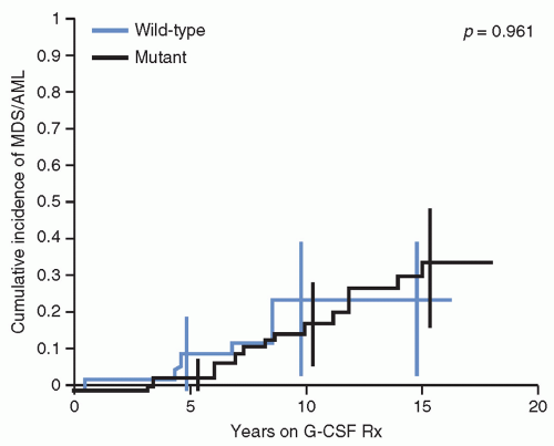
Figure 20.4 The cumulative incidence of MDS and AML in patients with SCN and either mutant or wild-type ELA2 mutations according to years on G-CSF cytokine treatment. (From Rosenberg PS, Alter BP, Link DC, et al. Neutrophil elastase mutations and risk of leukaemia in severe congenital neutropenia. Br J Haematol 2008;140(2):210-213.91)


Inherited Cytopenias of Selective Lineages

Severe congenital neutropenia (SCN), also called Kostmann syndrome, represents an inherited granulocytopenia with a 25% estimated 10-year cumulative risk of MDS/AML and with a long-term annual risk of 2.3% per year after 10 years.79,80 Children present during infancy with absolute neutrophil counts of less than 500/µL and have significant bacterial infections.

SCN is primarily caused by inherited mutations in neutrophil elastase (ELANE) gene (previously termed ELA2). However, there are cases of SCN that do not have mutations of ELA2, but still have an increased risk of developing MDS/AML (Fig. 20.4). Mutations of the HAX1 gene, which encodes a heat shock associate protein-linked LYN, a key SRC family tyrosine kinase that plays a key role in myelopoiesis, have been described in SCN. Neurological abnormalities are associated with patients with SCN due to HAX1 gene mutations.81 The mechanism(s) leading to neutropenia is unclear for these gene defects, but likely linked to apoptosis resulting from misfolded elastase proteins or to a role of HAX1 in mitochondria-regulated apoptosis.82

Further, as Next-Gen sequencing approaches to such disorders have increasingly been used, other mutations have been identified, such as those involving CXCR2, which encodes a chemokine receptor that in turn transduces interleukin 8 (IL8)-mediated signals through a G-protein-activated signal transduction cascade.83 Mutations of the VPS45 gene, which encodes a protein involved in endosomal membrane trafficking, have been reported in familial neutropenia, neutrophil dysfunction, bone marrow fibrosis, and nephromegaly.84 Mutations involving the ELANE gene, some of which overlap with those associated with SCN, have also been identified in cases of cyclic neutropenia, which, however, is not associated with an increased incidence of MDS/AML.85,86,87 Mutations in the glucose 6-phosphate catalytic subunit 3 (G6PC3) gene and the transcription factor encoding GFI1 gene have also been linked to cases of SCN.88 Mutations affecting the Wiskott-Aldrich syndrome gene (WAS) have also been linked to cases of SCN.88

The bone marrow of such patients reveals a decreased production of granulocytic lineage and usually a maturational arrest before or at the promyelocytic/myelocytic stage. In addition,
neutrophils that are produced in SCN have been reported to have abnormal granule maturation and decreased antimicrobial activity. The increased production of neutrophils associated with recombinant G-CSF exposure does not appear to abrogate some of these maturational, functional defects.89 Nevertheless, patients with SCN usually benefit from treatment with recombinant G-CSF, which increases neutrophil counts and reduces the risk of fatal infections. However, the use of G-CSF has been associated with an increased frequency of MDS/AML, estimated to be about 2.3% per year, in a dose-dependent manner.79,90 Of note, the risk of leukemia appears to be similar in patients with or without ELANE mutations (Fig. 20.4).79,80,91

G-CSF receptor (CSF3R) mutations have been identified in approximately 80% of cases of SCN and AML, strongly suggesting that these mutations contribute to the development of AML as a second genetic hit.92 In addition, coexistence of cooperating CSF3R and RUNX1 mutations has been reported in SCN.93

Thus, patients with SCN are usually clinically followed with both complete blood counts on a regular basis as well as with yearly bone marrow examinations to detect morphologic, chromosomal, and gene mutations associated with the evolution of MDS/AML. In such cases, early hematopoietic stem cell transplantation (HSCT) before progression to AML can be both curative of the SCN and prevent emergence of leukemia. It has been recommended that patients with SCN and a poor response to G-CSF should be referred for allogeneic HSCT during the first year of life.94,95,96,97,98

Neurofibromatosis Type 1 (NF1), along with Noonan, Costello, LEOPARD, and SPRED1 syndromes, all represent disorders considered part of the inherited Neuro-Cardio-Facio-Cutaneous (NCFC) disease family. They all are the result of inherited gene alterations that lead to activation of the RAS-BRAF-MAP-ERK pathway critical in hematopoietic cell proliferation and survival. These syndromes are also cancer predisposition syndromes that are associated with both MDS/AML (particularly LEOPARD, Costello, and SPRED1 syndromes) and with JMML (NF1 and Noonan Syndrome).99,100,101,102,103

CEBPA-mediated familial AML is a rare disorder with autosomal dominant inheritance of germ line mutations involving the CEBPA, which encodes a CCAAT/enhancer-binding transcription factor and master regulator of myelopoiesis.104 Somatic mutations of CEBPA are also commonly observed in sporadic cases of AML. The AML that arises as a result of CEBPA mutation is often characterized as differentiated with the presence of Auer rods and expression of the lymphoid antigen, CD7.105

Germ line mutations in the key hematopoietic transcription factor, GATA2, have also been reported in patients with familial CEBPA mutations, but not in those without CEBPA mutations, suggesting the need for cooperation for the development of AML.106 Germ line mutations of GATA2 have also been reported in patients with MDS/AML in conjunction with profound circulating monocytopenia, B- and NK-cell lymphopenia, pulmonary alveolar proteinosis, and susceptibility to multiple opportunistic infections. This autosomal dominant inherited syndrome, termed the “Mono-MAC” syndrome, has a median onset of 32 years of age, but has been reported in children as young as 7 years.83,107


Figure 20.5 Kaplan-Meier plots of the risk of severe cytopenia, nonmalignant cytopenias, and malignant cytopenias expressed in years since birth in patients with Shwachman-Diamond syndrome. (From Donadieu J, Fenneteau O, Beaupain B, et al. Classification of and risk factors for hematologic complications in a French national cohort of 102 patients with Shwachman-Diamond syndrome. Haematologica 2012;97(9):1312-1319.110)

Mutations of these genes in MDS/AML should alert clinicians to consider that such patients may have such inherited predisposition conditions.108

Shwachman-Diamond syndrome (SDS) is an autosomal recessive disorder characterized by exocrine pancreatic insufficiency, heart and skeletal abnormalities (e.g., short stature, osteopenia, metaphyseal dysostosis), and neurocognitive problems and BMF, particularly neutropenia, with a significant predisposition for MDS/AML.109 With a median follow-up of 11.6 years, a French national cohort study of 102 patients with SDS showed that there was a 20-year cumulative risk of severe cytopenia of 24.3%; first symptoms appearing before 3 years of age and significantly low peripheral blood counts were associated with the most severe hematologic complications.110 Out of a total of 102 patients, 12 patients developed what were considered malignant cytopenias; if only the 41 patients who had severe cytopenias were considered, then about 29% developed MDS/AML (Fig. 20.5).110 An NCI study from the United States observed AML in patients with FA and DC, but none in 17 patients with SDS.65 The difference is likely due to the smaller number of patients examined in this study compared with the National French Cohort. The evolution to MDS/AML in patients with SDS is also associated with characteristic accompanying chromosomal abnormalities, including those involving chromosome 20 and isochromosome 7q. The del20(q11) involves a region known to be involved in patients with MDS/AML without SDS. The isochromosome 7q defect cases more often had stable persistence of the abnormal clone that may be dependent on SBDS gene dosage variability located in this chromosomal region.111

SDS is caused in about 90% of diagnosed patients by mutations in the SBSD gene, which encodes for a critical cofactor associated with Elongation Factor 1 (ELF1). Together they catalyze the GTP-dependent release of EIF6 from the nascent 60S ribosomal subunit, allowing it to effectively combine with the 40S ribosomal subunit in order to effect protein synthesis. This defect in ribosomal biosynthesis results in altered mRNA utilization and protein synthesis, which in turn stimulates a p53 response and apoptosis.109,112

Diamond-Blackfan anemia (DBA) is another important BMF syndrome characterized by skeletal anomalies (triphalangeal thumbs, short stature), craniofacial defects (hypertelorism, flat nasal bridge, cleft palate), cardiac abnormalities, red blood cell aplasia, and an increased risk of malignancy.113,114,115 DBA is also another ribosomopathy in that the majority of the etiologically linked mutated genes encode for proteins that comprise the large and small ribosome subunits. While mutated ribosomal protein-encoding genes represent approximately 25% of patients with
DBA, approximately another 25% of patients may have segmental gene or chromosomal deletions involving ribosomal protein-encoding genes.116,117 The ribosomal protein-encoding gene, RPS14, has also been shown to cause the 5q2 MDS syndrome that can mimic DBA, but responds to lenalidomide therapy.118,119,120 Such defects in ribosome biosynthesis in DBA have been shown to result in increased TP53 activity and apoptosis, although it is unclear why such a defect results primarily in erythroid lineage aplasia in DBA and primarily granulocytic decreases in SDS. Mutations of the GATA1 transcription factor have also been reported to result in a DBA phenotype.121,122,123 These observations have led to the proposal that GATA1 defects result in altered expression of ribosomal protein genes, thus resulting in a similar phenotype to a primary ribosomal protein-encoding gene defect.122

How such ribosome haploinsufficiency syndromes lead to cancer, and particularly MDS/AML, is unclear. However, the observed to expected ratios of MDS and AML in patients with DBA have been reported as 287 and 28, respectively.124 Therefore, these patients need to be monitored clinically for management of their anemia but also for the emergence of MDS/AML and other neoplasias.

The familial platelet disorder (FPD) with the propensity to develop AML is a result of germ line mutations in the RUNX1 (previously termed CBFA2, AML, and PEBP2AB) gene that encodes for the Runt-related transcription factor 1.125,126,127 Patients usually present in infancy with mild-to-moderate thrombocytopenia and bleeding tendency, but without dysmorphia.125 Some cases with associated mental retardation, dysmorphic features, and short stature have been described and appear to be related to large deletions that involve RUNX1 and other genes.128 Germ line mutations of RUNX1 have also been associated with T ALL.129 A syndrome of congenital transfusion-dependent thrombocytopenia and anemia with MDS has been linked to germ line mutation of GATA1.130

Congenital amegakaryocytic thrombocytopenia (CAMT) usually presents at birth without any physical abnormalities but with isolated thrombocytopenia with decreased bone marrow megakaryocytes; it then progresses to BMF during the first few years of life. The disorder has been shown to be due to either homozygous or compound heterozygous mutations of the c-MPL gene, which encodes for the thrombopoietic receptor.131,132,133,134 The lifelong risk of developing MDS/AML has been estimated to be less than 10%. Patients usually undergo HSCT, which is curative. The molecular basis for this thrombopoietic receptor defect leading to trilineage BMF remains unclear, but may relate to the type of mutation affecting the receptor and its role in earlier hematopoiesis.135

Thrombocytopenia with absent radius (TAR) syndrome has also been associated through case reports with the development of MDS/AML. The syndrome is linked to deletions involving chromosome 1q21.1, and is associated with several low-level regulatory single nucleotide polymorphisms (SNPs) as well as a null mutation in the RBM8A gene, which is an RNA-binding protein involved in RNA splicing.136 Hematopoietic precursors have been reported to have defective thrombopoietin signaling, particularly in the fetal and neonatal period, so that the thrombocytopenia usually resolves within the first year of life.137

For all of these syndromes it is important to follow the patients closely for the development of MDS/AML. And while HSCT can be curative in terms of the bone marrow defect, it is important to screen all potential family donors for the presence of the mutations in question. As Next-Gen sequencing of exomes and whole genomes are applied to more cases of unknown cytopenias with a propensity to develop MDS/AML, it is quite likely that more mutated genes will be identified to explain the phenotypes.


Acquired Aplastic Anemia and MDS/AML

Severe aplastic anemia (SAA) presents with pancytopenia and a hypocellular bone marrow. Although SAA may be the consequence of environmental and/or drug exposures as well as postinfectious, often postviral, hepatitis, many cases remain idiopathic. In such cases, a detailed diagnostic workup for germ line mutations and disorders associated with key BMF syndromes should be initiated. In addition, an important part of the differential diagnosis is hypoplastic MDS or hypoplastic refractory cytopenia of childhood, which may be difficult to distinguish from SAA. One study of 100 cases of SAA in children from the European Working Group of MDS in Childhood (EWOG-MDS) and the German SAA study group classified cases as MDS/RCC if patchy areas of erythropoiesis with abnormal maturation were observed in hypoplastic bone marrow.138 Another report in children with SAA and MDS (RCC) noted that the histopathological pattern of MDS (RCC) was characteristically associated with islands of immature erythroid precursors and scattered granulocytic precursors along with the occasional micromegakaryocytes.139 Examination of T-lymphocyte subsets has suggested that patients with low-risk MDS have increased T-helper and cytolytic T lymphocytes compared with patients with SAA.140 Another potentially helpful discriminator is immunohistochemical staining for CD34, which has been considered to be higher in cases of hypoplastic MDS than in SAA.141,142,143 However, this may not always be a distinguishing characteristic in RCC.144 The existence of clonal, chromosomal abnormalities, such as monosomy 7, or specific gene mutations characteristic of MDS (see MDS section), can provide further evidence for a definitive diagnosis.

Approximately 10% to 15% of patients with idiopathic SAA who are not treated with allogeneic HSCT will develop MDS/AML. The frequency of developing MDS does not appear to vary when those patients with idiopathic versus viral-associated AA are compared.145,146 In addition, a prospective study from Japan demonstrated that children with very severe presenting blood counts (neutrophil counts less than 200/µL) had a higher complete response rate than those with less severe presenting neutrophil counts, suggesting that those with more severe disease may have a more immunologically responsive disease and be less likely to have a primary MDS-like disorder.147

Children with SAA appear to develop MDS sooner than adults with SAA, and usually within the first 3 years following their diagnosis; this also suggests a fundamental difference in the disorders in children compared with adults. Another key point in managing such patients is that disease that is less responsive or requiring more prolonged periods of G-CSF treatment has a higher frequency of MDS/AML development.148,149,150

Allogeneic HSCT from a matched sibling donor (MSD) is a curative approach for children with newly diagnosed SAA and is also effective following the development of MDS.151 Overall survival (OS) and event-free survival (EFS) for children with new-onset SAA transplanted with HLA-MSD have been reported to be approximately 90% and 86%, respectively.152,153 Outcomes for children who develop MDS/AML after treatment for SAA have been reported to show 5-year OS of approximately 50% to 70%.154,155

Acquired amegakaryocytic thrombocytopenia (AAMT) can occur at any age and has been associated with environmental toxins, drugs, viral infections, and autoimmune disorders.156,157,158 The disorder often will respond to a variety of immunosuppressive therapies.159,160 Cases have been reported to progress to AML following development of a chromosome 5q2 abnormality.

Paroxysmal nocturnal hemoglobinuria (PNH) is an acquired hemolytic anemia due to increased sensitivity to complement-induced intravascular hemolysis. This increased sensitivity is secondary to a defect in glycophosphatidylinositol (GPI) anchoring of membrane proteins with decay-accelerating factor (DAF/CD55) and protectin (CD59/MIRL/MAC-IP) being affected. These cell-surface proteins play key roles in protecting red blood cells but also hematopoietic stem cells from destruction. The PIGA gene, which encodes phosphatidylinositol glycan A, is the most commonly mutated enzyme. PNH may also evolve into SAA and then MDS/AML with the clonal evolution of additional chromosomal changes.161,162


Environmental Mediated Mutagenesis

A large amount of evidence exists that certain environmental exposures can result in gene mutations and chromosomal rearrangements in bone marrow stem cells and can predispose individuals to the development of MDS and AML. Most of the evidence, however, has been derived from studies in adults. The very low incidence of MDS and AML in children has prevented large, definitive studies from being done. Nevertheless, there are several environmental exposures, both prenatal and postnatal, that have been examined and proven to be potentially important.

Ionizing radiation has been strongly associated with the development of MDS/AML. For example, an approximately 20-fold increase in AML was observed following the exposure of individuals to the atomic bombs on Nagasaki and Hiroshima during World War II. The peak of AML occurred between 6 to 8 years after exposure. The lack of a documented increase in leukemia in children prenatally exposed to the atomic bomb radiation is of note, and consistent with the lack of a definitive association of increased leukemia risk secondary to prenatal exposure to x-rays. In addition, there are no compelling data to link exposure to ultrasound or living near high-voltage power lines to the development of leukemia.163,164,165,166

However, maternal ingestion of various foods rich in topoisomerase II inhibitors, such as flavonoids in soy products and catechins in green and black tea, during pregnancy has been reported to be associated with an increased risk of AML with MLL rearrangements in offspring.167,168,169 Maternal alcohol consumption during pregnancy has also been linked to increased risk of AML in offspring in a dose-dependent fashion.170 Maternal smoking during pregnancy has not been reported to increase the risk of AML in offspring,170 although an association of cigarette smoking and AML has been linked in adults, thus emphasizing an additional rationale for smoking cessation.171

Exposure of individuals to environmental carcinogens, including pesticides, petroleum products, benzenes, and heavy metals, has been documented to increase the risk of MDS and AML. Of these, benzene has been the most definitively linked to the development of MDS/AML.172,173 There is, however, evidence of an increased risk of leukemia in children in rural areas associated with increased exposure to pesticides174 as well as an increased risk of AML in offspring of mothers exposed during gestation to significant levels of pesticides.175 The observation of increased concentration of organophosphate pesticides in the children of farmworkers compared with adults suggests a possible reason for the increased leukemia risk in these children.176 These associations should alert pediatric oncologists and pediatricians to include a careful history of potential environmental exposures in patients.

While environmental exposures to specific mutagens have been linked to the development of MDS/AML, the exposure of patients with cancer to ionizing radiation and chemotherapeutic agents also increases the risk of developing secondary leukemias. The risk and time to develop secondary MDS/AML is different depending on the type of exposure. For instance, secondary leukemias with characteristic MLL gene rearrangements are primarily associated with exposure to drugs that function as topoisomerase inhibitors, including epipodophyllotoxins such as etoposide, and anthracyclines, such as doxorubicin, daunorubicin, idarubicin, and mitoxantrone.177 This group of secondary AML usually develops within the first 3 to 5 years following exposure, although some cases have been reported 10 to 12 years later.178 Both the cumulative dose and the schedule of such agents have been shown to be associated with the development of secondary AML179; however, even single exposures have been reported in patients with subsequent secondary AML.180,181

Alkylating agents (e.g., cyclophosphamide, temozolomide) and related drugs (e.g., cisplatin) used primarily to treat patients with solid tumors represent a second class of chemotherapeutic agents that can result in secondary MDS/AML.182,183 These secondary AML cases appear later, usually beyond 5 years of exposure, than those with MLL rearrangements, and are more commonly associated with karyotypic abnormalities, such as monosomy 7.

Attempts to reduce the cardiotoxicity of anthracyclines have led to the use of cardioprotectant agents, such as dexrazoxane. The use of this cardioprotectant in combination with chemotherapy that included etoposide for patients with Hodgkin lymphoma led to the report of an increased incidence of secondary AML.184 However, subsequent reports in patients with ALL and AML did not show an increased risk of secondary AML following the use of dexrazoxane.185,186,187,188 Possible explanations for the differing results have included the schedule of dexrazoxane used in the Hodgkin lymphoma study, a distinct host effect in those with Hodgkin lymphoma, or chance variance based on small numbers.189

An additional, important component of increased risk to exogenous mutagens is the ability of the exposed person to metabolize or detoxify such compounds based on their genomic background. For example, polymorphisms in cytochrome p450 and glutathione-S-transferase, both of which are involved in the detoxification of environmental mutagens and chemotherapeutic agents, have been linked to increased risk of developing MDS/AML.190,191,192 Specific polymorphisms of the thiopurine methyltransferase (TPMT) gene that are key for the metabolism of 6-mercaptopurine and 6-thioguanine, both drugs used in the treatment of patients with ALL, have been reported to increase the risk of secondary MDS/AML.193,194


METHODOLOGIES FOR CLASSIFYING MYELOID MALIGNANCIES

Classification of myeloid malignancies relies on morphological, cytochemical, and immunophenotypic characteristics to define the cell lineage (e.g., myeloid vs. lymphoid), the degree of maturation, and whether there is evidence for dysplastic changes. These methods are usually performed on bone marrow aspirates and/or biopsy specimens, although analysis of peripheral blood is being increasingly used for initial assessment of a patient with presumed leukemia. To further define AML/MDS, cytogenetic or karyotyping is used to identify characteristic chromosomal abnormalities, such as chromosome numbers, deletions, amplification, and rearrangements. The use of fluorescence in situ hybridization (FISH) is an added refinement of examining chromosome number and rearrangements. Additional studies examining specific gene mutations or alterations provide further diagnostic support as well as important prognostic features. Future approaches to defining subtypes of AML will likely involve RNA expression patterns, epigenetic characteristics, real-time phospho-flow cytometric analysis, and detailed signal transduction networks.


Morphology and Cytochemical Staining to Examine Lineage

The usual dyes for staining hematopoietic cells from bone marrow or peripheral blood are May-Grunwald-Giemsa or Wright Giemsa stains. These stains provide morphologic information, such as nucleus/cytoplasmic ratios, chromatin quality, and evidence of primary and secondary cytoplasmic granules, all of which are characteristic of progressively mature stages of maturation. Cytochemical stains have been utilized to further define cell subtypes. For example, the detection of myeloperoxidase (MPO) defines myeloid differentiation in the 2008 WHO classification system. However, very early myeloblasts as well as monoblasts do not tend to express MPO. In addition, erythroblasts, megakaryoblasts, and lymphoblasts do not express MPO. Sudan black is another stain that tends to track with MPO, but because of its being less specific, also tends to be less useful. Nonspecific esterases (alpha-naphthyl butyrate and acetate) are characteristically expressed at high levels in monoblasts and monocytes. Of note, megakaryoblasts and
erythroblasts may also express nonspecific esterases, but it is not fully inhibited by sodium fluoride as in monoblasts and monocytes. The specific esterase, naphthol-ASD-chloroacetate esterase, primarily stains cells of the neutrophil lineage as well as mast cells. Thus, using nonspecific and specific esterase detection may provide a rapid approach to identifying a leukemia as myelomonocytic. The periodic acid-Schiff (PAS) stain identifies intracellular glycogen and neutral mucopolysaccharides, is positive in both lymphoid and myeloid leukemia, but particularly intense staining, diffuse, and sometimes in larger globules, may be observed in erythroleukemia. Staining for leukocyte alkaline phosphatase (LAP) is positive in many different cell types, but is usually very low in CML, thus sometimes helping to distinguish CML from leukemoid reactions or other myeloproliferative disorders. Iron stains should be done on bone marrow specimens in order to detect ringed sideroblasts, the result of an abnormal accumulation of iron that localizes near and surrounds at least two-thirds of the perimeter of the nucleus and is characteristic of a subtype of MDS. Toluidine blue binds to mucopolysaccharides, and staining is characteristic of basophils and mast cells, although stains for tryptase may be more specific in identifying mast cell malignancies (Table 20.2).


Immunophenotyping

In addition to immunohistochemistry, the detection of cell-surface or intracellular proteins using specific monoclonal antibodies with laser-stimulated immunofluorescent tags provides the basis for flow cytometric immunophenotyping. Since the discovery of monoclonal antibody production methods were published in 1976 to detect specific antigens (referred to as cluster determinants or CD), hundreds of cell type and differentiation selective antigens have been identified. Although immunophenotyping has not been included in the standard FAB or WHO classification systems, it plays important roles in several aspects of myeloid malignancy characterization. For instance, it is an important adjunct in discriminating myeloid from lymphoid malignancies as well as between AML and chronic myeloproliferative disorders. In addition, myeloid leukemias characteristically aberrantly express a variety of differentiation antigens, sometimes including certain lymphoid selective proteins. Using several monoclonal antibodies simultaneously (multiparameter flow cytometry MFC), greater than 90% of myeloid malignancies can be shown to have a distinct immunophenotype compared with normal hematopoietic progenitors, thus providing a means to detect minimal, or, more appropriately, measurable, residual disease.

For example, MFC is a highly accurate approach to initially defining a patient’s leukemia, usually from a peripheral blood sample, as myeloid or lymphoid in origin. This can be important information in terms of speaking to parents and for planning initial therapy. AML-selective or specific markers typically include CD11b, CD13, CD14, CD15, CD33, CD36, and CD41 (also referred to as platelet antiglycoprotein IIb/IIIa), CD42 (glycoprotein Ib), CD61 (glycoprotein IIIa), CD64, CD117 (cKIT), CD163, lysozyme, and MPO. Most AML subtypes express human leukocyte antigens (HLA) Class II (mostly HLA-DR subgroup of the Class II antigen family), with the exception of APL (Table 20.3).


TABLE 20.2 Histochemical Staining of Hematopoietic AML Subtypes


HISTOCHEMICAL STAIN


AML FAB Subtype


M0


M1-M2


M3


M4


M5


M6


M7


Myeloperoxidase


+


+


+


−/+


NSE—Chloroacetate


+


+


+


+/−


NSE—Alpha−naphthol acetate


+a


+a


+/−a


Sudan Black


+


+


PAS


+/−


+/−


+


MPO, Myeloperoxidase; NSE, Nonspecific Esterase; PAS, Periodic Acid-Schiff Stain;


a Inhibited by fluoride; NOTE; ALL is negative for all of the above histochemical stains except PAS.


These myeloid-associated markers can be helpful and useful in distinguishing different types of morphologically or FAB-defined subtypes of AML. FAB M1 and M2 subtypes, which show evidence of early myeloid differentiation, typically express high levels of CD13, CD15, CD33, CD117, MPO, and often the stem cell marker CD34, but low levels of more monocytic-associated markers such as CD14. Probably representing the one example where defining the FAB subtype can have implications on the choice of initial therapy is FAB M3 or APL. In this subtype, HLA-DR (Class II) antigen expression is usually absent or very low, in contrast to the other, nearly 80% of subtypes of AML, which express high levels of HLA-DR. Myelomonocytic (FAB M4) leukemia characteristically expresses CD14, CD36, CD64, CD68, CD163, and lysozyme, all monocyte lineage-associated antigens, along with more typical myeloid markers, CD13, CD15, and CD33. FAB M4 AML also tends to express CD11b and c antigens. The presence of these two populations defined by flow cytometry can thus distinguish this subtype of AML. Similarly, monocytic leukemia (FAB M5) typically shows decrease or loss of expression of CD13, but retains expression of CD14, CD15, and CD33 as well as CD36, CD11b, CD11c, CD64, and CD68 often on a single population of leukemic blasts, although some heterogeneity of expression can be observed. Expression of stem cell marker CD133, when present, is useful in differentiating M5 monoblasts from mature monocytes. Additionally, NG2 (7.1) expression may be observed in MLL-translocated cases. FAB M6 or acute erythroid leukemias characteristically express the erythroid marker Glycophorin A on the erythroblasts. FAB M7 or AMKL typically express platelet glycoproteins, such as CD41, CD42, and CD61 along with CD13, CD33, and often CD36.

Certain lymphoid antigens are also expressed aberrantly in myeloid malignancies. For example, B-lymphocyte lineage-associated antigens, such as CD10, CD19, CD20, and CD22, may be expressed on AML blasts in 10% to 20% of cases. T-lymphocyte lineage-associated antigens, such as CD2, CD3, CD5, and CD7, are expressed in 20% to 40% of AML cases. Other studies have reported that nearly 60% of AML cases express either B- or T-lymphoid-associated markers. While this type of coexpression of myeloid and lymphoid-selective antigens is thus not uncommon, there has been no convincing associated prognostic significance. Nevertheless, the aberrant expression can be particularly useful in distinguishing leukemic from normal hematopoietic precursors during bone marrow recovery, for detection of measurable residual
disease, for the monitoring of leukemia stem or initiating cells, and for helping to define leukemias of ambiguous lineage.195,196,197,198


TABLE 20.3 Immunophenotypic Determinants During Hematopoiesis and in Myeloid Malignancies


MARKER


AML FAB Subtypes


M0


M1


M2


M3


M4


M5


M6


M7


Myeloid lineage


CD11b


+/−


+/−


+/−


++


++


CD13


++


++


+


+


++


++


+


+


CD14


++


++


CD15


+/−


+


−/+


++


+


CD33


+/−


++


++


++


++


+++


+


+


CD64


+/−


++


++


CD68a


++


++


Erythroid lineage


Glycophorin A


+


Hemoglobin A


+


Megakaryocytic


CD36


+/−


+


CD41


+


CD42


+


CD61


+


T Cell lineage


CD2


−/+


−/+


+/−


CD3


CD4


−/+


−/+


+


+


CD5


CD7


+/−


+/−


−/+


+/−


+/−


+/−


+/−


CD56


−/+


−/+


−/+


+/−


B−cell lineage


CD10


CD19


−/+


−/+


CD20


CD22


Stem cell


CD34c


++


+/−


+


+/−b


+


+/−


CD117


+


++


+


+


+


+


+/−


+


Non−lineage selective


HLA−DR


++


+


+


+/−


++


++


Scoring System: This reflects estimated relative expression of antigens: − is usually negative; + is usually low but clearly positive expression; ++ is strongly positive; +++ is very strongly positive; +/− represents occasional staining of cases; −/+ represents weak or positive staining in usually 30% of cases.


a The PGM1 MaB clone is macrophage restricted, whereas the other commonly used clones, such as KP1, show cross−reactivity with the granulocytic series.

b Microgranular subtype frequently expresses CD34 and CD2.205

c CD34 and/or CD117 expression often occurs on a subpopulation of blasts.205


While acute leukemias of ambiguous lineage (ALAL), also called mixed phenotype acute leukemias (MPAL), are prognostically important subtypes of leukemia, fewer than 5% of cases occur in children.199,200,201 These leukemias are distinguished from AML with aberrant expression of lymphoid markers by the absence of a recognizable predominant lineage by morphological, histochemical, or immunophenotypic criteria.202


Chromosomal and Genomic Alterations


Karyotypic Changes

Myeloid malignancies are often characterized by specific chromosomal abnormalities, and particularly translocations (Fig. 20.6). Many of these abnormalities help to both classify AML as well as to serve as important prognostic markers. Thus, every patient should have cytogenetic analysis performed on a bone marrow sample of their AML at diagnosis.


Figure 20.6 Distribution of karyotypic abnormalities in pediatric AML.

Standard karyotyping approaches on mitotically arrested leukemia cells can identify clonal chromosomal abnormalities in 75% to 80% of pediatric AMLs.203 For example, chromosomal translocations t(8;21) and inv(16) are observed in approximately 20% of pediatric AML and are referred to as CBF leukemias because of the transcription factors involved and their associated protein complexes; they also predict a better outcome. The t(15;17) translocation is characteristic of APL and occurs in about 12% of pediatric AML. Rearrangements of chromosome 11q involving the MLL gene occur in approximately 18% of pediatric AMLs, but in the majority of AMLs in patients under age 2 years. About 31% of pediatric AMLs have other chromosomal abnormalities, including monosomy 7 or 27q, del(9q), del(5q), and +8.


Subkaryotypic (Cryptic) Changes

As karyotyping relies on detection of alterations in G-banding patterns in metaphases (or interphases), genomic changes that are either small or located in centromeric or telomeric regions of chromosomes may not be amenable to detection by conventional methodologies, and detection of such alterations requires secondary methodologies, including FISH or polymerase chain reaction (PCR)-based methodologies. Such approaches complement the karyotype and can increase the sensitivity of identifying more subtle chromosomal alterations that cytogenetic methods miss. In addition, FISH allows for the analysis of a much greater number of cells. For example, FISH is able to identify t(15;17) in APL or the chromosomal t(9;22) involving the Philadelphia chromosome, abnormalities that are not identified by routine cytogenetics. Of note, the identification of the t(9;22) translocation in a patient with AML is most likely to represent CML in myeloid blast crisis. Thus, FISH is, like karyotyping, a standard analysis done in patients with newly diagnosed AML as well as inserial samples to assess response to therapy. PCR-based assays are not only able to detect translocations that are not detectable by karyotyping, given the extreme sensitivity of the assay, they can be used for monitoring response to therapy and have potential utility in prediction of relapse prior to morphologic emergence of disease. Several cryptic translocations include NUP98/NSD1, NUP98/KDM5A, and CBFA2T3/GLLIS2 fusions that appear to be uniquely pediatric AML-associated changes.


Gene Mutation and Copy Number

The introduction of DNA sequencing and the ability to amplify specific regions of chromosomes to increase the sensitivity of detection have contributed to identifying a significant number of key gene mutations. With the application of whole exome, genomic, and RNA sequencing approaches, a further refinement of key interacting pathways that are altered in AML is emerging.

The recurring gene mutations along with chromosomal abnormalities profoundly impact aspects of AML subtype classification, prognostication, and, potentially directly, treatment choices.

Another type of alteration that has been found to have increasing importance involves gene copy number, including both amplification and deletion of specific genomic sequences. Such studies have identified, for instance, loss of heterozygosity (LOH) associated with gene deletions is a mechanism to produce haploinsufficiency of a gene. For instance, when a gene undergoes homologous recombination with subsequent duplication of one copy of the remaining allele, the product is called Copy Neutral LOH (CN-LOH). This situation, also referred to as acquired uniparental disomy (aUPD), can result in only two copies of a mutant allele, such as has been observed for the FLT3-ITD and CEBPA mutations.204,205,206,207

Some of these molecular changes have started to be incorporated into AML/MDS classification systems. Their role in prognosis is discussed in detail under the section on Prognostic Factors.


DEFINING SUBTYPES: EVOLUTION OF DIAGNOSTIC CLASSIFICATION OF MYELOID MALIGNANCIES


AML

The French-American-Berlin (FAB) Classification of myeloid malignancies represented the first attempt of a comprehensive system for classifying myeloid malignancies and was based primarily on morphology and immunohistochemical staining for lineage-selective markers. The initial FAB-proposed classification published in 1976 was followed by revisions in 1985 and 2003.208,209 Although this system played an important role in the classification of AML, the ability of the system to provide key prognostic indicators as well as the emergence of key cytogenetic and molecular markers limited its ultimate impact.

The WHO classification of myeloid malignancies, initially proposed in 2002, has replaced the FAB classification. The 2002 classification incorporated cytogenetic information and appeared to correlate with outcome more than the previous FAB system.210 Some important distinctions between the two systems included the following. The WHO classification reduced the percentage of leukemic blasts to make the diagnosis of acute leukemia from the FAB 30% to 20%. However, because such percentages are in some respects arbitrary and may not reflect the biology of AML, the minimum blast percentage requirement was changed so that patients with chromosomal translocations characteristic of AML, such
as t(8;21)(q22;q22) RUNX1-RUNX1T1; formerly AML1-ETO, inv(16)(p13.1q22) or t(16;16)(p13.1;q22) CBFB-MYH11, and APL with t(15;17)(q22;q12) PML-RARα, were considered sufficient to make the diagnosis. The 2008 WHO revised classification included specific gene mutations for the first time, such as those involving CEPBA and NPM.211 Subsequent refinements to the WHO classification have included additional chromosomal rearrangements, such as t(9;11)(p22;q23) KMT2A-MLLT3; formerly MLL-AF9, t(6;9)(p23;q34) DEK-NUP21, inv(3)(q21q26.2) or t(3;3)(q26.2) RPN1-EV11 and t(1;22)(p13;q13) RBM15-MKL1.

Thus, on the basis of the above considerations, the 2008 WHO classification has enumerated several distinct subtypes of AML and MDS (Table 20.4).211 The different subclasses include several distinctive AML subtypes characterized by distinguishing chromosomal changes, as well as morphological, histochemical, immunophenotypic, and molecular changes. Important prognostic impact also is associated with some specific subtypes.


TABLE 20.4 WHO Classification of Acute Myeloid Leukemiaa,205


AML with characteristic genetic abnormalities


AML with t(8;21)(q22;q22) RUNX1-RUNX1T1


AML with inv(16)(p13q22) or t(16;16)(p13;q22) CBFB/MYH11


APL with t(15;17)(q22;q12) PML/RARA


Variant fusions associated with microgranule variants


AML with t(9;11)(p22;q23) MLLT3-MLL


AML with t(6;9)(p23;q34) DEK-NUP214


AML with inv(3)(q21q26.2) or t(3;3)(q21;q26.2) RPN1-EVI1


AML (megakaryoblastic) with t(1;22)(p13;q13) RBM15-MKL1


AML with gene mutations


AML with mutated NPM1 (Provisional Entity)


AML with mutated CEBPA (Provisional Entity)


AML with myelodysplasia-related changes


Criteria include: ≥20% peripheral blood or bone marrow blasts AND any of the following: previous history of MDS; MDS-related cytogenetic abnormalityb; multilineage dysplasia AND Absence of both: Prior cytotoxic therapy for an unrelated disease; recurring cytogenetic abnormality as described in AML with recurrent genetic abnormalities.


AML not otherwise specified


AML with minimal differentiation (FAB M0)


Myeloperoxidase (MPO) may not always be evident by light microscopy, but MPO positive granules can be detected by electron microscopy. Immunophenotyping may be helpful in demonstrating expression of myeloid markers or cluster determinant (CD) markers for CD13, CD33, and CD117 (also known as cKIT)


AML without maturation (FAB M1)


Expression of MPO is detected by immunohistochemistry or flow cytometry.


AML with maturation (FAB M2)


AMML (Acute myelomonocytic leukemia; FAB M4)


Peripheral blood or bone marrow has ≥20% blasts—neutrophils and monocytes and their precursors each comprise at least 20% of bone marrow cells.


Acute monoblastic leukemia (FAB M5)


≥80% of leukemic cells are of monocytic lineage


Acute erythroid leukemias


Type 1 (FAB M6a): ≥50% erythroid precursors in the entire nucleated cell population and >20% myeloblasts in nonerythroid cell population


Type 2 (FAB M6b): ≥80% of bone marrow cells are either undifferentiated or proerythroblastic and with no significant myeloblastic differentiation


Acute Megakaryoblastic Leukemia (FAB M7)


Acute Basophilic Leukemia


Acute Panmyelosis with Myelofibrosis


Myeloid sarcoma


Myeloid proliferations related to Down syndrome


Transient abnormal myelopoiesis (TAM)/Transient myeloproliferative syndrome (TMD)


Myeloid leukemia associated with Down syndrome


Acute leukemias of ambiguous lineage


aFAB classification notations indicated in parentheses. The classification is based on morphology as well as cytogenetic as well as increasingly gene mutations. Note that the FAB classification required the bone marrow to have 30% blasts to make the diagnosis of AML, while the WHO criteria require 20% or greater.

b Complex Karyotype: >3 unrelated abnormalities, none of which are included in the AML with recurrent genetic abnormalities subgroup. Unbalanced abnormalities: −7/del(7q); −5/del(5q); i(17q)/t(17p); −13/del(13q); del(11q); del(12p)/t(12p); del(9q); indic(X)(q13). Balanced abnormalities: t(11;16)(q23;p13.3), t(3;21)(q26.2;q22.1), t(2;11)(p21;q23)—these three translocations usually associated with treatment-related AML, which should be excluded before using these in diagnosis of AML with MDS-related changes; t(1;3)(p36.3;q21.1); t(5;12)(q33;p12); t(5;7)(q33;q11.2); t(5;17)(q33;p13); t(5;10)(q33;q21); t(3;5)(q25;q34)


WHO-Defined Specific Subtypes of AML


AML with Characteristic Genetic Abnormalities

AML with t(8;21)(q22;q22) RUNX1-RUNX1T1. The FAB M2 subtype is most closely associated with this class of AML. RUNX1 (for Runt-related transcription factor, previously termed AML1) is on chromosome 21 and encodes transcription factor CBF-alpha 2; RUNX1T1 (previously termed ETO for eight-twenty-one gene) functions as part of transcription factor complexes and helps to recruit gene transcriptional corepressors. The leukemic blasts have abundant basophilic cytoplasm and commonly azurophilic granules, which sometimes can be quite large. Increased eosinophilic
precursors are often present. The presence of the t(8;21) translocation is sufficient to make the diagnosis of AML even if the bone marrow blast count is less than 20%. The t(8;21) AML is also associated with an increased frequency of chloromas (granulocytic sarcomas) or extramedullary leukemia. This translocation, occurring in about 12% of children, is associated with a favorable outcome.

AML with inv(16)(p13q22) or t(16;16)(p13;q22) CBFB/MYH11. This subtype is morphologically usually myelomonocytic (FAB M4), and the eosinophils frequently have characteristic large and abnormal immature basophilic granules. Like t(8;21) AML, the presence of the inv16 alternation is sufficient to diagnose AML even if the bone marrow has less than 20% leukemic blasts. The chromosome inversion results in the fusion of the CBF Beta gene at 16q22 fused to the MYH11 (encodes the smooth muscle myosin heavy chain) gene at 16p13. Of note, this chromosome abnormality is frequently missed using standard karyotyping and only identified using FISH or RT-PCR to detect the fusion. This fusion, occurring in about 7% to 9% of children, is associated with a favorable prognosis with 5-year OS of 80% to 90%.

APL with t(15;17)(q22;q12) PML/RARA. This subtype is also referred to as FAB M3 or APL and is characterized by promyelocytes with often bilobed nuclei, abundant azurophilic granules, multiple cytoplasmic Auer rods, and is strongly positive for MPO. A microgranular variant presents with bilobed nuclei, scarce or absent cytoplasmic granules at the light microscopic level, but with Auer rods often present in small promyelocytes. MPO staining is strongly positive. The RARA gene, encoding the retinoic acid receptor alpha protein, is fused to the PML (promyelocytic leukemia) gene, which encodes a myeloid transcription factor. Other translocations, such as t(11;17)(Q23;q21), t(5;17)(q32;q12), and t(11;17)(q13;q21), are also characteristic of APL.

AML with t(9;11)(p22;q23) MLLT3-MLL. This AML subtype often has monocytic or myelomonocytic morphology (FAB M4, M5a, M5b), and less commonly, myelocytic without or with features of differentiation (FAB M1 and M2). The MLL gene (mixed lineage leukemia and the homologue of the Drosophila trithorax gene) on chromosome 11q23 is fused to over 20 other gene partners, with the t(9;11) involving the MLLT3 gene (also known as AF9) on chromosome 9p22, being the most common MLL translocation partner. The WHO classification system considered this subtype a distinct subtype of AML with an intermediate prognosis. An analysis of other MLL translocation partners in children showed a wide variance in survival depending on the type of translocation.212

AML with t(6;9)(p23;q34) DEK-NUP214. This subtype usually has monocytic features, multilineage dysplasia, and is associated with basophilia in about 50% of cases. The DEK gene encodes for a transcription factor, and the NUP214 gene encodes for a nuclear transport channel protein. Thus, the resulting fusion protein generates an abnormal transcription factor as well as altered nuclear protein transport. The t(6;9) translocation occurs in children with a median age of approximately 13 years and is associated with a poor outcome.213,214,215 There is also a high frequency of FLT3-ITD expression of approximately 70% and 80% in pediatric and adult cases, respectively.216,217 The data from COG demonstrate that those cases with t(6;9) translocations have a high relapse risk regardless of the presence or absence of FLT3/ITD.

AML with inv(3)(q21q26.2) or t(3;3)(q21;q26.2) RPN1-EVI1. This subtype commonly derives from preexisting MDS but can be a de novo leukemia; it occurs mostly in adults. Hypogranular neutrophils with Pelger-Huet nuclear changes along with giant and hypogranular platelets are commonly observed. The RPN1 gene on chromosome 3q21 acts as an enhancer of EVI1, located at 3q26. This results in increased proliferation of maturationally arrested early precursors. This subtype is associated with a poor outcome.

AML (megakaryoblastic) with t(1;22)(p13;q13) RBM15-MKL1. This leukemia subtype shows abnormal maturation of the megakaryocyte lineage (FAB M7). While the leukemia represents less than 1% of pediatric AML, most of the cases occur in infants with a median age of presentation of 4 to 7 months of age.218 This translocation is uncommon in children with DS.219 The RBM15 (RNA-binding motif protein, also called OTT or One-Twenty-Two gene) on 1p13 is fused to the MKL1 (for megakaryoblastic leukemia, also termed MAL for myelin AND lymphocyte encoding gene) on 22q132. RBM15 is involved in spliceosome binding and signal transduction, while MKL1 plays an important role in the formation and maintenance of glycosphingolipid-enriched membranes. Although one report described a poor outcome with a survival of about 30%, other reports of small series of patients with t(1;22) AMKL showed a more favorable outcome when patients were treated with intensive chemotherapy.218,219,220


AML with Gene Mutations

AML with Mutated NPM1 (Provisional Entity). This subtype of AML usually has myelomonocytic or monocytic features and primarily occurs in AML with a normal karyotype. For instance, NPM1 mutations occur in about 20% of pediatric AML cases with a normal karyotype, but in 2% to 8% of all childhood AML. NPM1 (Nucleophosmin) encodes for a phosphoprotein that functions as a molecular chaperone between the nucleus and cytoplasm. Mutations result in the protein’s aberrant cytoplasmic expression, a characteristic that can be used for the diagnosis of this AML subtype. There is also a significant overlap between NPM1 mutations and FLT3-ITD in approximately 30% of cases. AML with NPM1 mutations has a favorable prognosis, and its presence appears to be able to partially ameliorate the poor prognostic impact of FLT3-ITD mutations.221,222,223,224

AML with Mutated CEBPA (Provisional Entity). CEBPA (CCAAT/Enhancer-Binding Protein-Alpha), encoded by the CEBPA gene, is known to play a key role in granulocytic differentiation. Mutations of CEBPA occur in about 4% of pediatric cases of AML, and most of these are biallelic, occurring as a result of different mutations in each allele.225 These mutations also occur primarily in AML with a normal karyotype and with myelomonocytic or monoblastic (FAB M4 and M5) morphology. Of potential interest, this subtype of AML also has a high (greater than 50%) expression of the lymphoid antigen, CD7. CEBPA mutations are associated with improved outcomes, including decreased relapse rates and overall improved survival, similar to that observed in CBF leukemias.225,226,227


AML not otherwise specified

AML with Minimal Differentiation (FAB M0). This subtype shows no evidence of myeloid differentiation by morphology or histochemistry. Myeloperoxidase (MPO) may not always be evident by light microscopy, but MPO-positive granules can be detected by electron microscopy. Immunophenotyping may be helpful in demonstrating expression of myeloid markers or cluster determinant (CD) markers for CD13, CD33, and CD117 (also known as cKIT). Expression of lymphoid markers is common and includes CD2, CD7, or CD19.

AML without Maturation (FAB M1). This subtype, which occurs in approximately 20% of pediatric AML cases, shows minimal evidence of myeloid differentiation, but some leukemic blasts may show azurophilic or primary cytoplasmic granules. Expression of MPO is detected by immunohistochemistry or flow cytometry. Immunophenotyping typically shows expression of CD13, CD33, CD34, and CD117.

AML with Maturation (FAB M2). Comprising approximately 30% of childhood AML, this subtype displays leukemic blasts with and without primary granule expression; Auer rods are commonly
observed. There may be associated basophilia and some dysplasia. Immunophenotyping may show CD7 expression in about one-quarter of cases with CD2, CD19, CD4, and CD56 present in about 10% of cases. The t(8;21) translocation is observed in about 10% of these cases, while translocation t(6;9) and abnormalities of chromosome 12p may also be seen.

AMML (Acute Myelomonocytic Leukemia; FAB M4). This subtype accounts for 25% to 30% of childhood AML and usually occurs in patients younger than 2 years of age. Peripheral blood or bone marrow has >20% blasts, with neutrophils and monocytes and their precursors each comprising at least 20% of bone marrow cells. Thus, a minimal percentage of 20% of monocytes is necessary for making this diagnosis. The monocytic blasts express CD14 and sometimes other monocytic markers such as CD11b, CD11c, CD36, CD4, CD68, and CD163 along with lysozyme. They also commonly express the lymphoid marker CD4. The myeloid blasts usually express CD13, CD15, and CD33. As noted above, the chromosomal abnormality inv16 typically has FAB M4 morphology with the presence of dysplastic eosinophilic granules.

Acute Monoblastic Leukemia (FAB M5). In this subtype, >80% of leukemic cells are of monocytic lineage. About 15% of childhood AML is represented by this subtype, which occurs in approximately 50% of AML in children under 2 years of age. Further refinement of this subtype includes the FAB M5a with blasts with large and often multiple nuclei and cytoplasm without Auer rods. The FAB M5b type has blasts that display more signs of differentiation and often the presence of Auer rods. This subtype expresses at least two monocytic differentiation antigens, such as CD4, CD14, CD11b, CD11c, CD36, CD64, CD68, and lysozyme. As noted above, the WHO classification defines the t(9;11) translocation with FAB M5 morphology as a separate subtype.

Acute Erythroid Leukemia (FAB M6). This subtype occurs in about 5% of childhood AMLs. Type 1 (FAB M6a) shows ≥50% erythroid precursors in the entire nucleated cell population and >20% myeloblasts in nonerythroid cell population; Type 2 (FAB M6b) has ≥80% of bone marrow cells that are either undifferentiated or proerythroblastic and with no significant myeloblastic differentiation. Immunophenotyping shows the absence of myeloid-associated antigens, including MPO, but positive expression of Glycophorin. The blasts may be positive for CD117. The occasional expression of CD41, CD61, and CD71 may reflect the common precursor from which the erythroid and megakaryocyte lineages arise as a result of the differential expression of the GATA1 transcription factor. The name “acute Di Guglielmo syndrome” has been used to describe this subtype. Although the outcome of adults with this subtype is worse than those with other types of AML, the outcome of children with this subtype does not appear to be consistently worse than those with other subtypes classified as standard risk (see section on Prognostic Factors and Implications of Risk Group Stratification on Treatment).

Acute Megakaryoblastic Leukemia (FAB M7). This subtype occurs in about 5% to 10% of pediatric AML, but is the predominant subtype in children less than 2 years of age who also have DS. There is often myelofibrosis associated with this subtype, possibly secondary to the expression of PDGF and TGFBeta, which may result in inadequate (“dry”) bone marrow aspiration. In such instances, bone marrow biopsy can be helpful. Immunophenotyping shows expression of CD41, CD42, and CD61, the latter being more specific. In addition, other myeloid antigens are usually absent, although aberrant expression of Cd7 has been observed. When treated with intensive chemotherapy regimens and with optimal supportive care, outcome for children with this subtype appears about the same as others with standard-risk AML (see section on Prognostic Factors and Implications of Risk Group Stratification on Treatment).

Acute Basophilic Leukemia. This is an extremely rare leukemia that occurs in less than 1% of all cases of AML. The blasts usually have ovoid and bilobed nuclei with cytoplasmic basophilic granules. Immunophenotyping shows expression of CD13, CD33, and CD123; while expression of CD11b is observed, other monocytic markers are negative. Although nearly nonexistent in children, the subtype can be confused with other subtypes that are associated with basophilia, such as AML with a t(6;9) translocation.

Acute Panmyelosis with Myelofibrosis. This rare subtype of WHO-classified AML presents with pancytopenia but, interestingly, with severe bone pain, fever, and fatigue. While primarily a disease of adults, cases have been reported in children. The peripheral blood smear may show anisopoikilocytosis and a leukoerythroblastic picture. Fibrotic areas of the bone marrow are interspersed with hypercellular niches. Dysplastic megakaryocytes and immature hematopoietic precursors are evident. Of note, MPO is usually negative in the blasts, which typically express the stem cell marker, CD34, and sometimes one or more myeloid-associated antigens such as Cd13, CD33, and CD117. The detection of chromosomal abnormalities, such as 25/del(5q) or 27/del(7q), would lead to a diagnosis of AML with myelodysplasia changes and not panmyelosis. Response to chemotherapy is usually poor.228,229

Myeloid Sarcoma. This subtype is defined as a solid tumor consisting of abnormal myeloblasts that occurs outside the bone marrow. Other names for this entity include granulocytic sarcoma, chloroma (because of their often greenish color), or extramedullary myeloid tumor. This type of extramedullary involvement occurs frequently in skin (leukemia cutis), lymph nodes, gastrointestinal tract, bone, soft tissue, testis, and gingiva. Myeloid sarcomas are more common in infants than in older children and adults. The blast cells are usually MPO positive and express most frequently CD68. They may also express CD13, CD33, CD117, and MPO for myeloid antigens, but CD14, CD11c, and CD163 for those lesions with monocytic differentiation. AML with t(8;21) and with MLL rearrangements as well as those with NPM1 mutations are associated with the presence of myeloid sarcoma. Although the bone marrow in isolated myeloid sarcoma is negative for the presence of leukemia, patients should be treated with systemic AML therapies; with such treatment, outcomes are overall the same as for typical or standard-risk AML.230,231,232


Myeloid Proliferations related to Down Syndrome

Transient Abnormal Myelopoiesis/Transient Myeloproliferative Syndrome. This unique subtype occurs in 5% to 10% of infants with DS and is characterized by truncations of the GATA1 gene, which encodes a key transcription factor that regulates megakaryopoiesis and erythropoiesis.233 These infants present with a profound leukocytosis with a high percentage of blasts and often with thrombocytopenia. They also typically may have hepatosplenomegaly. The bone marrow in such infants mimics what the peripheral blood shows. Importantly, this disorder usually spontaneously remits with the first several months of presentation. Because of this and a presentation indistinguishable from acute leukemia, this syndrome has also been referred to as transient leukemia. TAM may also occur in infants who do not have the clinical signs of DS, but are genetically mosaic for the trisomy 21. Cases have been reported in which the trisomy 21 is present only in the hematopoietic lineage. Thus, careful examination of bone marrow samples or fibroblasts by FISH can be helpful in establishing such examples of mosaicism.234,235 TMD is fatal in 10% to 20% of affected infants, usually due to progressive organomegaly, bleeding, effusions, and liver dysfunction.49,50,236,237

Myeloid Leukemia Associated with Down Syndrome. Approximately 10% to 30% of patients with TAM will go on to develop true AML, usually of the megakaryoblastic subtype. The mean time
for the development of AML in patients with DS who have had a spontaneous remission of their TAM is approximately 16 months (range 1 to 30 months).49 The leukemia is almost exclusively of the megakaryoblastic type, but the course is commonly indolent with a prodrome of refractory cytopenia. The blasts in DS-associated AMKL share a similar phenotype to that observed in TAM,238 with expression in most cases of CD36, CD41, CD42, CD61, and CD71, representing mostly megakaryocytic differentiation, but also with CD13, CD33, and CD117. Outcome for patients with DS and AML under the age of 4 years shows an overall 5-year or more survival of 80% to 90% when treated with less-intensive regimens that are used for children without DS and AML.49,150,239,240,241,242,243,244


Acute Leukemias of Ambiguous Lineage (ALAL)

Fewer than 5% of cases of acute leukemia in children can be classified as being of ambiguous lineage, also sometimes referred to as MPAL, biphenotypic or bilineage leukemia. The key diagnostic consideration for leukemias is that they should express characteristics of both myeloid and lymphoid lineages, but distinct from cases of AML or ALL in which there is a predominant myeloid or lymphoid clone that coexpresses antigens of the alternative lineage. Acute leukemia of ambiguous lineage thus should not have clear-cut, predominant single lineage. Several criteria have been used to define this group of acute leukemias. The WHO classification breaks this group of leukemias into several subclasses based on requirements to define more than one lineage to a single blast population as well as taking into consideration different cytogenetic features (Table 20.5). While some of the requirements may appear somewhat arbitrary, they represent attempts to emphasize the lineage specificity of different characteristics. In another classification system, proposed by the European Group for the Immunologic Classification of Leukemia (EGIL), antigenic determinants are assigned specific scores based on an assessment of their lineage specificity. Unlike the WHO system, the EGIL classification does not require MPO to be expressed to define a lineage as myeloid; this system has also not addressed the issue of how much emphasis to give to specific antigens.245,246


TABLE 20.5 WHO Classification System Requirements of Acute Ambiguous Lineage Leukemias (2008)a, 245


Myeloid lineage


Myeloperoxidase positive by cytochemistry, immunohistochemistry, or flow cytometry


OR


Monocytic differentiation with at least two of the following markers being positive: NSE, lysozyme, CD11c, CD14, or CD64


T-lymphoid lineage


Presence of cytoplasmic CD3 using antibodies to CD3 epsilon chain and detection by flow cytometryb


OR


Surface CD3c


B-lymphoid lineage


High expression of CD19 with at least one of the following also being highly expressed: CD10, CD79a, cytoplasmic CD22


OR


Low expression of CD19 with at least two of the following being highly expressed: CD10, CD79a, cytoplasmic CD22


aFrom Borowitz MJ, Bene MC, Harris NL. Acute leukaemias of ambiguous lineage. In: Swerdlow SH, Campo E, Harris NL, et al., eds. WHO classification of tumours of haematopoietic and lymphoid tissues. Lyon, France: International Agency for Research on Cancer (IARC), 2008:150-155.245

b Note that detection by immunohistochemistry using polyclonal anti-CD3 antibody can detect CD3 zeta chain, which is not T-lymphoid specific.

c Surface CD3 expression is rare in mixed phenotype acute leukemia.


MDS/MPD

MDS and MPS myeloid malignancies are usually classified together. In this chapter, only MDS will be discussed, and MPS is discussed in Chapter

21. The FAB and WHO classification approaches for MDS have not been completely applicable to children. This is largely due to the differences observed in MDS in children compared with adults as well as the considerable challenges in making a definitive diagnosis (Tables 20.6 and 20.7). For example, these classification systems have not routinely included the unique presentation of patients with DS or the many other predisposition syndromes, such as Fanconi anemia, SCN, and SDS.

The WHO 2001 classification included JMML, but did not provide for the distinctive characteristics of childhood MDS or some of the key differences between MDS in adults and in children.247 For instance, approximately 30% of children with MDS have a predisposing condition.248 Thus, there is an important distinction between primary MDS and secondary MDS, the latter occurring in the context of a predisposition disorder, usually an inherited BMF syndrome. Approximately 50% of MDS in children can be classified as refractory cytopenias with usually bilineage involvement, unlike the isolated refractory anemia frequently observed in adults. In contrast, refractory anemia (RA) and refractory anemia with ringed sideroblasts (RARS) are quite rare in children. The presence of multilineage dysplasia, common in adult MDS, is less frequently observed in pediatric cases. The distinctive characteristics associated with MDS, TMD (TAM), and AML in patients with DS were not carefully considered. And while the importance of cytogenetic abnormalities was included, the unique features associated with MDS in children were not distinguished. For example, monosomy 7 is the most common chromosomal abnormality observed in childhood MDS, although it does not appear to define a unique subtype or predict a poor outcome as in AML.249 Other chromosomal abnormalities frequently observed in adult MDS, such as 3q2, 5q2, 20q2, and -Y, are rare in MDS in children.250 The presence of a complex karyotype (greater than three abnormalities) in conjunction with monosomy 7 has been reported to be a statistically significant adverse factor.251 The importance of an abnormal hematopoietic blast percentage of 20% versus 30% was also arbitrary in determining whether a patient had AML or not. For instance, the presence of chromosomal abnormalities characteristic of AML in a patient with refractory cytopenias and presumed MDS should lead to the diagnosis of AML regardless of the percentage of blasts, and the patient should be treated accordingly.250

Several approaches to classifying pediatric MDS have been proposed to improve on a more accurate and prognostic system. One system, the Category, Cytology, and Cytogenetics (CCC) system, was published in 2002 and attempted to address some of the discrepancies between MDS in children and adults.252 Although this system advanced thinking regarding MDS classification in children and appeared to be able to classify a higher percentage of patients than the FAB system, it did not include the myeloproliferative syndromes, such as JMML, provided no definitive guidelines on differentiating MDS from SAA or AML, and lacked key, minimal criteria for a diagnosis. In addition, while the CCC approach appeared to provide an improved prognostic appraisal of the MDS compared to the FAB system, the number of subgroups that were possible makes the system problematic in terms of standardization.

A pediatric adaption of the WHO MDS/MPD classification published in 2003 further refined the classification schema by distinguishing subcategories into JMML, DS-related disorders, and MDS, which was even further categorized into RC, RAEB, and RAEB-T. 8 This system, however, included the category of RAEB in transformation (RAEB-T), which was defined as RAEB with

peripheral or bone marrow abnormal blasts of 20% to 29%. The WHO classification system had omitted this category and classified MDS with greater than 20% abnormal blasts as AML.247


TABLE 20.6 WHO Classification of MDS205


Class


Peripheral Blood


Bone Marrow


Refractory cytopenias with unilineage dysplasia (RCUD)


Refractory anemia (RA)


Refractory neutropenia (RN)


Refractory thrombocytopenia (RT)


Uni- or Bicytopeniaa


Blasts (None or <1%)b


Unilineage dysplasia:


≥in one myeloid lineage


<5% blasts


±15% ring sideroblasts


Refractory anemia with ring sideroblasts (RARS)


Anemia


No blasts


>15% ring sideroblasts


Only erythroid dysplasia


<5% blasts


Refractory cytopenia with multilineage dysplasia (RCMD)


Cytopenia(s)


Blasts (None or <1%)b


Absent Auer Rods


<1 × 10⁹ Monocytes/L


Dysplasia in ≥10% of cells in ≥myeloid lineages


<5% blasts


No Auer rods


+15% ring sideroblasts


Refractory anemia with excess blasts-1 (RAEB-1)


Cytopenia(s)


<5% blastsb


No Auer rods


<1 × 10⁹ monocytes/L


Uni- or multilineage dysplasia


5-9% blastsb


No Auer rods


Refractory anemia with excess blasts-2 (RAEB-2)


Cytopenia(s)


< 5-19% blasts


Auer rods ±c


<1 × 10⁹ monocytes/L


Uni- or multilineage dysplasia


10-19% blasts


Auer rods ±c


MDS associated with isolated del(5q)


Anemia


Normal to Increased Platelet Count


Blasts (None or <1%)


Normal to Increased Megakaryocytes (Hypolobulated Nuclei)


<5% blasts


Isolated del(5q)


No Auer Rods


Myelodysplastic syndrome—unclassified (MDS-U)


Cytopenias


≤1% blastsb


Dysplasia in <10% of cells in ≥one myeloid cell lineage


Cytogenetic abnormality associated with diagnosis of MDSd


<5% blasts


Childhood myelodysplastic syndrome


PROVISIONAL ENTITY:


Refractory cytopenia of childhood (RCC)e


a Note that cases with pancytopenia would be classified as MDS-U.

b When the marrow has <5% myeloblasts, but the peripheral blood has 2%-4% myeloblasts, RAEB-1 should be diagnosed.

c If Auer rods are present and there are <5% myeloblasts in the peripheral blood and the marrow has <10% myeloblasts, the diagnosis should be RAEB-2.

d Recurring chromosomal abnormalities in MDS: Unbalanced: +8, -7 or del(7q), -5 or del(5q), del(20q), -Y, i(17q) or t(17p), -13 or del(13q), del(11q), del(12p) or t(12p), de(9q), idic(X)(q13); Balanced: t(11;16)(q23;p13.3), t(3;21)(q26.2;q22.1), t(1;3)(p36.3;q21.2), t(2;11)(p21;q23), inv(3)(q21q26.2), t(6;9)(p23;q34). The WHO classification notes that the presence of these chromosomal abnormalities in the presence of persistent cytopenias of undetermined origin should be considered to support a presumptive diagnosis of MDS when morphological characteristics are not observed.

e See Table 20.7.


TABLE 20.7 Childhood Myelodysplastic Syndrome.140 Refractory Cytopenia of Childhood (RCC)—Provisional Entity. Definitions of Minimal Diagnostic Criteria for Diagnosis


Erythroid Lineage


Myeloid Lineage


Megakaryocyte Lineage


Bone marrow aspirate


Dysplasia and/or megablastoid changes in >10% of erythroid precursorsb


Dysplasia in >10% of granulocytic precursors and neutrophils


<5% blastsc


Micromegakaryocytes plus other dysplastic featuresd


Bone marrow biopsya


Presence of erythroid precursors


Increased proerythroblasts


Increased number of mitoses


No additional criteria


Micromegakaryocytes plus other dysplastic featuresd


Immunohistochemistry positive for CD61 and CD41


Peripheral blood


Dysplasia in >10% of neutrophils


<2% blasts


Diagnostic Criteria: Persistent cytopenia with < 5% bone marrow blasts and <2% peripheral blood blasts


Dysplastic changes should be present


a Bone marrow trephine/biopsy may be required as bone marrow in childhood RCC is often hypocellular.

b Characteristics include abnormal nuclear lobulation, multinuclear cells, presence of nuclear bridges.

c Presence of pseudo-Pelger-Huet cells, hypo- or agranular cytoplasm, giant “band” forms.

d Megakaryocytes have variable size and often round or separated nuclei; the absence of megakaryocytes does not exclude the diagnosis of RCC.


The revised WHO classification published in 2008 retained JMML and included the distinct group of patients with DS (Table 20.4).236 This classification also included the key category of refractory cytopenia of childhood (RCC), which requires peripheral blood blasts less than 2% and bone marrow blasts less than 5%. RCC accounts for approximately 50% of cases of MDS in children. A patient with refractory cytopenias or anemia with ≥20% blasts is considered AML as well as patients with chromosomal translocations characteristic of AML regardless of the blast percentage. Of note, the diagnosis of RCC in children often requires a trephine biopsy of the bone marrow, which is hypocellular in about 75% of cases, thus making aspiration of the bone marrow often difficult to interpret.236

Another important system for classifying patients with MDS in terms of prognosis is the International Prognostic Scoring System (IPSS). This system was originally published in 1997 as a methodology to assess the prognosis of untreated adults with primary MDS. Four risk groups (low, intermediate 1, intermediate 2, and high risk) were predicted based on several criteria, including cytogenetics, percentage of blasts, and the number of cytopenias. Several modifications of this system have evolved to include an expanded number of cytogenetic subgroups, subdividing the lowest marrow blast percentage, and the depth of cytopenias.253 Application of the IPSS approach to 142 children with de novo MDS demonstrated that only a bone marrow blast count of less than 5% and a platelet count greater than 100,000/µL predicted a better OS.250 The OS of children with MDS was significantly better than that of adults, and the impact of monosomy 7 on outcome did not carry an adverse prognosis in children as it did in adults.254 However, older children with MDS and with monosomy 7 as well as high-risk MDS clinically behave more similar to MDS in adults.7,255 Such results underscore the biological differences between MDS in children and adults, but also a likely age-dependent transition. Table 20.8 summarizes some of the key characteristics of MDS in children versus adults.

As additional genomic changes are identified through Next-Gen sequencing efforts that define additional subtypes of AML and MDS with more refined prognostic accuracy, the WHO and IPSS classification systems will continue to evolve. In addition, the integration of other markers, such as proteomic changes and immunophenotyping should contribute to further refinements.


TABLE 20.8 Comparison of Childhood and Adult MDS


Characteristic


Children


Adults


Incidence per million


1-2


>30


Inherited predisposition


˜30%


<5%


RA with ringed sideroblasts


<2%


25%


Cytogenetics


-7/del(7q)


-5/del(5q)


30%


1-2%


10%


20%


Gene mutationsa


Mostly associated with inherited syndromes, Fanconi anemia associated genes, Severe congenital neutropenia (HAX1, ELANE ELA2, GFI1, WASP), Shwachman-Diamond (SBDS), Diamond-Blackfan anemia associated ribosomal genes and GATA1, Dyskeratosis congenita (DKC1, TERC, TERT, NOP10, NHP2), Amegakaryocytic thrombocytopenia (MPL), GATA2, RUNX1


TP53, DNMT3A, Splisosome (U2AF1, SF3B1), NRAS, EZH2, TET2, BCOR, STAG2, NF1, ASXL1, RUNX1)


Treatment


Supportive care with PRBC and platelet transfusions Immunosuppressive Agents, HSCT only curative treatment


Supportive care with PRBC and platelet transfusions, Hematopoietic Growth Factors such as erythropoietin, DNA demethylating agents, lenalidomide for 5q− MDS, Immunosuppressive Agents, HSCT for advanced cases


a These are some of the most commonly seen gene mutations; for a complete list please see Walter et al., 2013598; Holme et al., 2012599; and Zhang et al., 2014.600


AML IN CHILDREN


Clinical and Laboratory Presentation

AML may present with a myriad of clinical signs and symptoms that are the result of leukemic cell infiltration into key organs and tissues causing different degrees of dysfunction. Importantly, some subtypes of AML characteristically are associated with distinct clinical presentations.


Consequences of Bone Marrow Replacement by Leukemic Blasts

A primary consequence of leukemic blast replacement of the bone marrow compartment is the decreased numbers of normal precursor cells and consequential anemia, neutropenia, and thrombocytopenia. Normocytic, normochromic anemia with hemoglobin values are less than 9 g/dL in the majority of patients with associated pallor, fatigue, headache, dyspnea on exertion and, when severe, congestive heart failure. Anemia may also be a consequence of blood loss due to decreased platelets as well as disseminated intravascular coagulation (DIC) secondary to infection and/or directly from the release of proteins with anticoagulant activity from leukemic blasts, which is particularly characteristic of APL. Platelet counts are less than 100×109/L in approximately 75% of patients, and are associated not only with easy bruising, mucosal bleeding, and petechiae, but also sometimes gastrointestinal, pulmonary, and central nervous system hemorrhage. The white blood cell count (WBC) may be low, normal, or high; a median WBC has been reported to be about 20 × 109/L, and in about 20% of cases, above 100 × 109/L. Peripheral blood leukemic blasts may be present and commonly neutropenia, resulting in fevers as well as infection. Fevers may also be a result of pyrogenic molecules
released from leukemic blasts or a host inflammatory reaction to the leukemic cells. An absolute neutrophil count below 0.5 × 10⁹/L is often associated with life-threatening infections. Significantly increased WBC can be associated with hyperleukocytosis and severe complications (see later). Bone marrow infiltration can also result in the expansion of the marrow space resulting in bone pain in about 20% of patients.


Extramedullary Leukemia

Extramedullary, that is, outside the bone marrow, leukemia can present with hepatosplenomegaly in approximately half of patients, and lymphadenopathy in about 10% to 20% of patients, with these being more common in infants with monocytic leukemia subtypes associated with MLL gene rearrangements as well as high WBC.230 Involvement of the skin with purplish, firm, nontender nodules, termed leukemia cutis, as well as gingival hypertrophy with associated bleeding and pain, occur in about 10% to 15% of patients, again being more common in infants. Skin changes can also occur as a consequence of Sweet syndrome, also termed acute febrile neutrophilic dermatosis, which may be associated with AML; these changes are characterized by painful, erythematous eruptions mainly on arms, neck, face, and back.

Extramedullary myeloid leukemia masses, referred to as myeloid sarcomas, granulocytic sarcomas, or chloromas (due to their greenish color), may occur in any anatomic location, including skin, bone (causing fractures), retro-orbit (causing proptosis), testes (causing pain and swelling), paraspinal (causing spinal cord compression with pain, weakness, and bowel/bladder dysfunction), brain, and leptomeningeal involvement (causing increased intracranial pressure, headache, seizure, hemorrhage). Chloromas are more frequently associated with subtypes of AML characterized by MLL gene rearrangements as well as the chromosome abnormalities t(8;21) and inv(16).256,257,258 Patients with isolated granulocytic sarcomas without identifiable bone marrow involvement of the leukemia should receive the same treatment as patients with standard bone marrow involvement of AML.230,231,259,260


Hyperleukocytosis

Hyperleukocytosis is usually defined as a WBC above 100 × 109/L in a patient with leukemia who has symptoms, usually involving the respiratory and neurological systems, associated with hypoxia secondary to leukostasis.261,262 An analysis of 1,364 pediatric patients with de novo AML reported about a 20% incidence of hyperleukocytosis.263 Pulmonary signs and symptoms include tachypnea, increased respiratory rate, hypoxia, pulmonary edema, hemorrhage, and respiratory failure. Central nervous system manifestations may include headache, confusion, seizures, somnolence, and coma. Estimates have been reported of greater than 20% of patients with severe leukocytosis having fatal complications due to intracranial bleeding, pulmonary failure, or metabolic complications, although more contemporary analyses have reported between 1% and 11%, depending on the level of WBC.263 For instance, in a COG analysis for children with WBC between 100 ×109/L and 200 × 109/L, there was a 3.4% death rate during the first course of chemotherapy (termed Induction I) compared to 1.3% with WBC under 100×109/L, while there was a 10.5% induction I death rate for a WBC greater than or equal to 400×109/L.263

Most children with AML-related hyperleukocytosis do not develop significant symptoms until their WBC exceeds 200×109/L, although patients with monoblastic leukemia may show symptoms at lower WBC, possibly secondary to the large size and adherent characteristics of monoblasts. The COG study also reported that in a multivariable regression analysis, FAB subtypes M1, M4, and M5, inv(16), and FLT3-ITD positivity were independently associated with increased risk of hyperleukocytosis.263

There are also significant metabolic consequences of extreme hyperleukocytosis that include potentially life-threatening hyperkalemia, hypercalcemia, hyperuricemia, and hypophosphatemia, although examples of severe hypokalemia and hypophosphatemia have been reported as a presumed result of leukocyte metabolism.264 These complications constitute what is referred to as spontaneous tumor lysis syndrome (TLS), which can complicate hyperleukocytosis. DIC can also result from hyperleukocytosis due to APL besides other subtypes secondary to the release of anticoagulant proteins from leukemic blasts.265,266

Decisions regarding therapeutic interventions for patients with AML and hyperleukocytosis are usually best made in terms of whether a patient is symptomatic or not and the level of the WBC.267 Conservative, supportive care measures are usually effective for initial reduction of WBC and metabolic correction.

Only gold members can continue reading. Log In or Register to continue

Stay updated, free articles. Join our Telegram channel

Aug 25, 2016 | Posted by in ONCOLOGY | Comments Off on Acute Myeloid Leukemia and Myelodysplastic Syndromes

Full access? Get Clinical Tree

Get Clinical Tree app for offline access