V.G.001 Alcohol Poisoning
V.G.001
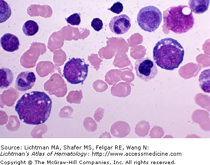
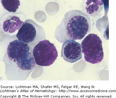
Alcohol poisoning. Marrow cells of a patient with chronic and acute alcoholism. Note dyserythropoiesis. Small erythroblasts with scant cytoplasm. Large orthochromatic erythroblast with lobulated nucleus. Three large erythroblasts (one proerythroblast and two basophilic erythroblasts) with vacuoles characteristic (but not specific for) of acute alcoholism. The vacuoles usually occur in early erythroid cells as seen here and very occasionally in myeloid cells. They may be observed after abstinence for about 3 to 14 days, suggesting the vacuoles persist that long despite cell division and maturation. The vacuoles often appear to overlie the nucleus as well as the cytoplasm, but in electron micrographic studies, they form from endocytotic vesicles and are localized only to the cytoplasm.
V.G.002 Arsenic Poisoning
V.G.002
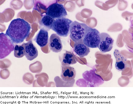
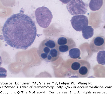
Arsenic poisoning. Marrow cells of patient with severe arsenic intoxication. Note severe dyserythropoiesis with megaloblastic features. Erythroblasts are mildly increased in size with bizarre nuclear abnormalities including multilobulated nuclei, open chromatin pattern simulating megaloblastic appearance. Note two very coarsely stippled erythroblasts.
V.G.003 Arsenic Intoxication
V.G.003
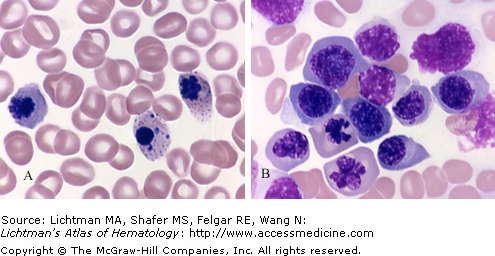
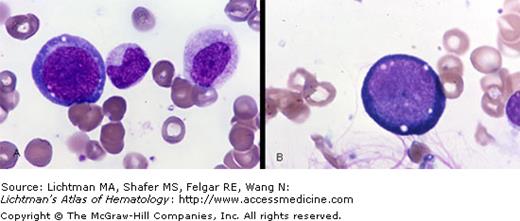
Arsenic Intoxication. Marrow Films. (A)Erythroblasts with nuclear fragmentation and nuclear remnants in cytoplasm. (B)Large erythroblasts with nuclear lobulation and with chromatin patterns similar to that seen in megaloblastic anemia. The dyserythropoiesis of arsenic poisoning mimics that seen in megaloblastic anemia and has morphologic features similar to erythropoiesis in myelodysplastic syndrome.
V.G.004 Congenital Dyserythropoietic Anemia, Type I
V.G.005 Congenital Dyserythropoietic Anemia, Type I
V.G.006 Congenital Dyserythropoietic Anemia, Type II
V.G.006
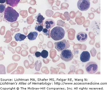
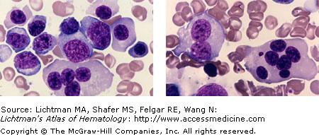
Congenital dyserythropoietic anemia, type II. Hereditary Erythroblastic Multinuclearity associated with a Positive Acidified Serum test (HEMPAS). Multinucleated erythroblasts. Note binucleate erythroblast and one cell with five nuclei. This variant of congenital dyserythropoietic anemia has erythroblasts with two to seven nuclei.
V.G.007 Congenital Dyserythropoietic Anemia, Type II
V.G.008 Congenital Dyserythropietic Anemia
V.G.008
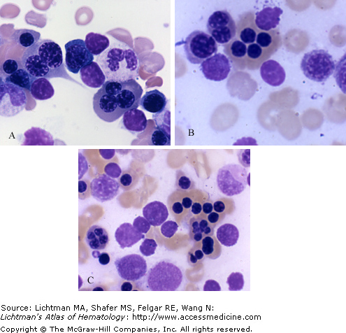
Congenital dyserythropoietic anemia. Marrow films. (A) Trinucleated late polychromatophilic erythroblast in lower right and binucleated earlier polychromatophilic erythroblast in upper left. (B) A cluster of orthochromatic erythroblasts, appearing to be a tetralobed giant erythroblast, but more likely one is bilobed(right) and two are monolobed(left). An adjacent smaller bilobed erythroblast. Upper left. (C) A tetranucleated (far left) and a cluster of several binucleated orthochromatic erythroblasts. A hexalobed small orthochromatic erythroblast in the center of the ring of bilobed and monolobed cells. Note in (A) and (B) megaloblastoid nuclear appearance in several of the erythroblasts.
V.G.009 Erythroid Aplasia
V.G.009
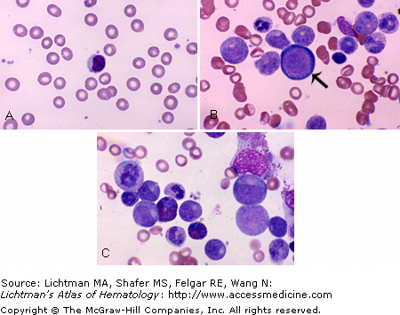
Erythroid aplasia. (A) Blood film. Extremely low frequency of red cells corresponds to the very low red cell count in this circumstance. In erythroid aplasia red cell size, shape, and hemoglobin content is usually normal. (B and C) Cellular marrows composed almost entirely of myeloid cells, macrophages, lymphocytes and plasma cells. A selective absence of erythroid precursors. This is reflected in a reticulocyte count of 0 to <0.5%. Note a rare very large basophilic erythroblast (arrow) in image B. Marrow red cell precursors were markedly reduced and no later forms were evident.
V.G.010 Erythroid Aplasia
V.G.010
Erythroid aplasia. (A, B) Marrow films. Complete lack of erythroblasts with normal number and sequence of granulocyte precursors. Plasma cell in (A). In cases of erythroid aplasia the reticulocyte count is usually less than 0.5 and may be zero, reflecting the virtual total absence of erythropoiesis. White cell counts and platelet counts are normal as is granulopoiesis, lymphopoiesis, and megakaryocytopoiesis in the marrow.
V.G.011 Human Immunodeficiency Virus Infection
V.G.011
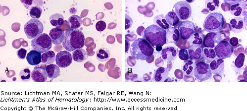
Human immunodeficiency virus infection. Marrow film. There are many effects of HIV infection on the hematopoietic system. Here we show the vacuolization of basophilic erythroblasts in (A) and (B). Dyshematopoiesis simulating myelodysplasia may occur. Blood cell changes may also occur from certain drugs administered to patients with HIV, such as azidothymidine. These changes in erythroblasts simulate megaloblastic anemia.
Stay updated, free articles. Join our Telegram channel

Full access? Get Clinical Tree


