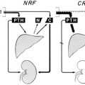
TECHNIQUES FOR LOCALIZATION OF ABNORMAL PARATHYROID TISSUE IN PATIENTS WITH PERSISTENT OR RECURRENT HYPERPARATHYROIDISM
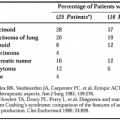
Several noninvasive techniques have been developed for localizing hyperfunctioning parathyroid tissue. Barium swallow, cine-esophagography, and thyroid scanning are rarely helpful, whereas other procedures such as computed tomographic (CT) scans, magnetic resonance imaging, 99mTc sestamibi scanning, and high-resolution real-time ultrasonography identify enlarged parathyroid glands in 40% to 60% of patients studied.18,19 and 20 CT scans, 99mTc sestamibi scans, and sestamibi/iodine subtraction single photon emission computed tomography (SPECT)20a are most useful for identifying lesions in the neck and mediastinum (Fig. 62-1). Sometimes, these techniques are coupled with needle aspiration of an identified mass followed by assay of the aspirate for PTH. Markedly increased PTH values, compared with an aspirate from muscle, document the presence of parathyroid tissue.
Related posts:

Stay updated, free articles. Join our Telegram channel

Full access? Get Clinical Tree


