9 Carcinoid and Neuroendocrine Tumors: Intra-arterial Therapies
9.1 Introduction
Neuroendocrine tumors (NETs) are rare, typically slow-growing tumors that are frequently found only when the disease becomes metastatic, with the liver being the most common site of involvement. 1 , 2 , 3 The incidence in the United States in 2004 was 1.25% of all malignancies and has been rising 3 to 10% per year since then. These tumors may be functional or nonfunctional, and disease-specific survival ranges from 70 to 95% at 5 years. 4 If functional, these tumors may secrete a variety of vasoactive or hormonal substances, such as serotonin, glucagon, gastrin, or insulin. The development of hepatic metastases from functional NETs is accompanied by release of these hormonal substances into the systemic circulation that lead to a constellation of symptoms, such as flushing, hypertension, diarrhea, and electrolyte disorders, known as the carcinoid syndrome. The development of these symptoms often brings the patient to medical attention. Symptoms can result in significant morbidity and can negatively impact the patient’s quality of life. Nonfunctional NETs metastatic to the liver may first be diagnosed incidentally on an imaging study obtained for something unrelated or when the metastases cause “bulk-related” symptomatology, such as pain, discomfort, and dyspnea, usually in the latest stages of the disease, due to significant hepatomegaly.
Similar to other liver metastases, such as colorectal adenocarcinoma, surgical resection of NET liver metastases has been associated with improved survival in carefully selected patients. Five-year survival rates of 60 to 80% have been reported. 5 , 6 , 7 , 8 , 9 Unfortunately, < 10% of patients are initially seen with resectable tumors. 10 , 11 , 12 , 13 For untreated patients, 5-year survival ranges from 17 to 54%. 14 Inoperable tumors can be managed by the administration of pharmacological antagonists of tumor metabolites, nonsurgical liver-directed therapies, or a combination of these treatments. 1 , 2 , 3 , 10 , 11 , 12 , 13 , 15 , 16 More than 80% of patients express somatostatin receptors and increased availability of long-acting somatostatin analogue (SSA) has allowed relatively good medical management of NETs, with SSA being a cytostatic agent as well as controlling hormonal symptoms. 17 A 5-year overall survival rate of 75% has been reported in a recent retrospective study that comprised 146 patients with midgut NETs who had received SSA treatment. 18 Unfortunately, over time patients frequently become refractory to this treatment. At this stage, transarterial therapies (hepatic arterial embolization [HAE], transarterial chemoembolization [TACE], and radioembolization [RAE]) are useful for treating both hormone-producing and nonfunctional tumors, thus reducing hormonal symptoms and pain. 19 , 20 , 21 , 22 , 23 , 24 , 25 , 26 , 27
NET metastases derive ~ 90% of their blood supply from the hepatic artery, whereas nutritional supply to normal hepatic parenchyma originates predominantly from the portal venous system. Therefore, transarterial therapies (TATs) allow for selective treatment of the tumor while preserving the blood supply to the organ. 28 , 29 In 1983, Moertel demonstrated that 80% of patients with carcinoid syndrome responded to hepatic artery ligation alone. 11 During HAE with particles alone, occlusion of the intratumoral blood supply results in ischemia and selective tumor necrosis. 28 , 29 , 30 This is achieved with the use of spherical embolic agents (particles) small enough to reach and occlude such vessels. TACE combines the effects of HAE with chemotherapeutic agents, based on the theoretical advantage of delivering concentrated drug to a tumor sensitized by embolization-induced ischemia. Chemotherapeutic dwell time in the tumor is thought to be prolonged, and the systemic effects are minimized. 31 Drug-eluting bead transarterial chemoembolization (DEB-TACE) has been more recently used as a form of chemoembolization in which embolic microspheres are loaded with anthracycline drugs, such as doxorubicin, acting as a direct carrier and resulting in a more favorable drug-release profile, 32 , 33 , 34 despite the fact that doxorubicin has never demonstrated activity against NETs. Unfortunately, higher complication rates have been reported with DEB-TACE for metastatic NETs 35 , 36 ; we do not use DEB-TACE for metastatic NETs and do not recommend it. Radioembolization using yttrium-90 (90Y) microspheres allows targeted radiotherapy to be delivered to the tumor. It is not intended as an embolic treatment but rather a means of delivering internal radiation to the tumor.
Until recently there has not been any effective systemic therapy for NETs metastatic to the liver, and external beam radiation is not useful for diffuse hepatic metastases; thus TAT has formed the mainstay of therapy. This is changing, and appropriate treatment for the patient is now based on knowledge of the site of the primary as well as the grade of the tumor. Tumors with the primary arising in the pancreas are known as pancreatic neuroendocrine tumors (pNETs), whereas those arising outside the pancreas—typically in the aerodigestive tract—are called carcinoids. They are further classified by degree of differentiation and grade. Poorly differentiated high-grade tumors are considered aggressive and treated with platinum-based therapy, so histology is important, and biopsy should be performed before initiating TAT if tissue has not already been obtained.
9.2 Indications
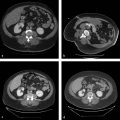
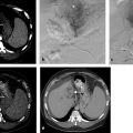
Due to the “hypervascular” nature of hepatic NET metastases and the dual vascular supply of the liver, TATs have been used for symptomatic relief with the hope of improving survival in unresectable patients. 19 , 20 , 21 , 22 , 23 , 24 , 25 , 26 , 27 , 30 , 37 , 38 , 39 , 40 , 41 , 42 , 43 , 44 , 45 , 46 , 47 , 48 , 49 , 50 Indications for embolization include rapid progression of liver disease in the face of stable or absent extrahepatic disease, progression of liver disease while on SSA ( Fig. 9.1 ), and symptoms related to hepatic tumor bulk or to hormonal excess that is refractory to SSA. 51 Control of hepatic metastases may allow prolonged survival when compared with patients who are not treated. 1 , 52 For the uncommon situation of a relatively small (< 5 cm) solitary lesion or not more than three NET deposits, TAT can be combined with radiofrequency ablation or other ablation techniques to maximize tumor necrosis and improve local disease control. 25
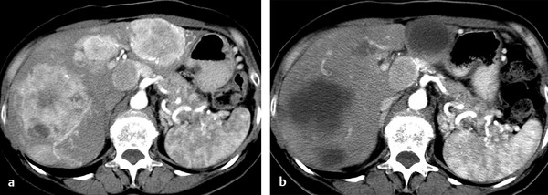
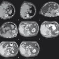
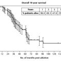
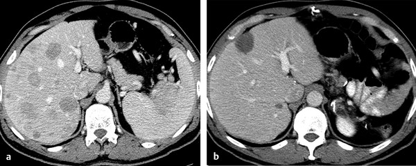
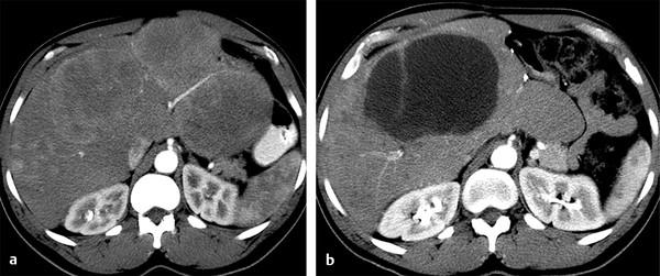
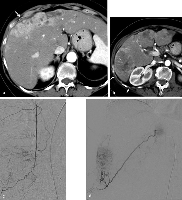
Rarely, embolization can be used in patients with large hepatic deposits to decrease the tumor load and render a previously inoperable patient a surgical candidate. With tumor reduction the previously inoperable or borderline patient may become a surgical candidate, 1 or, based on response to embolization, a decision may be made to resect the patient’s primary tumor ( Fig. 9.2 ). It is probably more common, at least at our institution, for surgery to precede embolization for patients with bulky metastases that can be resected, leaving behind a lower volume of metastases to be controlled with HAE ( Fig. 9.3 ).
9.3 Contraindications
There is a limited number of relative contraindications to hepatic embolization for the treatment of hepatic NET metastases. Involvement of > 75% of the liver parenchyma by tumor renders a patient vulnerable to embolization and at high risk for the development of liver failure. Any treatment should proceed with caution, and embolization should be performed in stages. 22 , 38 , 51 Instead of treating a hemi-liver, either the anterior or the posterior division of the right liver, or the lateral segment of the left liver could be treated initially, and after seeing how this treatment is tolerated by the patient, further embolization could be performed.
Renal insufficiency is a contraindication unless the patient is already undergoing hemodialysis and plans have been made for hemodialysis before and after the procedure. Mild renal insufficiency (creatinine > 1.5 but < 2 mg/dL) can be managed with good hydration; we also infuse sodium bicarbonate (3 mL/kg/h of a solution of sodium bicarbonate, 154 mEq/L administered over a 1-hour period before the procedure and 1 mL/kg/h infused for 6 hours during/after the procedure) in an attempt to prevent deterioration in renal function. The volume of contrast material should be kept to a minimum. Patients with allergies to contrast agents are premedicated with 50 mg of prednisone administered orally 13, 8, and 1 hour before the procedure.
Patients with elevated bilirubin, leukopenia, thrombocytopenia, or coagulopathy should be carefully evaluated because liver dysfunction is uncommon in this group of patients and abnormalities here should make one consider the possibility of underlying cirrhosis. Patients with metastatic NET are as likely to suffer from nonalcoholic fatty liver disease as the rest of the U.S. population, or they may have hepatitis B, hepatitis C, or drink alcohol excessively. If the laboratory abnormalities are due to replacement of the liver by tumor, the disease is probably too advanced to consider TAT. Additional considerations apply to RAE. Absolute contraindications include significant hepatopulmonary shunting and uncorrectable extrahepatic shunting to the gastroduodenal tract, which may result in extrahepatic deposition of microspheres and cause complications related to nontarget radiation. At our institution, patients are treated with RAE only if they fail HAE.
9.4 Patient Selection and Preprocedure Workup
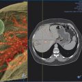
Prior to transarterial therapy, patients undergo hepatic and renal function tests, complete blood count, and coagulation studies. It is critical that, prior to any embolization procedure, optimal imaging of the liver be performed to depict the anatomy and the characteristics of the tumor to be embolized. Multiphase computed tomography (CT) or magnetic resonance imaging (MRI), ideally obtained within 1 month of treatment, is essential for documenting the extent of disease, demonstrating arterial anatomy, evaluating the portal venous system, and looking for nonhepatic blood supply to the tumor ( Fig. 9.4 ). We prefer multiphase CT because we find it easier to assess the nonhepatic blood supply to the tumor, as well as to identify landmarks that might be helpful during embolization, such as metallic clips from previous surgery. This study serves as the basis for a treatment plan. The extent and distribution of the tumor are laid out, arterial blood supply to the tumor is evident, and contribution from the nonhepatic vasculature, such as the phrenic or internal mammary arteries, can be seen. On the day of the procedure, the plan need only be executed.
When RAE is chosen for treatment, preprocedure “mapping” is performed 2 to 4 weeks before treatment to (1) delineate the hepatic vasculature, including any variant anatomy, and to assess the tumor vascular supply; (2) prophylactically embolize any extrahepatic vessels with hepatofugal flow to prevent inadvertent delivery of 90Y in extrahepatic sites; and (3) inject technetium-99m macroaggregated serum albumin (99mTc-MAA) to vascular territories to be treated in order to detect extrahepatic activity (e.g., extrahepatic shunting to the gastrointestinal tract) and to estimate the percentage of shunting to the lungs.
Patients are to receive nothing by mouth after midnight. They are admitted to the hospital the morning of the procedure and have an intravenous line started. Patients with impaired renal function receive sodium bicarbonate (see earlier mention). Hydration is begun in all patients with normal saline. Patients with allergies to contrast receive 50 mg of prednisone orally 1 hour before the procedure (they have already received two other doses of prednisone). All patients are given 4 mg of ondansetron (Zofran, GlaxoSmithKline) and 1 g of cefazolin (Ancef, GlaxoSmithKline) intravenously and 250 µg of octreotide subcutaneously. The SSA is administered to all NET patients, even those without hormonal symptoms. We have found these tumors capable of making low levels of vasoactive or hormonal substances that may be clinically silent but become clinically significant when cells undergo uniform and rapid cell death and release intracellular contents. 38 For this reason we also have 250 µg of octreotide available in the procedure room to be administered intravenously during the procedure if necessary. It is very important in this group of patients to screen for bilioenteric bypass because patients with pNET may have undergone a Whipple for treatment of a pancreatic primary. These patients are at extremely high risk for developing a liver abscess. In a review of almost 1,000 patients undergoing more than 2,000 embolization procedures, the risk of liver abscess in patients with a contaminated biliary tree was found to be up to 30 times higher than the baseline risk. 53 Patients who do not have an intact sphincter of Oddi receive cefotetan (B. Braun), 2 g intravenously. Patients without an intact sphincter of Oddi are treated prophylactically with an antibiotic expected to cover biliary flora, such as cefotetan (Cefotan 2 g), before embolization, rather than the customary Ancef. The cefotetan is given intravenously for as long as the patient is in the hospital. Oral metronidazole (Flagyl, Pfizer) and ciprofloxacin (Cipro, Bayer) are continued for 1 week postdischarge. Despite this, liver abscess may still occur.
9.5 Technique
Visceral angiography is always performed before embolization, studying the celiac axis and superior mesenteric artery (SMA). Imaging is carried out into the portal venous phase, not as much for assessing portal vein patency, which is rarely an issue in this patient population, as for determining direction of flow. Common or proper hepatic angiography is then performed using 4 or 5F selective catheters, such as Cobras (Terumo Medical Corporation) and reverse-curve catheters (Simmons or Sos catheters, AngioDynamics), through a vascular sheath placed in the common femoral artery. Coaxial microcatheters are used when necessary for subselective embolization. Conventional nonionic contrast material is typically used.
When treating patients with multifocal, bilobar disease, we usually treat the lobe with the most disease at the first sitting. Initially, arteriography is performed with the catheter positioned selectively in the right or left hepatic artery, followed by embolization. Coaxial catheters might be used to avoid nontarget embolization, or in other situations when necessary. For HAE, spherical embolic agents are used, typically tris-acryl gelatin microspheres (Embosphere Microspheres, Merit Medical Systems Inc.), beginning with the smallest microspheres (40–120 µm) unless patients are felt to be at risk for nontarget pulmonary embolization. 54 Those would be patients with large (≥ 10 cm) very vascular tumors, particularly high in the dome of the liver adjacent to the hemidiaphragm, or with systemic blood supply (phrenic). In such cases we begin embolization with 100 to 300 µm tris-acryl gelatin microspheres. When TACE is used, antineoplastic agents such as doxorubicin, cisplatin, gemcitabine, and/or mitomycin are used in combination with Lipiodol (Guerbet) and embolic agents. In DEB-TACE, embolic beads loaded with doxorubicin are infused to a total dose of no more than 150 mg, with 100 mg being more widely used today.
RAE is performed selectively based on preprocedure planning. Most patients have bilobar disease, and the side with the most lesions is treated first, with administration to the contralateral lobe performed 4 to 6 weeks later in a separate session. Patients who have undergone right or left hepatectomy receive treatment to the whole remnant liver in a single session. Targeted therapy is performed in a sublobar, selective manner whenever feasible for patients with disease limited to one segment to further minimize toxicity to uninvolved liver parenchyma. This is relatively uncommon in this patient population, who generally have more widespread disease.
For HAE the desired end point of the procedure is stasis in the target vessel defined as absence of antegrade flow, such that even slow administration of contrast material results in reflux, or retrograde flow. For TACE the end point is a bit more variable, with some authors advocating a “pruned tree” appearance and many stating that complete stasis should be avoided. When embolization is complete, a final angiogram is obtained to document occlusion of the target vessel and identify any supply to the target area from other vessels, which may then be selected and embolized. Finally, arteriography will show stasis in the embolized vessels, with preservation of antegrade blood flow to nontarget vessels, such as the gastroduodenal and the cystic artery.
RAE is not intended to be an “embolic” therapy and is not intentionally performed to stasis. Radiation-induced cell death requires normal oxygen tension 55 ; therefore transarterial delivery of the 90Y loaded spheres is concluded when the calculated dose has been administered or when stasis occurs. Stasis that limited the total administered activity has been documented to occur in up to 38% of the cases in one series. 56
Stay updated, free articles. Join our Telegram channel

Full access? Get Clinical Tree


