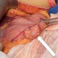
##
Adrenal tumors >4 cm in diameter represent 17% of adrenal tumors seen in the tertiary endocrine center. The proportion of malignant adrenal tumors and pheochromocytomas is high in large adrenal tumors, representing 31% and 22%, respectively. Hounsfield unit (HU) measurement of adrenal mass on unenhanced computed tomography (CT) is an important diagnostic step that may distinguish benign adrenal mass from malignancy or pheochromocytoma.
Case Report
The patient was a 45-year-old woman with history of nephrolithiasis who was incidentally discovered with a 4.9 cm left adrenal mass on abdominal CT scan performed for abdominal pain. Past medical history included migraines, fibromyalgia, and gastric bypass performed 10 years prior. At the time of presentation, her weight was normal and she did not have hypertension or diabetes mellitus type 2. She denied symptoms consistent with catecholamine excess or Cushing syndrome.
INVESTIGATIONS
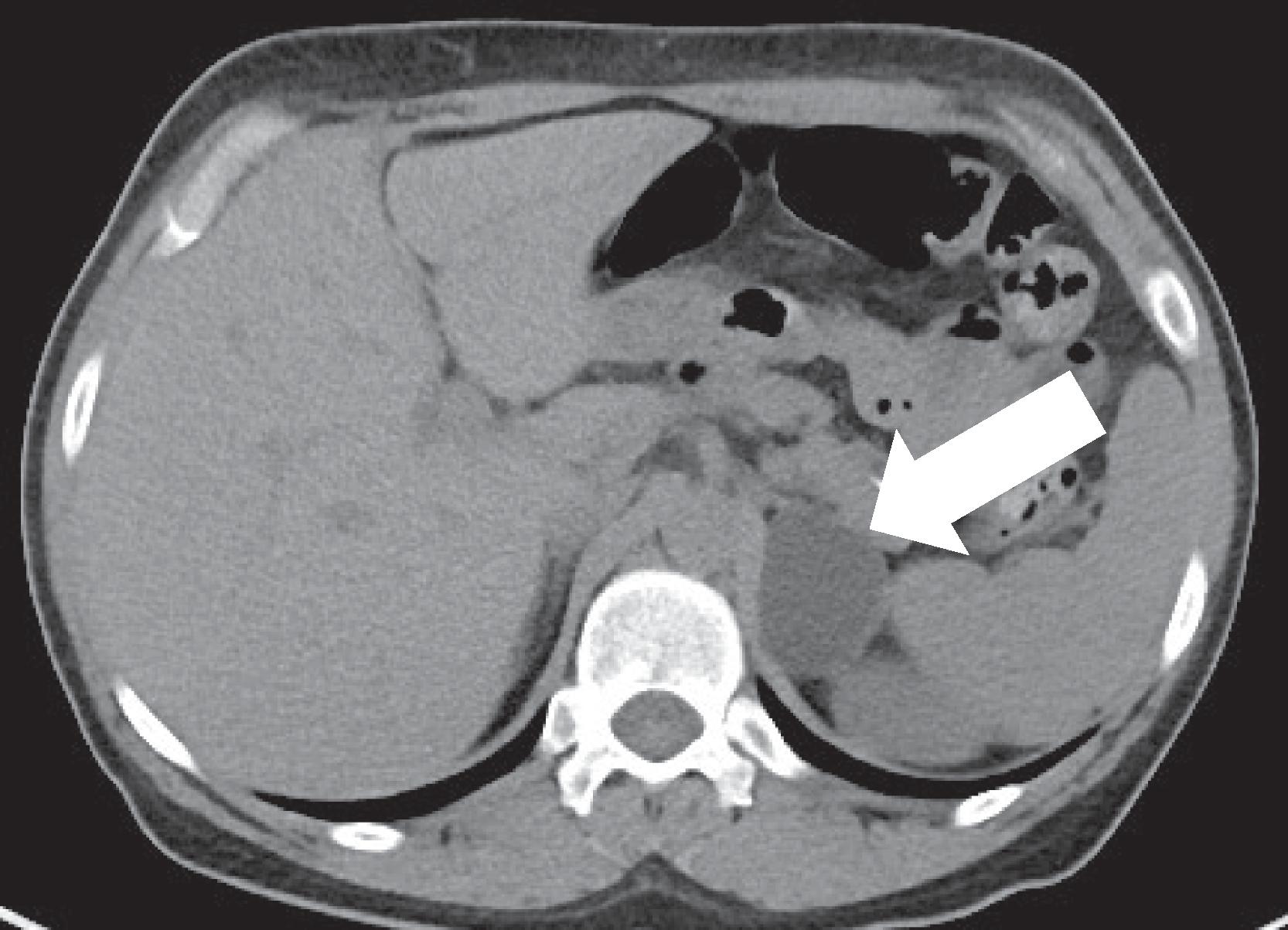
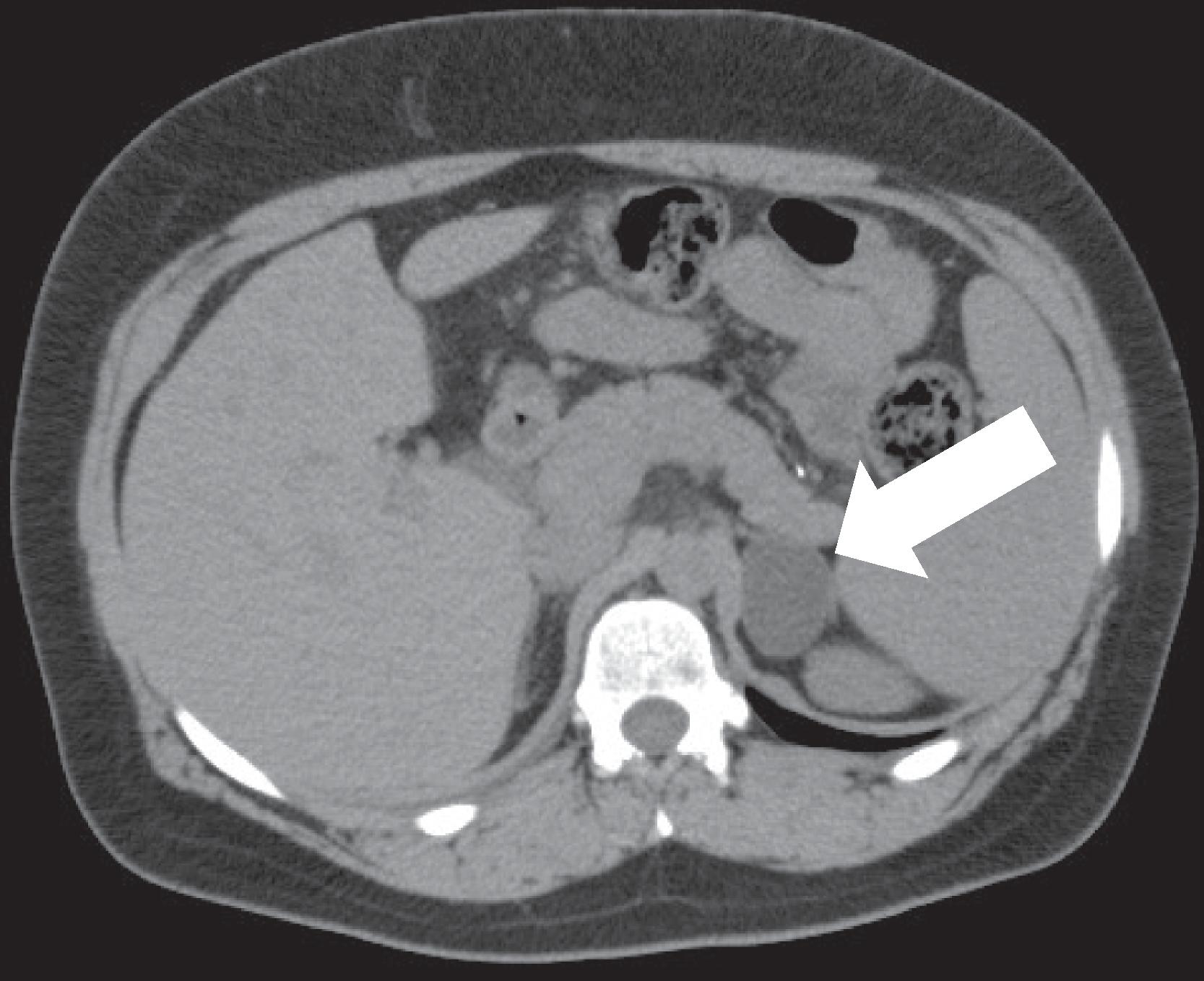
On unenhanced CT the left adrenal mass was homogeneous and lipid rich, measuring 4.9 cm in the largest diameter and demonstrating a CT attenuation of −13 HU ( Fig. 1.1 ). The right adrenal gland appeared normal. Prior imaging from 8 years ago was obtained for comparison and revealed a 3.7 cm adrenal mass (−14 HU) ( Fig. 1.2 ).

Stay updated, free articles. Join our Telegram channel

Full access? Get Clinical Tree


