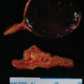
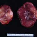
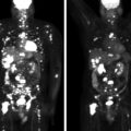
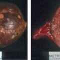
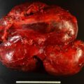
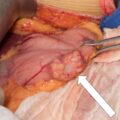
Composite adrenal tumors occur in approximately 4% of adrenal tumors. Imaging characteristics may be suggestive of different tumor etiologies or present as one heterogeneous adrenal mass. Assessment of malignancy potential and hormonal excess should be performed in all adrenal tumors, and treatment should be guided by imaging characteristics and biochemical workup.
Case Report
The patient was a 42-year-old woman who presented for evaluation of an incidentally discovered left adrenal mass on a computed tomography (CT) scan performed for abdominal pain after hernia repair surgery. Medical history was positive for depression. She denied hypertension, diabetes mellitus type 2, or history of malignancy. She did note new symptoms over the preceding 5 months, including night sweats (multiple times per night), weight gain of 20 pounds, fatigue, worsening depressive symptoms, and episodes characterized by anxiety and hyperventilation. On physical examination, her blood pressure was 127/85 mmHg, body mass index 31 kg/m 2 , and heart rate 82 beats per minute. She demonstrated mild supraclavicular pads and abdominal obesity but no striae, skin thinning, or proximal myopathy.
INVESTIGATIONS
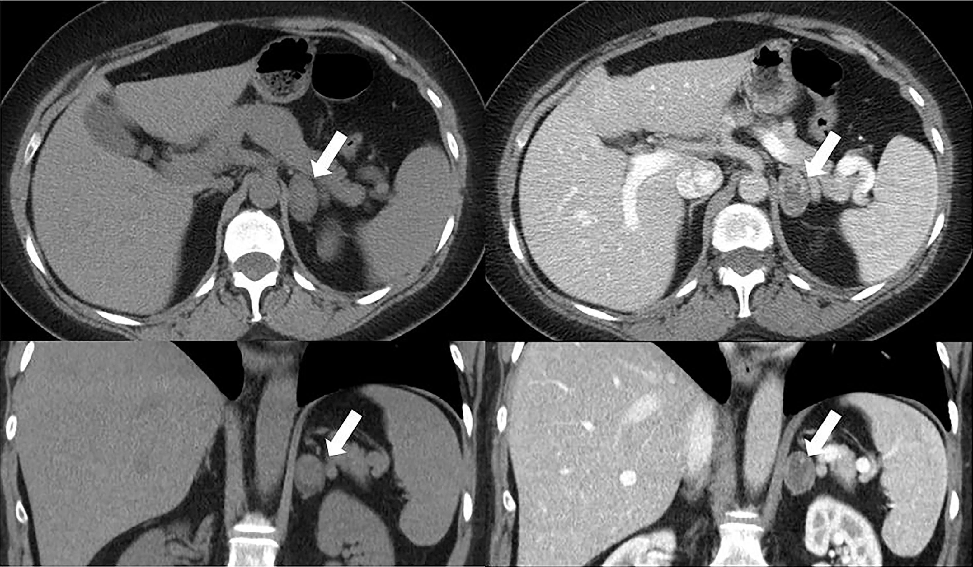
Review of an outside CT of the abdomen revealed a heterogeneous 3.4 × 2.3–cm left adrenal mass with two components, one measuring −8 Hounsfield units and the other measuring 22 Hounsfield units on unenhanced CT ( Fig. 75.1 ). No prior imaging was available for comparison. Hormonal workup included evaluation for catecholamine excess and autonomous cortisol secretion ( Table 75.1 ). Workup for catecholamine excess was negative, with normal 24-hour urine metanephrine and normetanephrine levels. However, the overnight 1-mg dexamethasone suppression test was abnormal. In addition, low serum concentrations of corticotropin (ACTH) and dehydroepiandrosterone sulfate further corroborated the diagnosis of autonomous cortisol secretion. Adrenalectomy was recommended.
| Biochemical Test | Result | Reference Range |
| Cortisol after overnight 1-mg DST, mcg/dL | 5.9 | <1.8 |
| Cortisol, mcg/dL | 10 | 7–21 |
| ACTH, pg/mL | <5 | 7.2–63 |
| DHEA-S, mcg/dL | 27 | 18–244 |
| Aldosterone, ng/dL | Not performed | <21 |
| Plasma renin activity, ng/mL per hour | Not performed | 2.9–10.8 |
| 24-Hour urine metanephrines, mcg/24 h | 71 | <400 |
| 24-Hour urine normetanephrines, mcg/24 h | 200 | <900 |
Stay updated, free articles. Join our Telegram channel

Full access? Get Clinical Tree


